Separate and combined multi-modality diagnostic imaging system

a multi-modality, diagnostic imaging technology, applied in the field of diagnostic imaging, can solve the problems of compromising the value of these image combinations and comparisons, difficulty in registering the various scans for comparison and/or combination, and difficulty in accessing the different imaging subsystems and maintaining thereof, so as to improve clinical productivity and patient care. the effect of reducing patient anxiety

Inactive Publication Date: 2007-10-11
SIEMENS MEDICAL SOLUTIONS USA INC
View PDF11 Cites 57 Cited by
  • Summary
  • Abstract
  • Description
  • Claims
  • Application Information

AI Technical Summary

Benefits of technology

[0019] One advantage of the present invention is the provision of a new and improved separate and combined multi-modality diagnostic imaging system that provides CT anatomical imaging with nuclear medicine functional imaging in one clinical study episode (location and time period) and in such a manner that clinical productivity and patient care are improved. Further, patient anxiety is reduced since they are moved during the scanning episodes.
[0020] Another advantage of the present invention is the provision of a new and improved separate and combined multi-modality diagnostic imaging system where a patient is imaged on the exact same table, and in a single imaging episode. This permits the subject to be in the same external and internal imaging states during both imaging studies.
[0021] Yet another advantage of the present invention is the provision of a new and improved separate and combined multi-modality diagnostic imaging system that allows the combination functional images (i.e. nuclear) with anatomical images (i.e. CT) via image fusion and side by side image registration since various scans are performed in a single episode.
[0022] Still further advantages of the present invention will become apparent to those of ordinary skill in the art upon reading and understanding the following detailed description of the preferred embodiments.

Problems solved by technology

However, the value of these image combinations and comparisons is compromised by having been obtained in separate study episodes.
This horizontal movement of the patient causes patient anxiety and may inadvertently change the placement of the patient and their internal organs from scan to scan causing difficulties in the registration of the various scans for comparison and / or combination.
Further, combination systems, for example, as that disclosed in U.S. Pat. No. 5,391,877, make access to the different imaging subsystems and maintenance thereof difficult.

Method used


Image

  • Separate and combined multi-modality diagnostic imaging system
  • Separate and combined multi-modality diagnostic imaging system
  • Separate and combined multi-modality diagnostic imaging system

Examples


Embodiment Construction

)

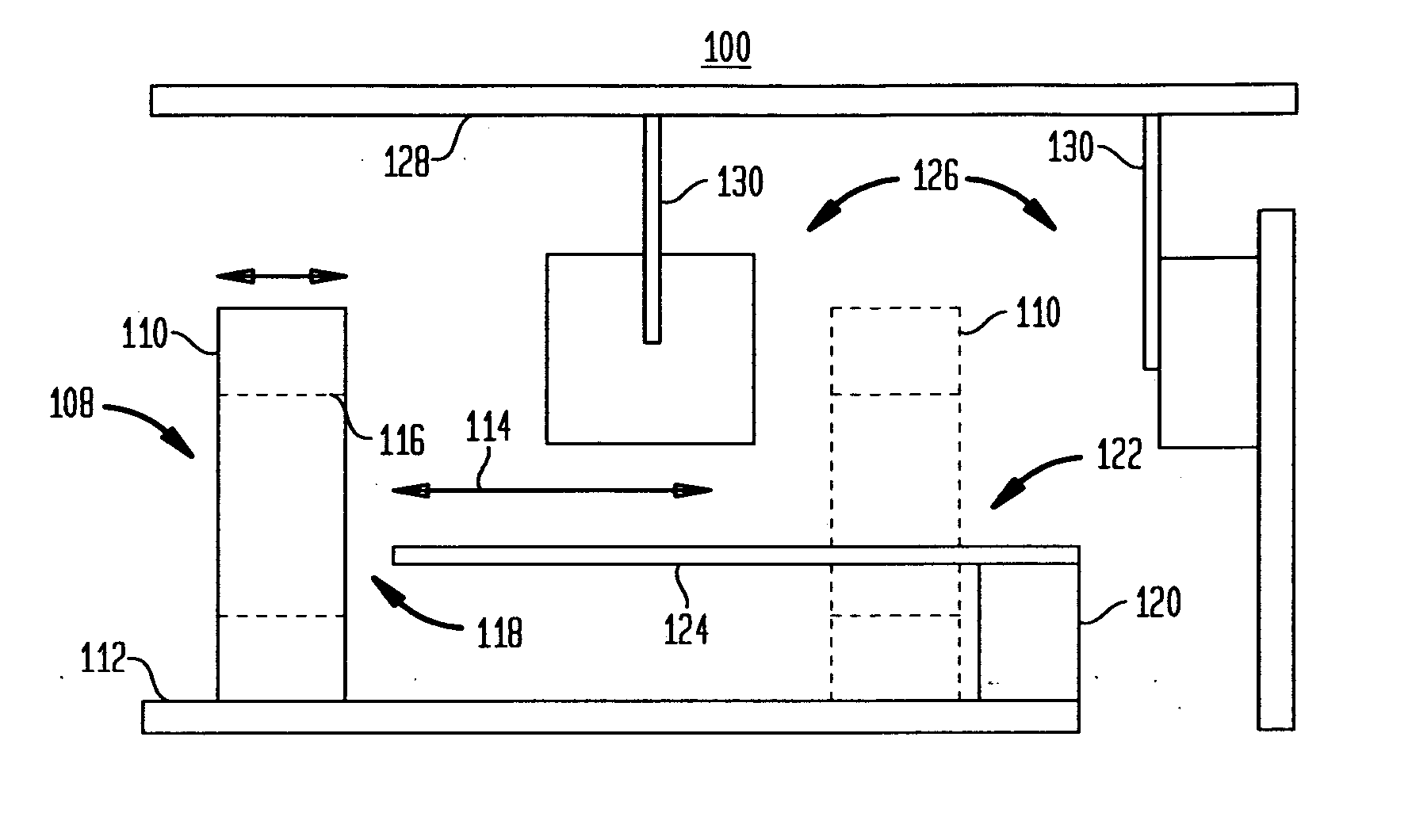
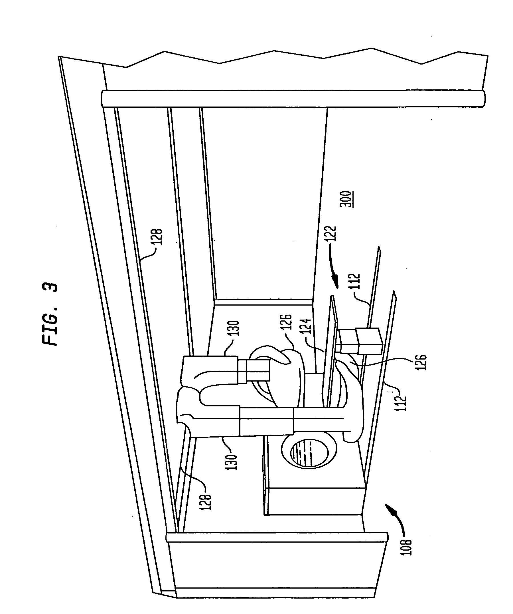
[0030] With reference to FIG. 1, a first embodiment of a separate and combined multi-modality medical imaging system 100 according to the present invention is shown. The imaging system has a computed tomographic (CT) scanner 108 with a non-rotating, sliding gantry 110 mounted on tracks 112 that extend parallel to the longitudinal axis 114. This allows the gantry 110 to be moved parallel to the longitudinal axis 114 and placed at the desired location during data collection. An x-ray tube (not shown) is rotatably mounted on a rotating gantry (not shown). The gantry 110 includes a cylinder or bore 116 that defines a patient examination region 118. An array of radiation detectors (not shown) is disposed concentrically around the patient receiving region 118. The x-ray detectors can mounted on the gantry 110 such that an arc segment of the detectors receives radiation from the x-ray tube (not shown) which has traversed the examination region 118. Alternatively, an arc segment of radiati...


PUM


Abstract

A multi-modality diagnostic imaging system includes a first imaging subsystem, such as a computed tomographic (CT) system, for performing a first imaging procedure on a subject. The first imaging system has a gantry that slides along rails. A second imaging subsystem, such as a nuclear medicine system (NUC), performs a second imaging procedure on a subject. The second imaging subsystem is separate from the first imaging system, but in the same room. The second imaging subsystem has a gantry or is gantryless. A patient couch supports a subject. The patient couch is supported on a couch support having vertical adjustment and horizontally fixed. In this manner, the patient is maintained in the same position for multiple types of scans using the same couch support without horizontal translation of the patient couch and patient.

Description

BACKGROUND OF THE INVENTION [0001] 1. Field of the Invention [0002] The present invention relates to the art of diagnostic imaging. It finds particular application in conjunction with a separate and combined multi-modality diagnostic imaging system, and more particularly, a separate and combined computed tomographic / nuclear medicine (CT / NUC) diagnostic imaging system. [0003] 2. Description of the Background Art [0004] Nuclear medicine imaging, such as single photon emission computed tomography (SPECT), is used to study radionuclide distribution in subjects. Typically, one or more radiopharmaceuticals are injected into a subject. The radiopharmaceuticals are commonly injected into the subject's blood stream for imaging the circulatory system or for imaging specific organs which absorb the injected radiopharmaceuticals. One or more gamma or scintillation camera heads are placed closely adjacent to a surface of the subject to monitor and record emitted radiation. [0005] In single photo...

Claims


Application Information

Patent Timeline
Patent Type & Authority Applications(United States)
IPC IPC(8): A61B5/05
CPCA61B6/032A61B6/037A61B6/04A61B6/4441A61B8/5261A61B6/5235A61B6/0407A61B6/5247A61B8/13A61B6/4464
Inventor VIJA, A. HANSHAHN, GUENTERQUAM, STEVE D.LUSSER, MARKUS
Owner SIEMENS MEDICAL SOLUTIONS USA INC
Why Eureka
Try Eureka