Quantitative optoacoustic tomography with enhanced contrast

a tomography and contrast technology, applied in the field of optoacoustic tomography, can solve the problems of reducing the resolution and clarity of the image, prior art deficient in effective methods applying algorithms, and prior art deficient in using the maximum angular amplitude probability reconstruction algorithm

Inactive Publication Date: 2007-10-11
SENO MEDICAL INSTR
View PDF3 Cites 30 Cited by
  • Summary
  • Abstract
  • Description
  • Claims
  • Application Information

AI Technical Summary

Benefits of technology

[0008] The present invention is directed to an optoacoustic imaging system for producing images of one or more objects in a body. The optoacoustic imaging system is configured to perform the steps of illuminating a body using pulses of electromagnetic radiation and generating distribution of absorbed thermal energy under illumination conditions of temporal pressure confinement within a volume of a resolution of interest, detecting temporarily resolved profiles of optoacoustic signals resulting therefr

Problems solved by technology

Image blurring and the presence of artifacts in form of radiating arcs decrease resolution and clarity of the image.
Specifically, the prior art is deficient in methods applying algorithms effective to significantly improve the (1) accuracy of quantitative information displayed on optoacoustic images and (2) contrast of op

Method used


Image

  • Quantitative optoacoustic tomography with enhanced contrast
  • Quantitative optoacoustic tomography with enhanced contrast
  • Quantitative optoacoustic tomography with enhanced contrast

Examples


example 1

Image Reconstruction Algorithm Based on MAAP

[0061] A detailed maximum angular amplitude probability algorithm described as it is implemented in the LOIS is provided in this example.

[0062] Data Inherited from the Current Image Reconstruction:

[0063] Ui—integrated signal from i-th transducer (i=1 . . . Ntr);

[0064] I—m×n matrix (old image) reconstructed using radial back-projection;

[0065] MAAP Image Reconstruction:

[0066] 1. Create an m×n matrix I′ of ZEROS.

[0067] 2. For each temporal sample j (j=1 . . . Ns) of a signal Ui find corresponding elements of the matrix I (physical positions (on a real probe) of those matrix I elements should have the same distance to i-th transducer equal to cs(j−1)Δt; where cs−speed of sound, and Δt is the temporal resolution of a transducer).

[0068] 3. Find the maximum of those elements and all elements around maximum, which satisfy the relationship: {Imax}:{I⁡(k)>I⁡(k+2),k>kmaxI⁡(k)>I⁡(k-2),k<kmax⁢k=1⁢ ⁢…⁢ ⁢ARC⁡[s,t]max.

Where kmax is t...

example 2

Comparison of MAAP and RBP Reconstructed Images (Spherical Models)

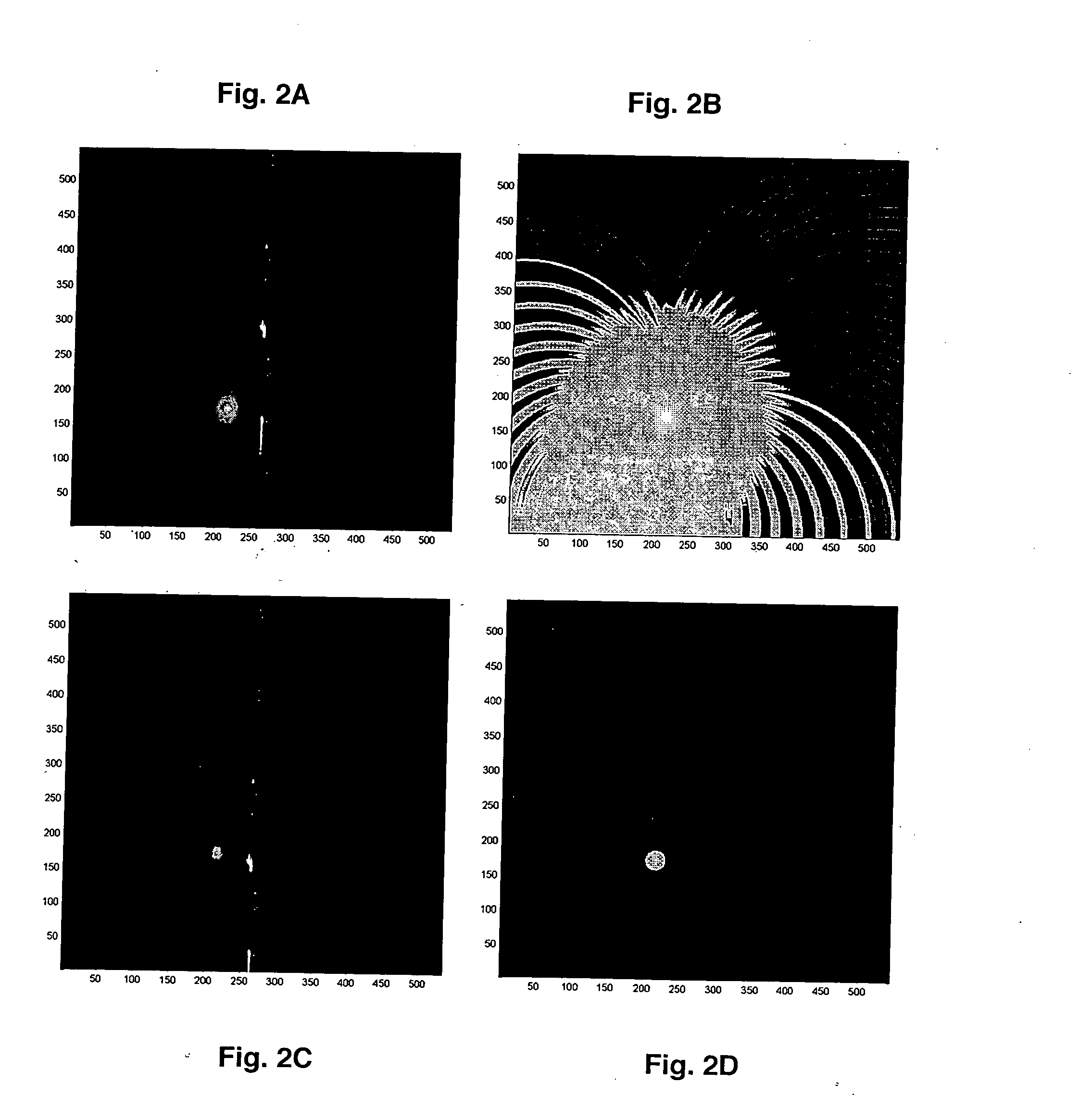
[0074] A radial back projection and maximum angular amplitude probability reconstructed optoacoustic images were compared using computer simulated signals from spherical absorptive objects. FIGS. 2A and 2B show a radial back projection image of a 7 mm in diameter absorptive object plotted using grayscale and triple-color threshold (hot metal palette shifted all the way to the cold colors) palettes. Image blurring as well as the artifacts in form of radiating arcs are clearly seen on FIGS. 2A-2B, respectively. Although linear grayscale maximum angular amplitude probability images (FIG. 2C) shrink and blur the object, triple-color threshold palette (FIG. 2D) reveals the true size of the object and recreates a high-contrast artifact-free image. Improvement of the optoacoustic image after maximum angular amplitude probability reconstruction is seen even more clearly when several objects of different size are simulated (F...

example 3

Examples of Quantitative OA Tomography Using Spherical Models

[0075] To demonstrate the accuracy of the offered approach for quantitative estimation of absorption coefficients, the algorithm is used that is described in Example 2 with signals obtained from spherical models, which had different diameters and the same unit absorption. FIGS. 4A-4D shows the histograms of the absorption coefficients found for single objects of 4 different diameters. The histograms show normally distributed K with a fairly small standard deviation. Considering the very large number of measurements, even for an object as small as 2 mm in diameter, the approach gives a very accurate estimation of the absorption coefficient where median values Kmed, are close to the original value K=1. For several objects of interest the least squares solution to equation (18) still gives a very good estimate of absorption coefficients. FIGS. 5A-5B are examples of this for 2 and 3 objects.


PUM


Abstract

Provided herein is an optoacoustic imaging system configured to produce images of one or more objects in a body using at least a maximum angular amplitude probability algorithm to reconstruct the optoacoustic images of the body. In addition the optoacoustic imaging system may be configured to produce 3D maps from the reconstructed optoacoustic images of the body. Also, provided is a method for diagnosing a pathophysiological condition characterized by abnormal optical properties of tissues in a body from maps so produced.

Description

CROSS-REFERENCE TO RELATED APPLICATIONS [0001] This application claims benefit of priority of provisional application U.S. Ser. No. 60 / 760,846, filed Jan. 20, 2006, now abandoned.BACKGROUND OF THE INVENTION [0002] 1. Field of the Invention [0003] The present invention relates to the field of optoacoustic tomography. More specifically, the present invention relates to a method of enhancing contrast during quantitative optoacoustic tomography and removing image artifacts using maximum angular amplitude probability (MAAP) reconstruction of an image. [0004] 2. Description of the Related Art [0005] Optoacoustic tomography is used in biomedical applications for in vivo and in vitro imaging of animal and human tissues and organs based on differences in tissue optical properties. Optoacoustic tomography has the potential to become valuable modality of functional molecular imaging. The essence of functional molecular imaging is to provide quantitative information (maps) of distributions and ...

Claims


Application Information

Patent Timeline
IPC IPC(8): A61B8/00A61B5/05
CPCA61B5/0073A61B5/0095A61B8/0825A61B5/7239A61B8/483G01N21/1702G01N2021/1787A61B8/406
Inventor ORAEVSKY, ALEXANDERERMILOV, SERGEY
Owner SENO MEDICAL INSTR
Why Eureka
Try Eureka