

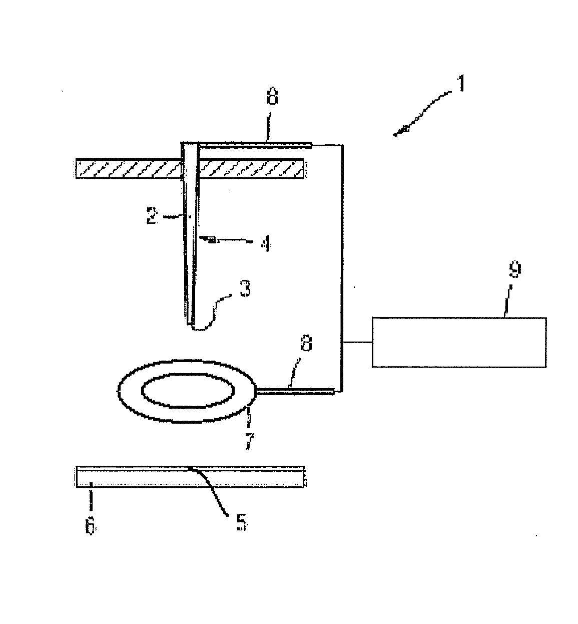
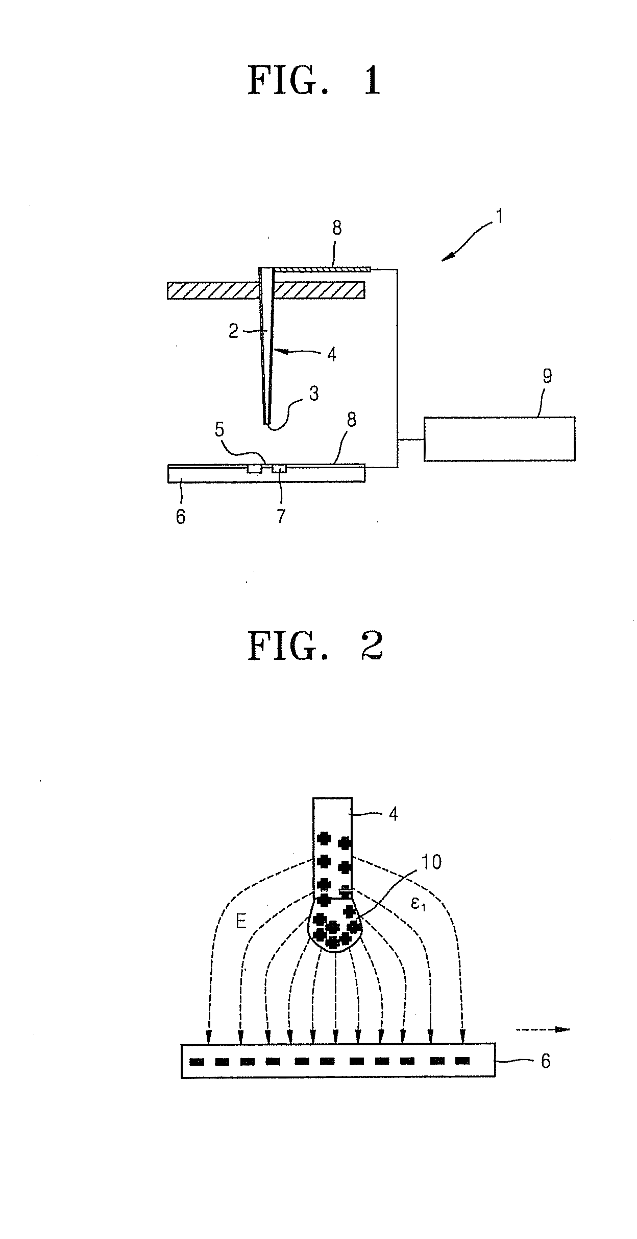
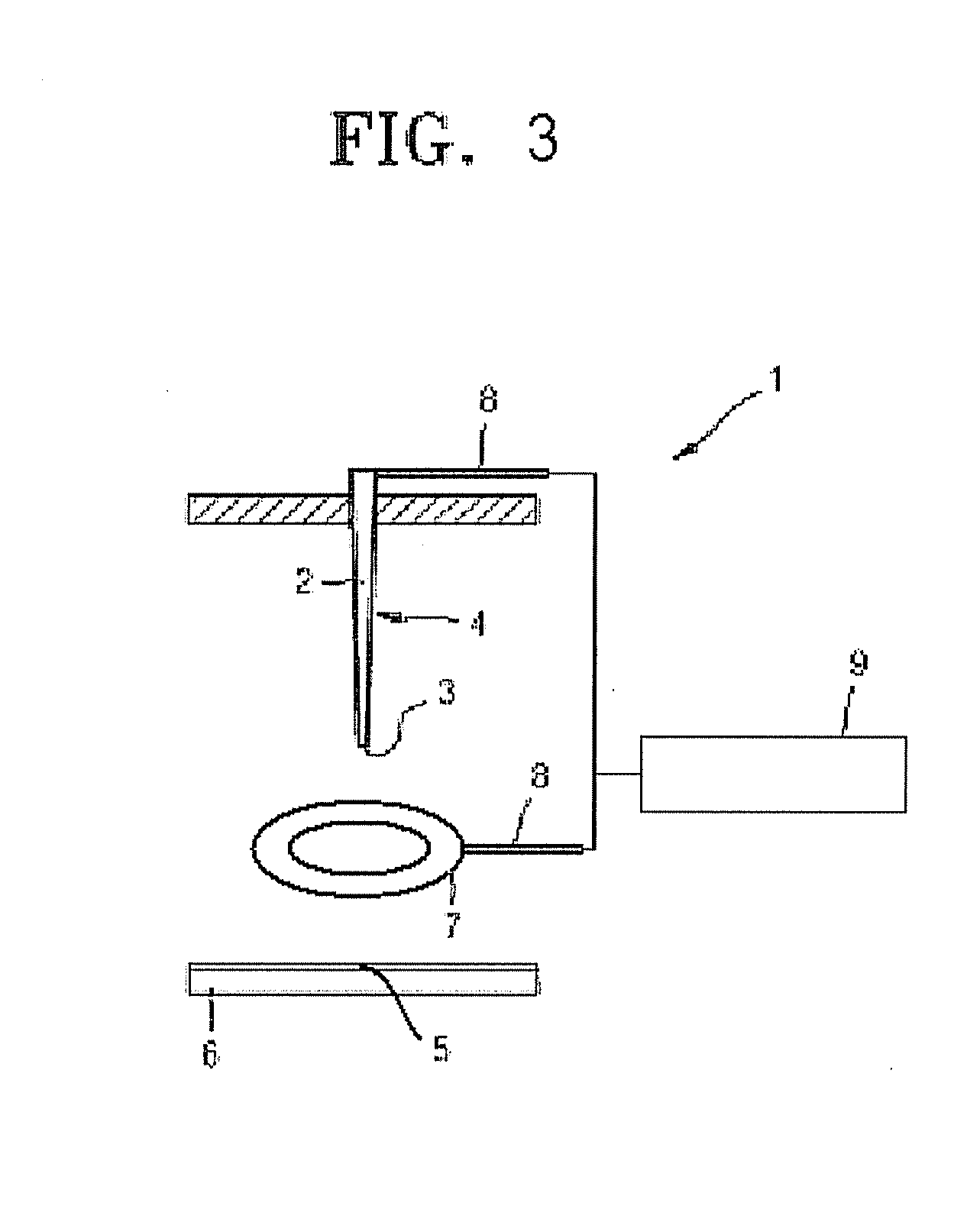
Apparatus and method for printing biomolecular droplet on substrate
a biomolecular and substrate technology, applied in the field of apparatus and method for printing biomolecular droplets on substrates, can solve the problems of inability to consistently eject biomolecular droplets onto the desired position of the substrate, the complexity of the sanger method, and the inability to analyze a large number of genes using the sanger method,
- Summary
- Abstract
- Description
- Claims
- Application Information
AI Technical Summary
Benefits of technology
Problems solved by technology
Method used
Image

Examples
experiment 1
[0100]A biomolecular droplet comprising 3% of an aqueous glycerol solution including 6.7×10⁸ beads / ml of Dynabeads and M-280 streptavidin (2.8 μm) was used to fill an accommodating area of an electric field forming electrode. A control solution of 3% of an aqueous glycerol solution was prepared to provide a comparison result. An electric field forming electrode having a diameter of 0.46 mm and a substrate made of glass were prepared. An electric field of 3 kV and 4 kHz was applied to the electric field forming electrode to measure a diameter of the biomolecular droplet ejected onto the substrate. Such a process was repeated 30 times to calculate an average diameter and a coefficient of variation (“CV”) of a biomolecular droplet ejected onto the substrate. The results are shown in FIG. 14. As shown in FIG. 14, when a micro magnetic bead is introduced, an average diameter of the printed biomolecular droplet is 59.7 μm and the coefficient of variation of the printed biomolecular drople...
example 2
[0101]Next, to confirm whether an exemplary embodiment of an apparatus for printing the biomolecular droplet according to the present invention as described above can print 6 or fewer cells when using cells as a biomolecule, the following experiment was performed using the exemplary apparatus for printing the biomolecular droplet used in the above experimental example.
[0102]A biomolecular droplet comprising 3% of an aqueous glycerol solution including 3×10⁶ cells / ml of HeLa cell (ATCC® Number: CCL-2) and 3.4×10⁸ beads / ml of Dynabeads M-280 streptavidin (2.8 μm) was used as a biomolecular droplet to fill an accommodating area of the electric field forming electrode. A control droplet was prepared in the same manner as above, except that Dynabeads M-280 streptavidin was not added to the biomolecular droplet. A substrate made of glass was also prepared. An AC voltage of 3 kV and having a frequency of 4 kHz was applied to the electric field forming electrode to print the biomolecular dr...
example 3
[0104]Next, a comparison was performed using a non-magnetic micro bead and the exemplary embodiment of a micro magnetic bead according to the present invention. The following experimental example was performed using the exemplary embodiment of an apparatus for printing the biomolecular droplet used in the previous examples described above.
[0105]A biomolecular droplet comprising 3% of an aqueous glycerol solution including 6.7×10⁸ beads / ml of Dynabeads M-280 streptavidin (diameter: 2.8 μm), 3% of an aqueous glycerol solution, 3% of an aqueous glycerol solution including 6.7×10⁸ beads / ml of a silicon bead (diameter 3.0 μm) was used to fill the accommodating area of the electric field forming electrode. When the silicon bead was included, experiments were performed when the magnet was included and excluded respectively.
[0106]A substrate made of glass was prepared, and an AC voltage of 3 kV and having a frequency of 4 kHz was applied to the electric field forming electrode to measure a ...
PUM
| Property | Measurement | Unit |
|---|---|---|
| diameter | aaaaa | aaaaa |
| diameter | aaaaa | aaaaa |
| AC voltage | aaaaa | aaaaa |
Abstract
Description
Claims
Application Information