Supercharge Your Innovation With Domain-Expert AI Agents!

Novgel methods for displaying (POLY)peptides/proteins on bacteriophage particles via disulfide bonds

a technology of disulfide bonds and peptides, which is applied in the direction of viruses/bacteriophages, peptide sources, fused cells, etc., can solve the problems of undeired artefacts, loss of most interesting binders which bind with high affinity to the target, and toxic expression products of gene iii to the host cell

Inactive Publication Date: 2007-12-20
MORFOZIS AG
View PDF8 Cites 0 Cited by
  • Summary
  • Abstract
  • Description
  • Claims
  • Application Information

AI Technical Summary

Benefits of technology

This approach simplifies the display and screening of peptides / proteins, reduces the complexity of vector construction, and allows for direct coupling to other moieties, enhancing the efficiency of peptide / protein production and characterization.

Problems solved by technology

When screening phage display libraries in biopanning the problem remains how best to recover phage which have bound to the desired target.
However, the most interesting binders which bind with high affinity to the target might be lost by that approach.
Especially in the case of using gene m as partner for peptides / proteins to be displayed, this leads to several problems.
First, the expression product of gene III is toxic to the host cell, which requires tight regulation of gene III fusion proteins.
And finally, recombination events between gene m fusion constructs and wild type copies of gene III lead to undesired artefacts.
Furthermore, since at least the C-terminal domain of the gene III protein comprising about 190 amino acids has to be used in order to achieve incorporation of the fusion protein into the phage coat, the size of the vectors comprising the nucleic acid sequences is rather larger, leading to a decrease in transformation efficiency.

Method used


Image

  • Novgel methods for displaying (POLY)peptides/proteins on bacteriophage particles via disulfide bonds
  • Novgel methods for displaying (POLY)peptides/proteins on bacteriophage particles via disulfide bonds
  • Novgel methods for displaying (POLY)peptides/proteins on bacteriophage particles via disulfide bonds

Examples


example 1

Display of (Poly)Peptides / Proteins on the Surface of Non-Engineered Filamentous Bacteriophage Particles via Formation of Disulfide Bonds

[0135] In the following example, all molecular biology experiments are performed according to standard protocols (Ausubel et al., 1999).

[0136] Construction of Vectors Expressing scFvs

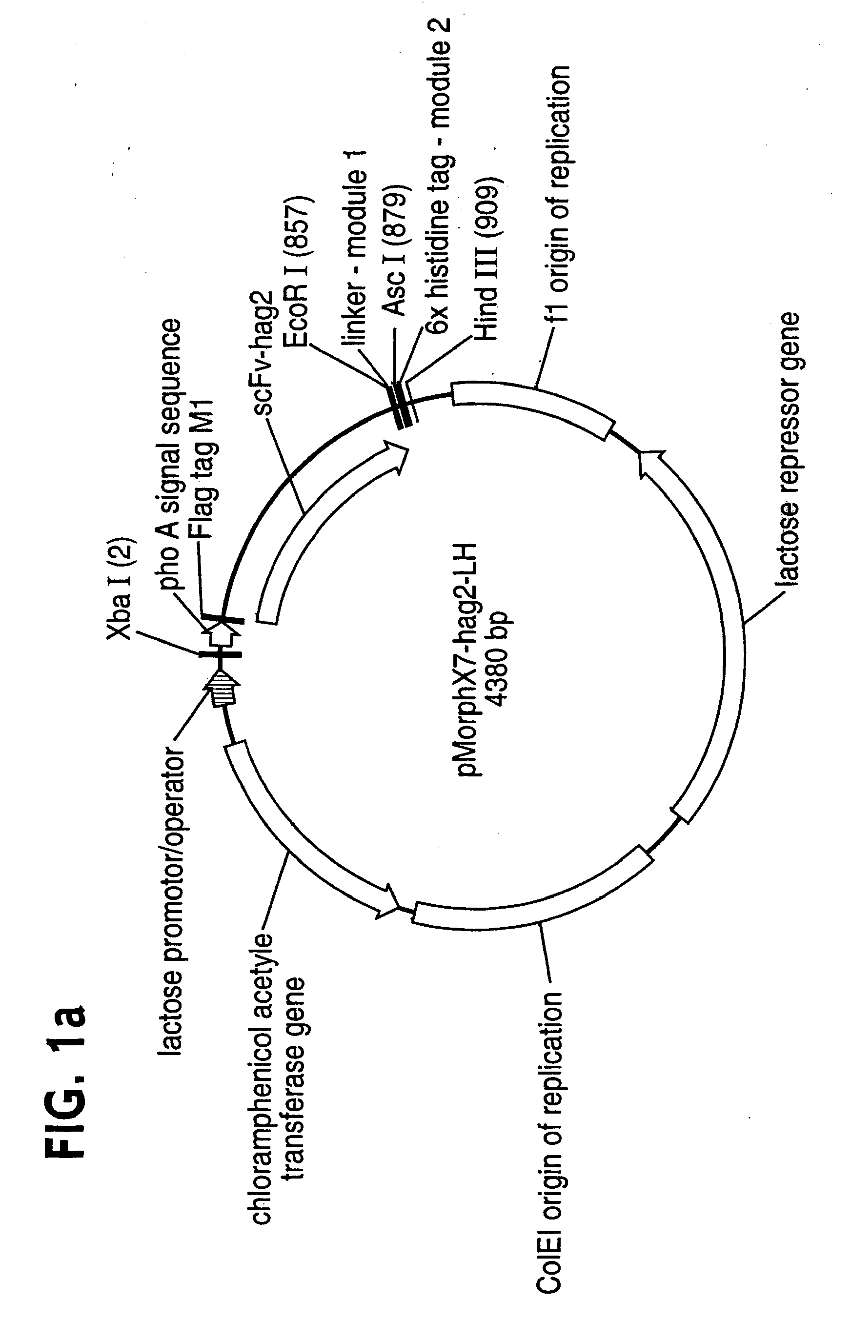
[0137] All vectors used are derivatives of the high copy phagemid pMorphX7-LH (FIG. 1a+b), a derivative of the pCAL vector series (WO 97 / 08320; Knappik et al., 2000). The expression cassette comprises the phoA signal sequence, a minimal binding site for the monoclonal antibody (mab) anti-FLAG M1 (Sigma #F-3040) (Knappik and Plückthun, 1994), a single chain fragment (scFv), a short linker (PGGSG) and a 6x histidine tag (6His; Hochuli et al., 1988) (FIG. 1a). pMorphX7-LCH and pMorphX7-LHC have been generated by inserting oligonucleotide cassettes coding for Cys-6His and 6His-Cys, respectively, between the unique AscI and HindIII sites of pMorphX7-LH (FIG. 1a, Table 1)....

example 2

Display of (Poly)Peptides / Proteins on the Surface of Engineered Filamentous Bacteriophage Particles via Formation of Disulfide Bonds

example 2.1

Display of scFvs

[0146] Example 1 described above shows that functional scFvs can be displayed on non-engineered phages via disulfide bonds. This system can be further improved, e.g. via engineering an exposed cysteine on a phage coat protein. One candidate phage coat protein is protein III (pIII) which is composed of three domains N1, N2 and pMCT. Possible sites for positioning an unpaired cysteine residue are the linker regions between the domains or the exposed N-terminus of the domain or the pIIICT in a truncated pIII version. A further example would be phage coat protein IX (pIX) where the cysteine could e.g. be linked to the N-terminus of the full length protein. In principle the cassettes for expression of such engineered proteins can be placed on the vector which is providing the scFv (one-vector system), or on a separate vector (two-vector system).

[0147] In the following we will describe experiments in which we engineered both a full length and a truncated pIII version as ...


PUM

PropertyMeasurementUnit
pHaaaaaaaaaa
pHaaaaaaaaaa
pHaaaaaaaaaa

Abstract

The present invention relates to methods for displaying (poly)peptides / proteins on the surface of bacteriophage particles by attaching the (poly)peptide / proteins via disulfide bonds.

Description

CROSS REFERENCE TO RELATED APPLICATIONS [0001] This application is a divisional of U.S. application Ser. No. 09 / 809,517, filed Mar. 15, 2001, the entire contents of which are expressly incorporated herein by reference. This application is based upon, and claims priority to, European patent applications EP 99 11 4072.4 and EP 00 10 3551.8, which are incorporated herein by reference in their entirety.BACKGROUND OF THE INVENTION [0002] The present invention relates to methods for displaying (poly)peptides / proteins on the surface of bacteriophage particles by attaching the (poly)peptide / proteins via disulfide bonds. A number of documents are cited throughout this specification. The disclosure content of these documents is herewith incorporated by reference in their entirety. [0003] Smith first demonstrated in 1985 that filamentous phage tolerate foreign protein fragments inserted in their gene III protein (pIII), and could show that the protein fragments are presented on the phage surfa...

Claims


Application Information

Patent Timeline
Patent Type & Authority Applications(United States)
IPC IPC(8): C12N15/11C12N1/00C12N1/20C12N5/06C12N5/04C12N15/00C12N15/09C07K14/01C07K16/46C12N1/15C12N1/19C12N1/21C12N5/10C12N7/00C12N7/01C12N15/10C12N15/33C12Q1/70C40B40/02
CPCC07K14/005C40B40/02C12N2795/10022C12N15/1037
Inventor LOHNING, CORINNA
Owner MORFOZIS AG
Features
  • R&D
  • Intellectual Property
  • Life Sciences
  • Materials
  • Tech Scout
  • Unparalleled Data Quality
  • Higher Quality Content
  • 60% Fewer Hallucinations
Learn More