
Apparatus and method for guiding insertion of a medical tool

a technology for medical tools and guiding devices, which is applied in the direction of ultrasonic/sonic/infrasonic diagnostics, applications, and therapy. it can solve the problems of only being used manually, difficult if not impossible to accurately place seeds in target locations, and not being particularly accurate in seed placement, etc., to achieve convenient alignment and more control over the procedure.

Inactive Publication Date: 2008-01-03
THE JOHN P ROBARTS RES INST
View PDF15 Cites 109 Cited by
  • Summary
  • Abstract
  • Description
  • Claims
  • Application Information

AI Technical Summary

Benefits of technology

[0017]The armature of the motor may be eccentric relative to the gear shaft and the set of enmeshed gears may comprise at least two eccentrically mounted gears. The apparatus may comprise a position encoder for determining the rotational position of the gear shaft and this rotational position may be fed back to the computer for use in determining any adjustments required in attaining the desired angle of insertion. The motor is designed such that the apparatus may be manually adjusted while the motor is in operation without backdriving or overdriving the motor. This advantageously permits the apparatus to be coarsely adjusted by a surgeon into the approximate position to facilitate proper alignment and / or allows a fine adjustment to be made by the surgeon using the manual adjustment knob, thereby giving the surgeon more control over the procedure. The manual adjustment feature also provides a safety override in the event of failure of the apparatus.

Problems solved by technology

This approach relies considerably upon the surgeon's skill in locating the prostate and is not particularly accurate in terms of seed placement.
However, limitations in the array-based needle guidance technique make it difficult if not impossible to accurately place the seeds in their target location.
This device can only be used manually.
The angle of the needle is fixed relative to the probe axis, and thus cannot be adjusted in three-dimensional space to reach an occluded target within the human body.
However, the devices disclosed in these documents are neither automatically operable, nor compatible with trans-rectal ultrasound probes.
The first robot described has inordinate flexibility of movement; the robot's operating envelope can extend into the patient, creating significant risk, and requiring a complex control system with complicated redundancy.
It also consists of only a single positioning means, which limits the degrees of freedom that are selectively available in choosing a desired insertion angle.
The second robot can position a needle, but has no mechanism to allow release of the needle once it is inserted.
Furthermore, the structural supports of the robot's stage mechanism inhibit the inclusion of any kind of needle release mechanism.
Since most brachytherapy procedures require the insertion of multiple needles, this makes the robot essentially un-useable in a clinic setting.
This robot cannot be manually controlled, and the design also prohibits sterilization, a necessity for surgery or invasive therapy.
Prior art needle guides have suffered from limited positional accuracy, do not allow needle release for multiple needle placement and / or are either only manually adjustable or only robotically adjustable.

Method used


Image

  • Apparatus and method for guiding insertion of a medical tool
  • Apparatus and method for guiding insertion of a medical tool
  • Apparatus and method for guiding insertion of a medical tool

Examples


first embodiment

[0071]Referring to FIG. 10, a tool release mechanism includes a stand-off 100 fixedly mounted to the first positioning means 20 proximal the hook joint 21 and extending towards the front of the apparatus. At the anterior end of the stand-off 100 is fixedly mounted a release block 101 having a pair of curved fingers 102 provided at one end thereof. The curved fingers 102 partially enclose an aperture 103 that is oversized compared with the tool (in this case, the needle 2) that is to be inserted therethrough. The non-enclosed portion of the aperture 103 provides a slotted chordal opening that is large enough to permit the needle 2 to be removed from the aperture in a direction perpendicular to the axis of insertion 4. A pair of side brackets 110 is secured to either side of the release block 101. Each side bracket 110 includes a crescent shaped slot 111 with an open end that is roughly aligned with the slotted chordal opening. When inserted within the aperture 103, the needle 2 seats...

second embodiment

[0073]Referring to FIG. 11, in a tool release mechanism a mounting block 220 is provided that permits the telescoping guide 6 to move therewithin along the guide axis 5. The mounting block 220 includes a T-shaped slot for receiving a complementary T-shaped mounting bar 222. The T-shaped mounting bar 222 is able to slide within the slot parallel to the guide axis 5. At the anterior end of the mounting bar 222 is fixedly mounted a release block 201 having a pair of curved fingers 202 provided at one end thereof. The curved fingers 202 partially enclose an aperture 203 that is oversized compared with the tool (in this case, the needle 2) that is to be inserted therethrough. The non-enclosed portion of the aperture 203 provides a slotted chordal opening that is large enough to permit the needle 2 to be removed from the aperture in a direction perpendicular to the axis of insertion 4. A pair of side brackets 210 is secured to either side of the release block 201. Each side bracket 210 in...


PUM


Abstract

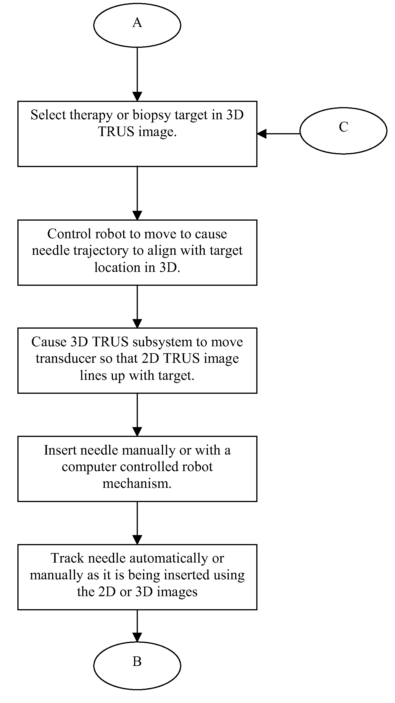
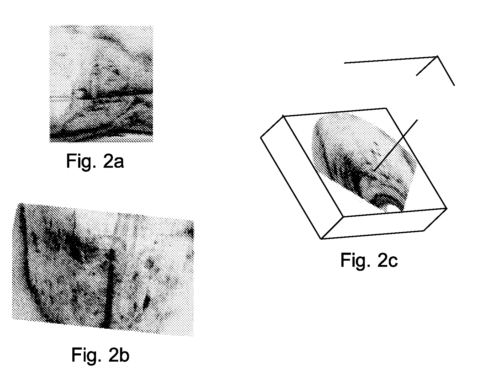
An apparatus and method for the insertion of a medical tool, for example a needle, within the human body. The apparatus and method are particularly useful in prostate brachytherapy and in prostate biopsy. The apparatus comprises a telescoping guide universally coupled to a first and second positioning means that are used to automatically and / or manually position the guide at a desired needle insertion trajectory. Automatic positioning of the guide is accomplished with reference to three-dimensional transrectal ultrasound images that can also be used to show needle insertion in real-time. The apparatus may be manually positioned in the approximate insertion trajectory and then a computer interconnected with the apparatus may be used to achieve the final trajectory based upon the ultrasound images. The apparatus is particularly useful in cases where multiple needles are to be inserted into a small target area and in cases where pubic arch interference prevents direct access to the target area.

Description

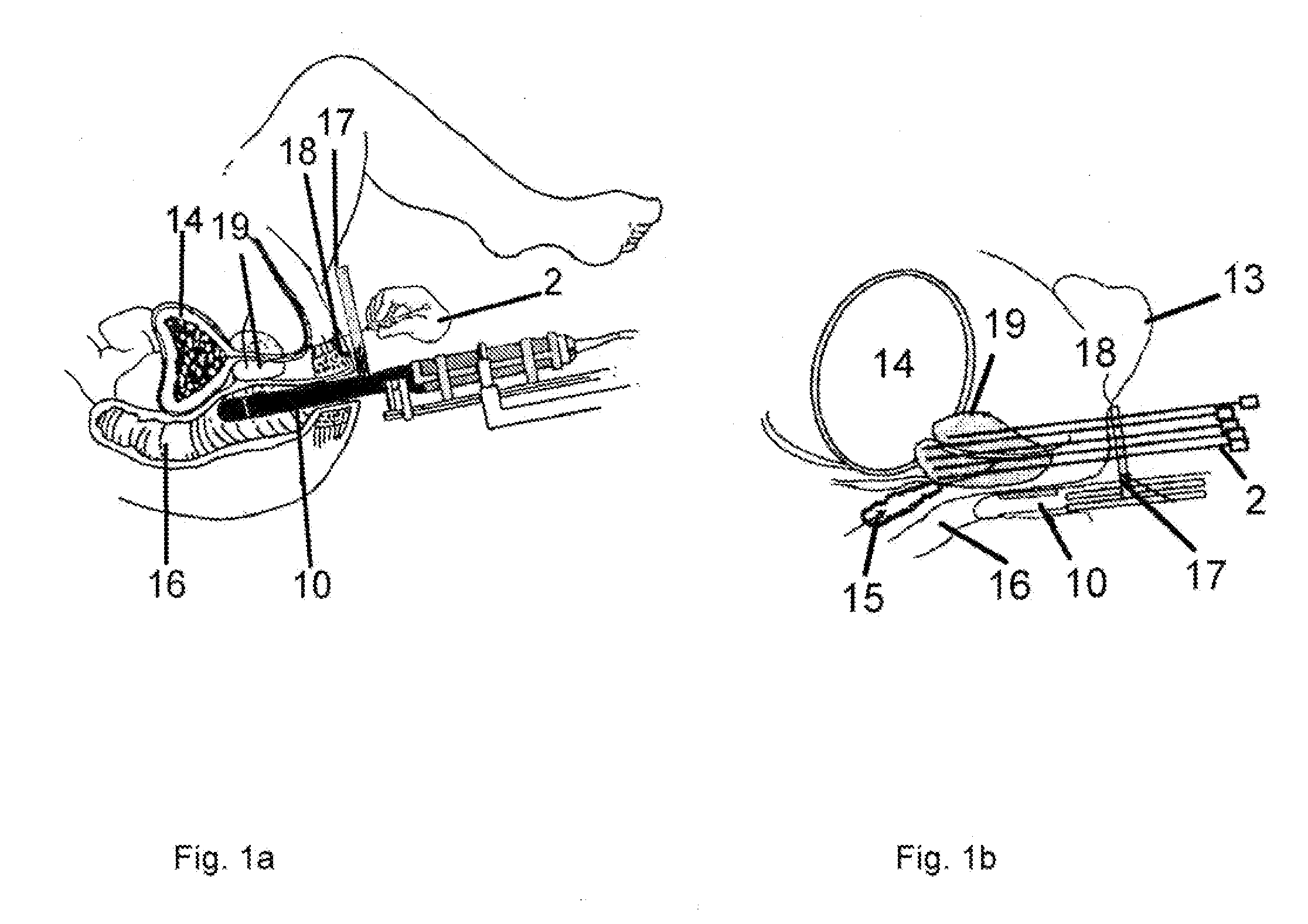
FIELD OF THE INVENTION[0001]The invention relates to the guiding of medical tools for insertion within the body of an animal. More particularly, the invention relates to an apparatus and method for positioning a guide for use in manually inserting a medical tool, such as a needle, within a human body at a particular angle in three-dimensional space, usually determined with reference to a medical image of an interior of the body. The guide may be automatically positioned by a computer and / or manually positioned. The apparatus and method are particularly useful as an aid in needle placement during the treatment of prostate cancer by brachytherapy or during prostate biopsy procedures.BACKGROUND[0002]There are a number of medical procedures where it is desirable to insert a tool within a human body to a precise position based upon medical imagery. An example of such a procedure is prostate brachytherapy, where radioactive seeds are implanted within a cancerous prostate using a long need...

Claims


Application Information

Patent Timeline
IPC IPC(8): A61M36/00
CPCA61B19/201A61B2019/5276A61B2018/00547A61B2017/00274A61B90/11A61B2090/378A61B17/3403A61B2017/3413A61B2017/3405A61B8/0833A61B8/0841A61B8/085A61N5/1007A61N5/1001A61N5/04A61N5/045A61N2005/0608A61N2005/0612A61N2005/1012A61N2005/1024
Inventor BAX, JEFFREYFENSTER, AARONMONTREUIL, JACQUESGARDI, LORISMITH, DAVID MARTIN
Owner THE JOHN P ROBARTS RES INST
Learn More