Method for Evaluating Regional Ventricular Function and Incoordinate Ventricular Contraction

a regional ventricular function and ventricular contraction technology, applied in the field of medical diagnostic methods, can solve the problems of radiation exposure, radiation exposure, radiation exposure, etc., and achieve the effect of reducing the impact of the procedure on the patien

Inactive Publication Date: 2008-01-10
ST JUDE MEDICAL ATRIAL FIBRILLATION DIV
View PDF99 Cites 39 Cited by
  • Summary
  • Abstract
  • Description
  • Claims
  • Application Information

AI Technical Summary

Benefits of technology

[0010]The various embodiments provide methods for evaluating the various chambers of the heart muscle through a percutaneous method to minimize the impact of the procedure on the patient while permitting a comprehensive review and evaluation of heart function.

Problems solved by technology

The pitfall of the echocardiogram is that it is prone to noise and patient's lungs, ribs, or body tissue may prevent the sound waves and echoes from providing a clear picture of heart function sufficient to assess atrial function.
Downsides of the cardiac blood pool scan test include the potential allergic reactions to the radioactive tracer and the risks associated with exposure to radiation.
Currently no percutaneous method exists for evaluating the function and ejection characteristics of the heart.
In addition, no percutaneous method currently exists in which regional ejection fraction during the systolic period can be monitored in a phased analysis of the regional wall motion to give a temporal sequence of regional ventricular ejection.

Method used


Image

  • Method for Evaluating Regional Ventricular Function and Incoordinate Ventricular Contraction
  • Method for Evaluating Regional Ventricular Function and Incoordinate Ventricular Contraction
  • Method for Evaluating Regional Ventricular Function and Incoordinate Ventricular Contraction

Examples


Embodiment Construction

[0024]The various embodiments will be described in detail with reference to the accompanying drawings. Wherever possible, the same reference numbers will be used throughout the drawings to refer to the same or like parts.

[0025]As used herein, the terms “about” or “approximately” for any numerical values or ranges indicates a suitable dimensional tolerance that allows the part or collection of components to function for its intended purpose as described herein. Also, as used herein, the terms “patient”, “host” and “subject” refer to any human or animal subject and are not intended to limit the systems or methods to human use, although use of the subject invention in a human patient represents a preferred embodiment.

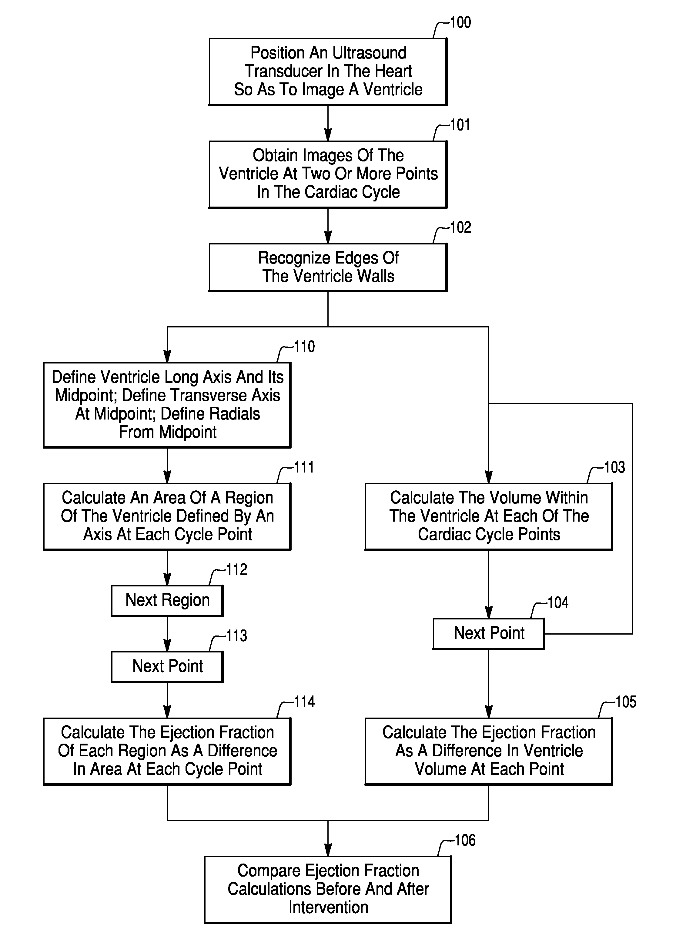
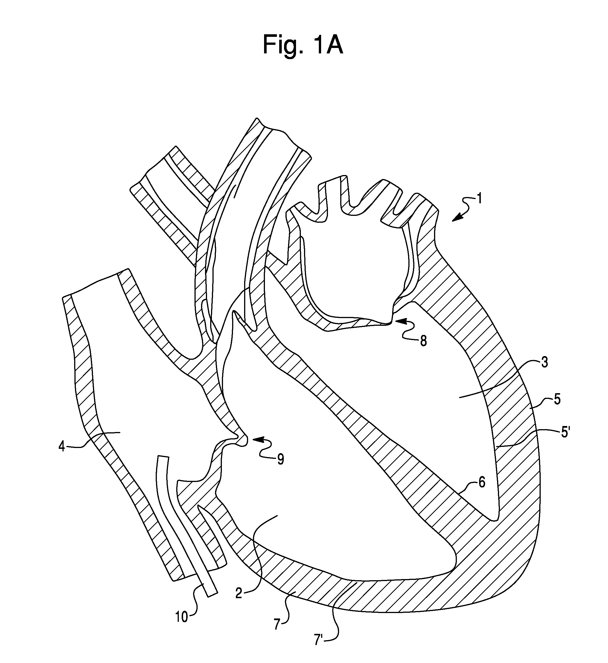
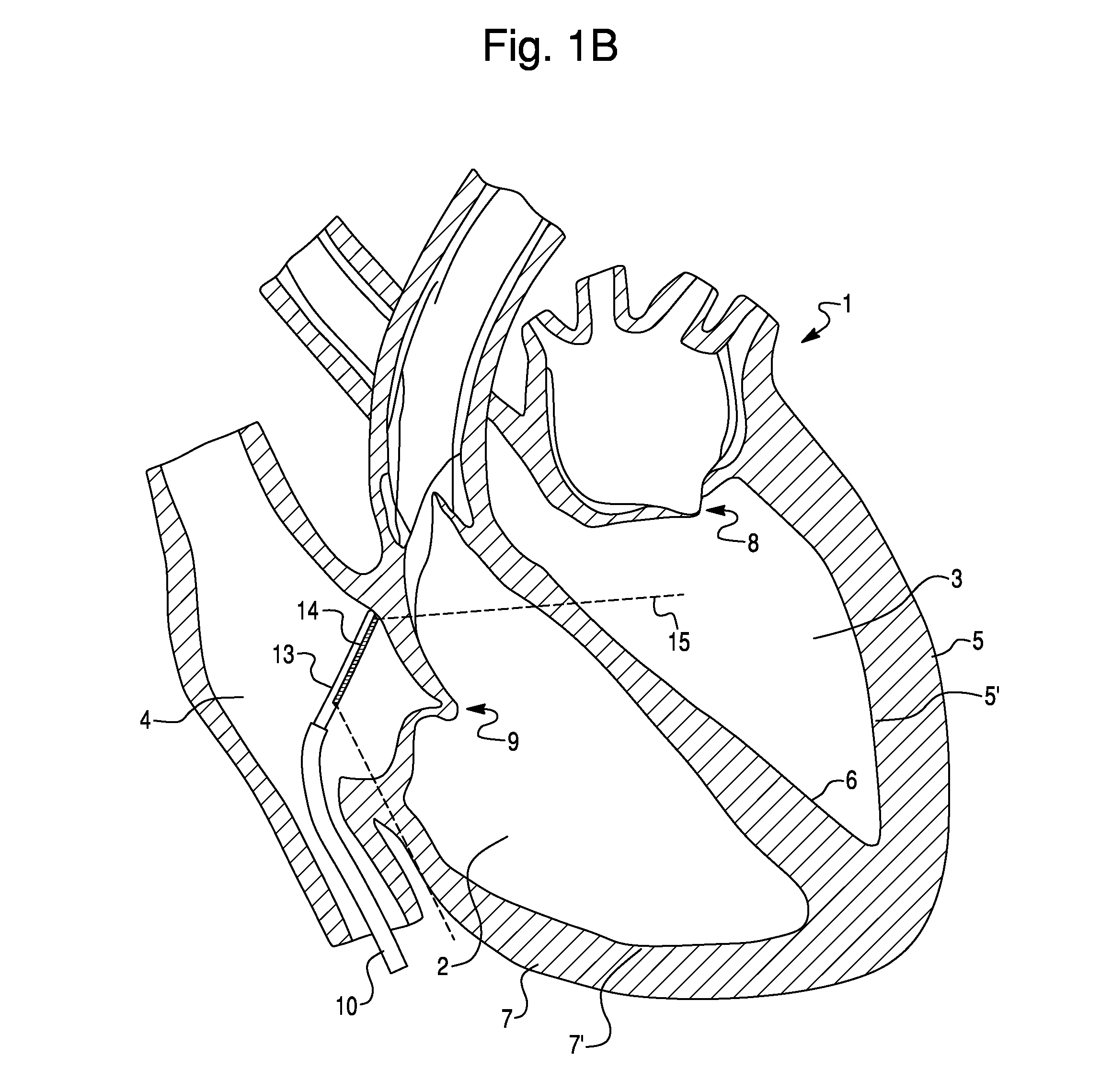
[0026]The methods of the various embodiments enable physicians to obtain more complete and comprehensive visualizations of the function and status of the ventricles of the heart. In the various embodiments, ultrasonic imaging of the chambers of the heart via intracardiac e...


PUM


Abstract

A method for assessing cardiac function using an ultrasound imaging catheter system includes positioning an ultrasound catheter so the ultrasound transducer can image a ventricle, obtaining images of the ventricle at two or more times within the cardiac cycle, recognizing an edge of the endocardium, measuring dimensions of the ventricle, calculating a volume or area of the ventricle at the two or more points in the cardiac cycle, and calculating the ejection fraction based upon the difference in volume or area at the two or more times in the cardiac cycle. The method can be used to determine a location for an intervention, such as placement of a pacemaker pacing lead, and may be performed before and after an intervention to assess the impact of the treatment on cardiac function.

Description

FIELD OF THE INVENTION[0001]This invention relates to medical diagnostic methods, and more particularly to methods for evaluating ventricular function and incoordinated ventricular contraction using intracardiac echocardiography.BACKGROUND OF THE INVENTION[0002]Evaluating the ejection function and ejection characteristics of the heart is important to the proper diagnosis of cardiovascular health and selecting optimum therapies for treating cardiac disorders. A common measure of the ejection function is ejection fraction, which is the fraction of blood taken into the ventricle that is ejected in each contraction cycle. The higher the ejection fraction, the more of the blood volume is ejected from the ventricle in each beat. Current methods for evaluating heart function and ejection characteristics involve using an echocardiogram or cardiac blood pool scan test. During an echocardiogram a transducer is placed on the patient's ribs near the breast bone and directed toward the heart. Th...

Claims


Application Information

Patent Timeline
Patent Type & Authority Applications(United States)
IPC IPC(8): A61B8/00
CPCA61B5/029A61B8/12A61B8/0883A61B5/411
Inventor SAKSENA, SANJEEV
Owner ST JUDE MEDICAL ATRIAL FIBRILLATION DIV
Why Eureka
Try Eureka