
Echo particle image velocity (EPIV) and echo particle tracking velocimetry (EPTV) system and method

a technology of echo particle and image velocity, applied in the field of echo particle image velocity and echo particle tracking velocity (eptv) system and method, can solve the problems of difficult flow measurement near the blood-wall interface, cumbersome use, and relatively poor temporal resolution

Inactive Publication Date: 2008-01-17
UNIV OF COLORADO THE REGENTS OF
View PDF26 Cites 96 Cited by
  • Summary
  • Abstract
  • Description
  • Claims
  • Application Information

AI Technical Summary

Problems solved by technology

Magnetic resonance imaging (MRI) velocimetry provides multiple components of velocity with good spatial resolution; however, the method is cumbersome to use since it requires breath-holds of the patient, collection of data over multiple cycles for ensemble averaging, and possesses relatively poor temporal resolution.
Although this method provides greater temporal resolution, it is dependent on the angle between the ultrasound beam and the local velocity vector, only provides velocity along the ultrasound beam (1-D velocity), and has difficulty in measuring flow near the blood-wall interface.
However, current PIV methods are limited to the measurement of flows in transparent media due to the requirement for optical transparency.
However, this method is limited by the requirement for extremely fast acquisition systems, heterogeneous signals caused by polydispersed particles, and high noise induced by high concentration of scattering particles.
The inherent necessity of very high scatterer particle concentrations in particular, seriously limits the application of USV in hemodynamics measurement in living creatures.
These early-stage studies demonstrated that, for both the velocity range and spatial resolution (thus limiting the dynamic range and maximum value of measurable velocities, the ability to capture transient flow phenomena, and the density of the resulting PIV vector field), early-stage Echo PIV was insufficient for full range vascular blood flow imaging.
Several issues, including poor signal-to-noise ratio when using blood cells for backscatter or requirement of excessive contrast particle seeding density when using contrast agents for backscatter, and loss of correlation in regions of high flow shear, have precluded this method from clinical use.
Since vascular anatomy is frequently oriented parallel to the superficial skin surface, the vascular ultrasound Doppler imaging is inherently limited by the less-than-ideal imaging window where the ultrasound beam is directed almost perpendicularly to the flow direction, making flow measurements highly inaccurate.
Even if one were able to gather accurate angle corrections using Doppler, it cannot resolve multi-component velocity vectors.
Determining multi-dimensional velocity fields within opaque fluid flows has posed challenges in many areas of fluids research, ranging from the imaging of flows in complex shapes that are difficult to render in transparent media, to the demanding constraints of flow in the aerated surf zone.
In the context of blood flow measurement in human body, the added requirements of measurements made in living creatures has traditionally limited the options and capabilities of flow field instrumentation, even further.
However, currently there is no direct means, with sufficient accuracy, of obtaining such information.
Conventional Doppler has the problem of angulation error, which limits the measurement to only the component of velocity along the ultrasound beam, thus requiring parallel alignment of the ultrasound beam to the flow direction.
While MRI is an attractive method for blood velocity measurements, since it provides multiple components of blood flow and has high spatial resolution, MRI is cumbersome by nature to obtain useful images, quite expensive, and has poor temporal resolution.

Method used


Image

  • Echo particle image velocity (EPIV) and echo particle tracking velocimetry (EPTV) system and method
  • Echo particle image velocity (EPIV) and echo particle tracking velocimetry (EPTV) system and method
  • Echo particle image velocity (EPIV) and echo particle tracking velocimetry (EPTV) system and method

Examples


example 1

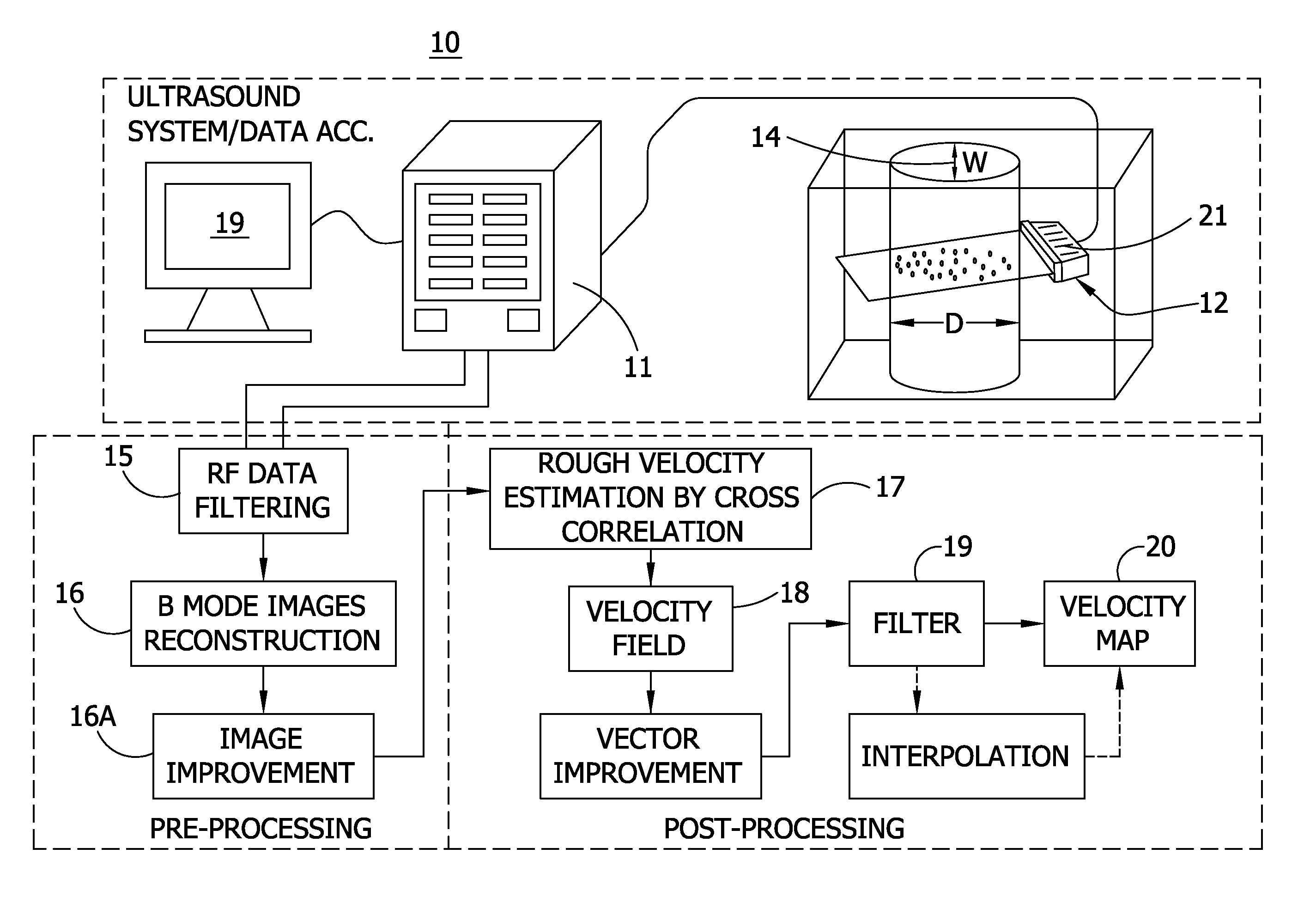
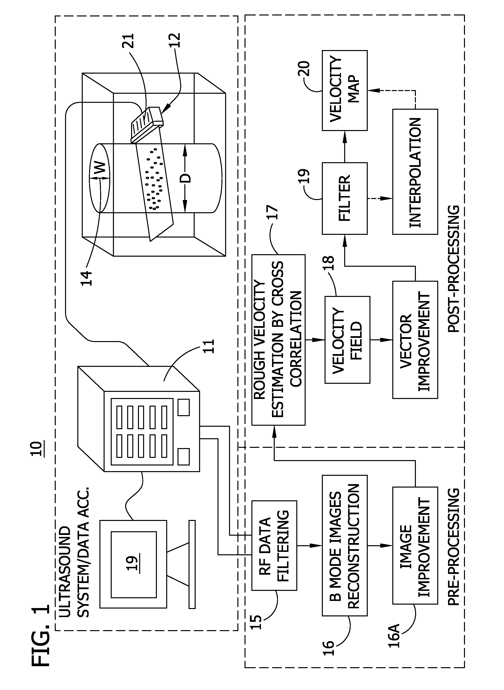
[0085] Imaging limits of conventional commercial systems revolve around spatial accuracy and resolution, as well as inherently low frame rates, in turn, limiting the range of measurable velocities, the ability to capture transient flow phenomena, and the density of the resulting PIV vector field. An overall schematic of the Echo PIV system is shown in FIG. 1, and includes host of aspects: automatic-control (via computerized device, such as a PC, for example) of firing sequences; a 7.8 MHz 128-element linear array transducer with element pitch of 0.3 mm and bandwidth of 73%; RF data acquisition; B-mode image generation; and velocimetry algorithms for analyzing the RF-derived B-mode data. The Echo PIV system provides freedom in selecting a much higher range of frame rates (up to 2000 fps) than that of conventional medical ultrasound systems, as well as more freedom in selecting field of view (FOV) (30˜90 mm), number of transducer elements used to create ultrasound beams (6˜48 elements...

example 2

[0088] The system of FIG. 1 focuses on two components of Echo PIV of interest: uniformly fine spatial resolution over the entire field of view (FOV) and wide dynamic velocity range. Good spatial resolution prevents bubble images from appearing smeared in the B-mode image 16 (FIG. 1), and maximizes the quality of individual bubble images. This improves the quality of cross-correlation during PIV analysis and increases the accuracy of velocity vector calculation to produce the map 18. However, the exact value of spatial resolution that will be optimal for a particular imaging application will depend on specifications such as the range of velocities to be measured, the diameter of the vessel, and the purpose of the measurement, such as whether shear will be derived. Based on these values, certain transducer operating parameters can be set, such as imaging frequency, pulse length, depth of focus, number of elements used for transmit and receive, order of element firing, etc.

[0089] The ...


PUM


Abstract

A system and method for detecting fluid flow. An ultrasound system comprises a signal generator providing ultrasound firing sequences applied to a linear array transducer. The transducer generating ultrasound energy applied to the fluid flow. A pre-processor comprises a digital RF data acquisition component receiving an RF signal from the transducer of back-scattered ultrasound energy and a B-mode image generation component for reconstructing images form the RF data. A post-processor executes particle image velocity (PIV) algorithms for generating velocity vectors indicative of the fluid flow. The sequences may have triangular waveforms.

Description

GOVERNMENT RIGHTS [0001] This invention disclosed herein was made with U.S. government support awarded by National Science Foundation (NSF) award No. CTS-0421461 and National Institute of Health (NIH-HLBI) award R21 HLO79868. Accordingly, the U.S. Government has certain rights in this invention.BACKGROUND OF THE INVENTION [0002] A majority of all cardiovascular diseases and disorders is related to hemodynamic dysfunction. For example, in congenital heart disease and subsequent surgical palliations, fluid shear stresses at the endothelial surface play a critical role in progression of diseases such as pulmonary hypertension. The focal distribution of atherosclerotic plaques in regions of vessel curvature, bifurcation, and branching also suggests that fluid dynamics and vessel geometry play a localizing role in the cause of plaque formation. Accurate measurement of fluid shear stress at the endothelial border in arteries and veins is essential in cardiovascular diagnostics both as a p...

Claims


Application Information

Patent Timeline
IPC IPC(8): A61B8/00
CPCA61B8/06A61B8/0883A61B8/0891A61B8/13G06T7/20A61B8/5238G01S7/52071G01S15/8984A61B8/481
Inventor SHANDAS, ROBINZHENG, HAIRONGZHANG, FUXINGLIU, LINGLIHERTZBERG, JEAN R.
Owner UNIV OF COLORADO THE REGENTS OF
Learn More