Left atrial appendage closure

Inactive Publication Date: 2008-02-07
INTUITIVE SURGICAL OPERATIONS INC
View PDF101 Cites 222 Cited by
  • Summary
  • Abstract
  • Description
  • Claims
  • Application Information

AI Technical Summary

Benefits of technology

[0014] Once the imaging hood has been advanced into the left atrium of the heart, it may be articulated into apposition against the opening of a left atrial appendage (LAA). Once suitably positioned, the imaging hood and the cavity of the left atrial appendage may be purged with the transparent displacing fluid such that the tissue region and cavity may be

Problems solved by technology

However, such imaging balloons have many inherent disadvantages.
For instance, such balloons generally require that the balloon be inflated to a relatively large size which may undesirably displace surrounding tissue and interfere with fine positioning of the imaging system against the tissue.
Moreover, the working area created by such inflatable balloons are generally cramped and limited in size.
Furthermore, inflated balloons may be susceptible to pressure changes in the surrounding fluid.
For example, if the environment surrounding the inflated balloon undergoes pressure changes, e.g., during systolic and diastolic pressure cycles in a beating heart, the constant pressure change may affect the inflated balloon volume and its positioning to produce unsteady or undesirable conditions for optimal tissue imaging.
Accordingly, these types of imaging modalities are generally unable to provide desirable images useful for sufficient diagnosis and therapy of the endoluminal structure, due in part to factors such as dynamic forces generated by the natural movement of the heart.
Moreover, anatomic structures within the body can occlude or obstruct the image acquisition process.
Also,

Method used


Image

  • Left atrial appendage closure
  • Left atrial appendage closure
  • Left atrial appendage closure

Examples


Embodiment Construction

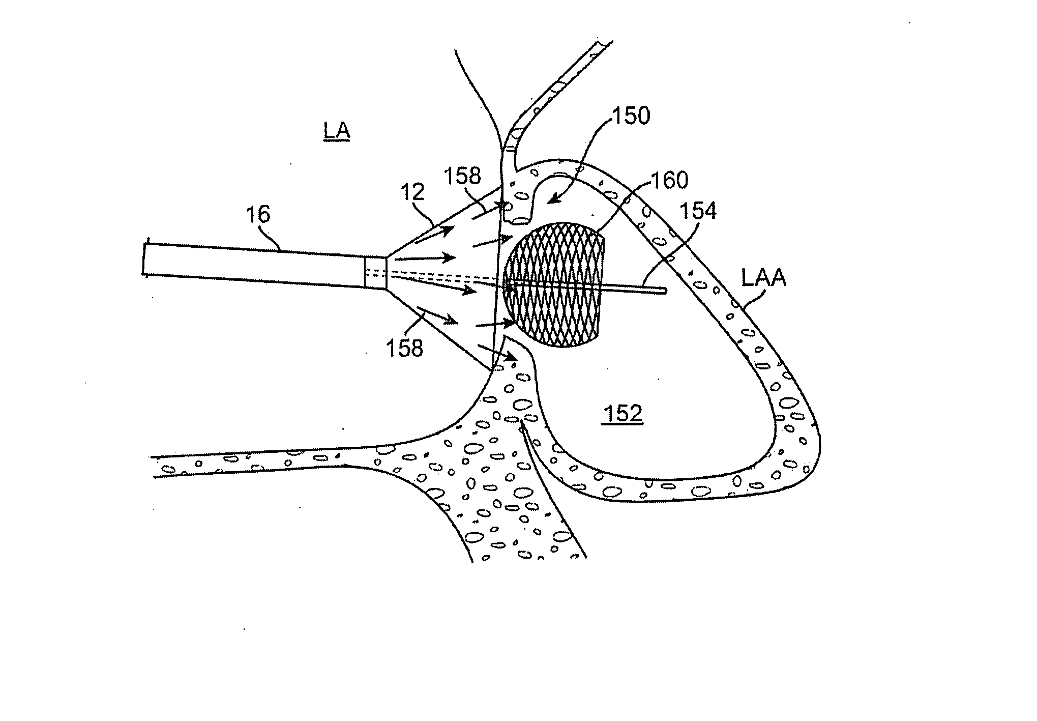
[0041] A tissue-imaging and manipulation apparatus described below is able to provide real-time images in vivo of tissue regions within a body lumen such as a heart, which is filled with blood flowing dynamically therethrough and is also able to provide intravascular tools and instruments for performing various procedures upon the imaged tissue regions. Such an apparatus may be utilized for many procedures, e.g., facilitating trans-septal access to the left atrium, cannulating the coronary sinus, diagnosis of valve regurgitation / stenosis, valvuloplasty, atrial appendage closure, arrhythmogenic focus ablation, among other procedures. Details of tissue imaging and manipulation systems and methods which may be utilized with apparatus and methods described herein are described in U.S. patent application Ser. No. 11 / 259,498 filed Oct. 25, 2005 (U.S. Pat. Pub. No. 2006 / 0184048 A1), which is incorporated herein by reference in its entirety.

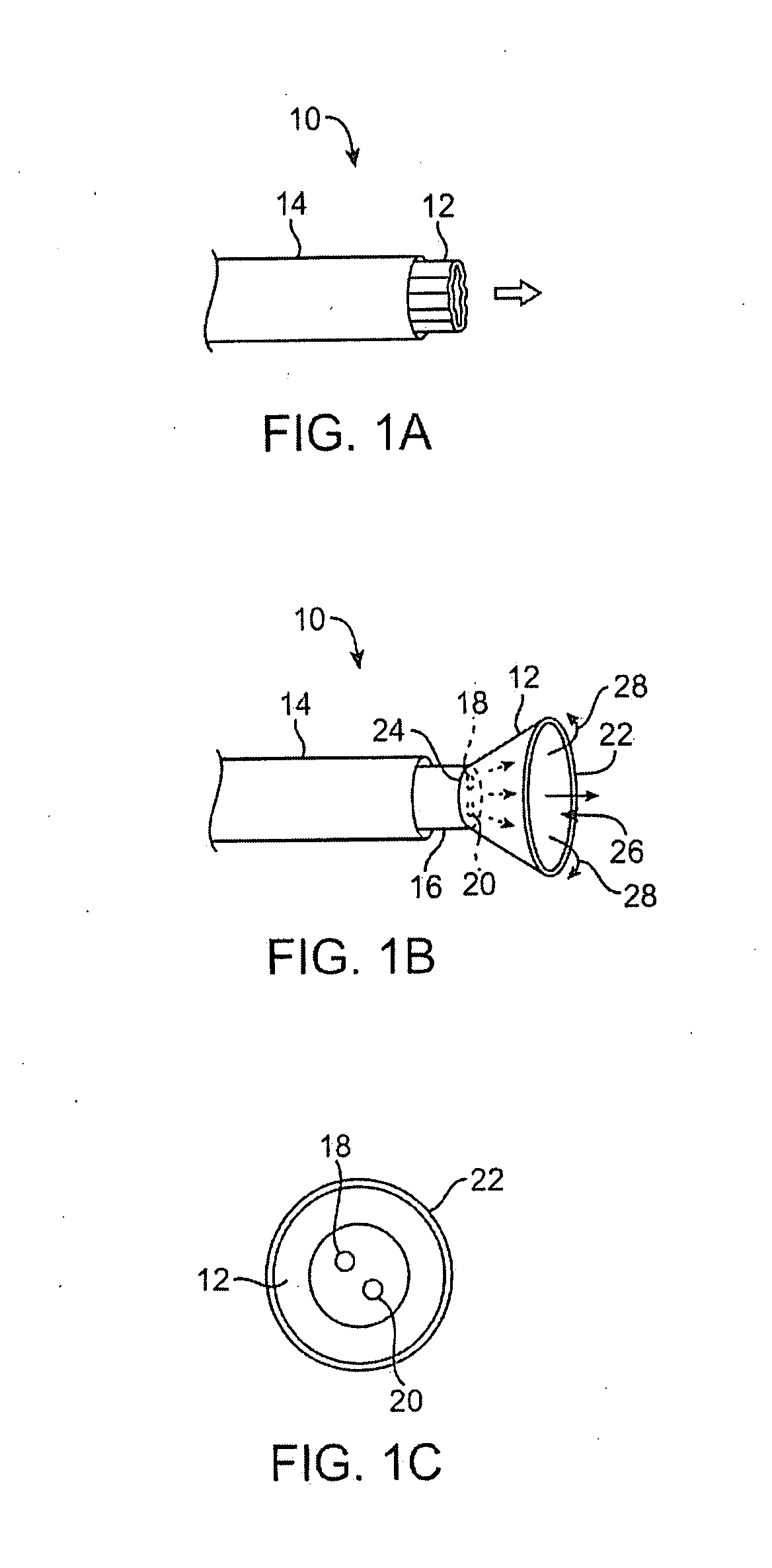
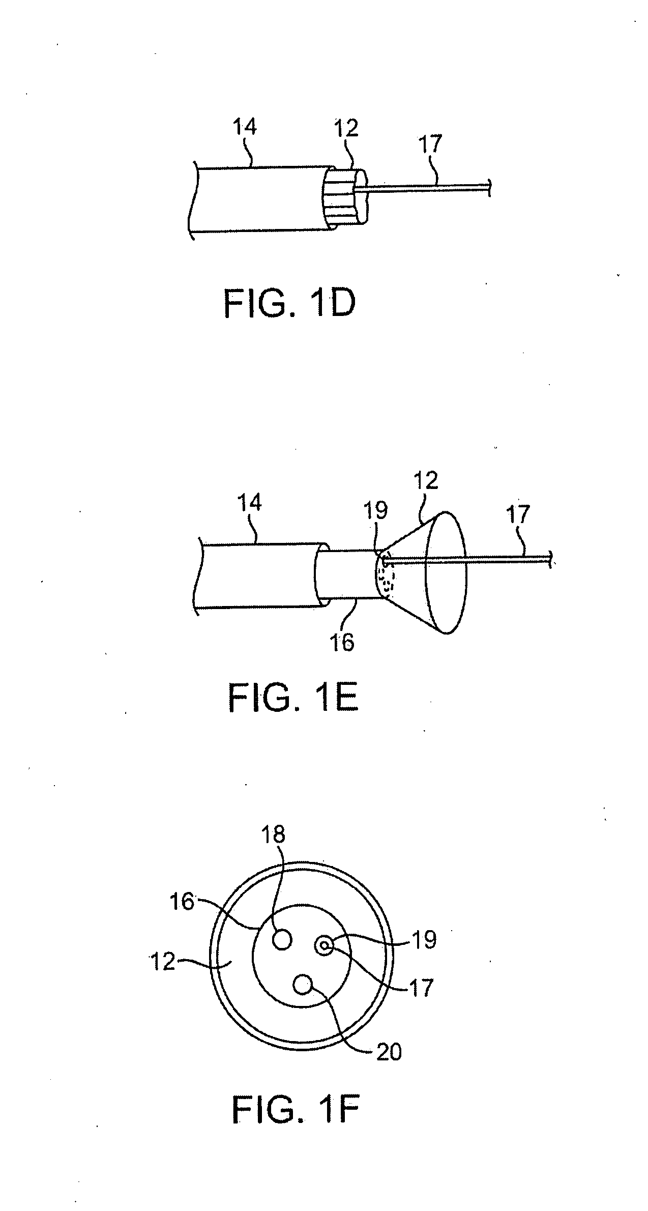
[0042] One variation of a tissue access and imagi...


PUM


Abstract

Methods and apparatus for intraluminally or transluminally closing a left atrial appendage while under direct visualization are described herein. Such a system may include a deployment catheter and an attached imaging hood deployable into an expanded configuration. In use, the imaging hood is placed against or adjacent to a region of tissue to be imaged in a body lumen that is normally filled with an opaque bodily fluid such as blood. A translucent or transparent fluid, such as saline, can be pumped into the imaging hood until the fluid displaces any blood, thereby leaving a clear region of tissue to be imaged via an imaging element in the deployment catheter. Additionally, any number of therapeutic tools can also be passed through the deployment catheter and into the imaging hood for performing any number of procedures on the tissue for accessing and closing the left atrial appendage.

Description

CROSS-REFERENCE TO RELATED APPLICATIONS [0001] This application claims the benefit of priority to U.S. Provisional Patent Application No. 60 / 821,113 filed Aug. 1, 2006, which is incorporated herein by reference in its entirety.FIELD OF THE INVENTION [0002] The present invention relates generally to medical devices used for accessing, visualizing, and / or treating regions of tissue within a body. More particularly, the present invention relates to methods and apparatus for accessing, treating, and closing a left atrial appendage within a patient heart. BACKGROUND OF THE INVENTION [0003] Conventional devices for accessing and visualizing interior regions of a body lumen are known. For example, ultrasound devices have been used to produce images from within a body in vivo. Ultrasound has been used both with and without contrast agents, which typically enhance ultrasound-derived images. [0004] Other conventional methods have utilized catheters or probes having position sensors deployed w...

Claims


Application Information

Patent Timeline
IPC IPC(8): A61B1/00A61B17/128
CPCA61B1/0008A61B2017/306A61B1/00089A61B1/00179A61B1/3137A61B5/6882A61B17/0057A61B17/0469A61B17/0644A61B17/12122A61B2017/00345A61B2017/00575A61B2017/0409A61B2017/0443A61B2017/0454A61B2017/0458A61B2017/0464A61B2017/0649A61B1/00082
Inventor PEH, RUEY-FENGSAADAT, VAHID
Owner INTUITIVE SURGICAL OPERATIONS INC
Why Eureka
Try Eureka