

Expandable cartilage implant
a cartilage implant and expandable technology, applied in the field of medical technology, can solve the problems of limited regenerative capacity compared to other tissues, pain, cartilage damage,
- Summary
- Abstract
- Description
- Claims
- Application Information
AI Technical Summary
Problems solved by technology
Method used
Image

Examples
example 1
[0063] Osteosponge™ (Bacterin) was used as the graft material in all examples.
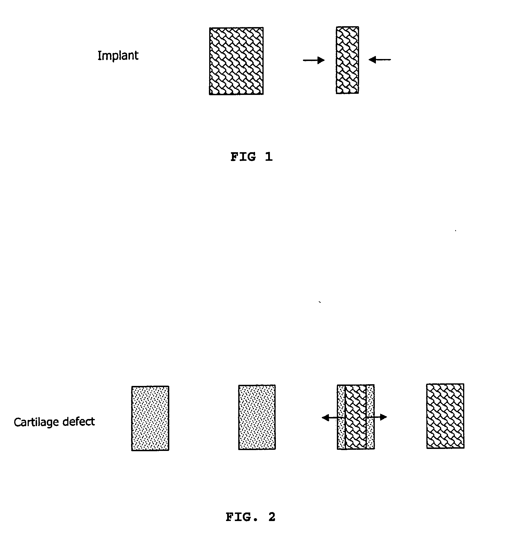
[0064] In this example, an in vitro study was performed to quantify the expansion of a demineralized bone matrix sponge when hydrated with commercially available 1× phosphate buffered saline (PBS).
[0065] Hydration was conducted by manually compressing and submerging the sponge in PBS, until pliable. In an effort to reproduce surgical conditions, the sponge was hydrated at room temperature.
[0066] The diameter, thickness and volume of the sponge were measured 3 times. Measurements were taken when the sponge was dry, immediately after being hydrated for 1 hour, and immediately after being hydrated for 2 hours.
[0067] The percent change in the diameter, thickness and volume were calculated by comparing both the 1 hour measurements and 2 hour measurements to the dry measurements. The sponge expanded ˜15% in diameter, ˜11% in thickness and ˜45% in volume. The measurements taken at 1 hour and 2 hours were stat...
example 2
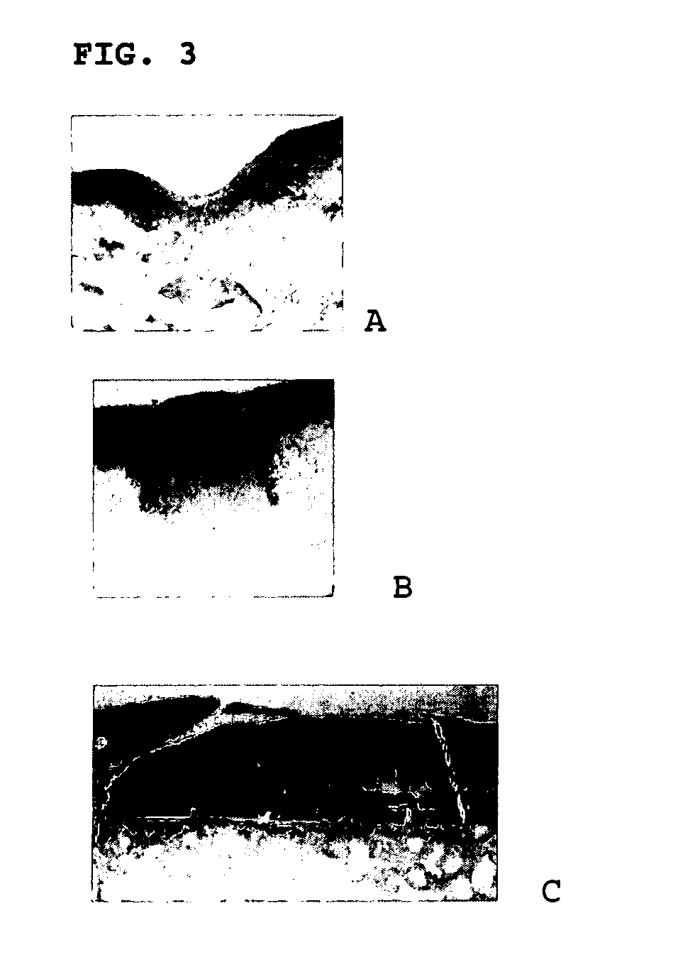
[0068] In vitro studies were also conducted to demonstrate the ability of demineralized bone matrix (DBM) sponges to support chondrogenesis. The sponges were divided into two groups: sponges containing cells and sponges without cells.
[0069] Chondrocytes were harvested from the rear joints of goats under the age of 3 months old. The articular cartilage was harvested within 24 hours of death. Articular cartilage was collected from the patellar groove, femoral condyle, and patella.
[0070] Throughout harvesting, the tissue was bathed in PBS containing gentamicin (25 ug / mL). Cartilage tissue was digested using 0.2% collagenase (Worthington collagenase type 22, 2 mg collagenase per mL culture medium) for approximately 18 hours at 37° C. while shaking in an orbital shaker. The resulting cells were pelleted by centrifugation at 200g for 10-15 minutes and strained through a 70 um cell strainer to separate the cells from cell debris and tissue fragments.
[0071] Following harvesting, the chon...
example 3
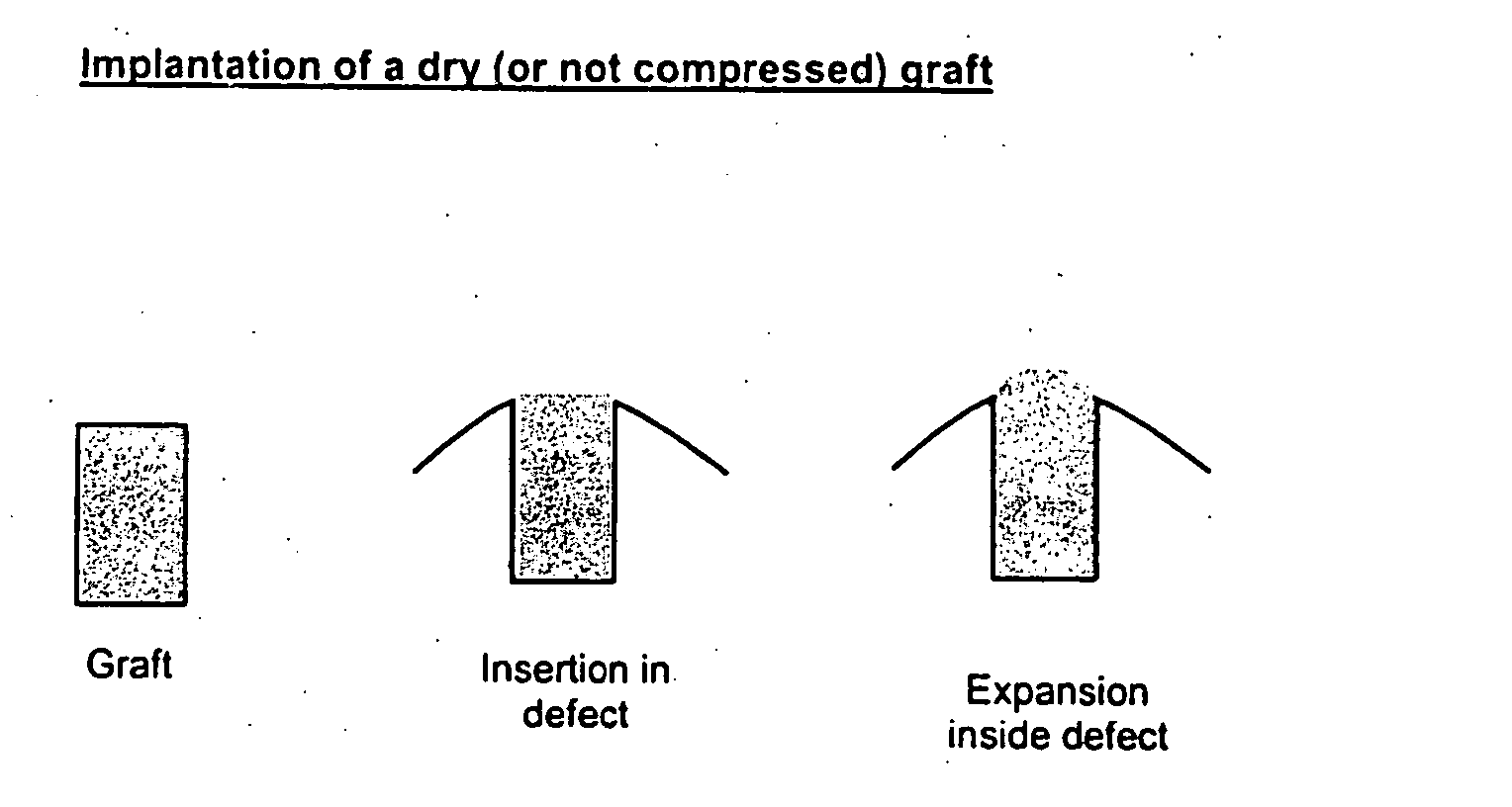
[0077] In the first in vivo study, the grafts were successfully implanted into defects created in the lateral and femoral condyle and trochlear grooves of goats. The femoral condyle was chosen because of its heavy weight bearing characteristics while the lateral groove was chosen because it is a lesser weight bearing site.
[0078] Tubular chisels were used to create and remove chondral and osteochondral cores measuring 4.5 mm in diameter. The remaining defects served as the implantation sites for grafts.
[0079] One graft consisting of DBM was hydrated with saline and implanted into each defect. Some grafts were combined with approximately 100-300 ul of fibrin glue according to manufacturer's instructions. Success was determined based on the ease of implantation, and whether the implanted grafts remained in the defect for the duration of the study.
PUM
| Property | Measurement | Unit |
|---|---|---|
| Fraction | aaaaa | aaaaa |
| Time | aaaaa | aaaaa |
| Size | aaaaa | aaaaa |
Abstract
Description
Claims
Application Information