Brain-Localizing Bone Marrow Progenitor cells

a bone marrow progenitor cell and brain-localizing technology, applied in the field of cells with brain-localizing activity, can solve the problems of difficult effective brain treatment and disruption of the blood brain barrier

Inactive Publication Date: 2008-03-27
ACTGEN
View PDF0 Cites 55 Cited by
  • Summary
  • Abstract
  • Description
  • Claims
  • Application Information

AI Technical Summary

Benefits of technology

[0017] Although many of the cells observed in the brain after bone marrow transplantation grow in the brain, the cells of the present invention arrest in their G0 / G1 phase in the cerebral parenchyma and exist in a growth-arrested state. The cells maintained relatively undifferentiated properties, and did not transdifferentiate into neural cells. The cells were confirmed to be stable in the cerebral parenchyma for several months, but the number of cells decreased with time. For example, GFP-expressing cells were also found in mouse brains 18 weeks after transplantation, but the number of cells was considerably less than in mouse brains up to four weeks after transplantation. Thus, the cells that translocated to the brain are considered to exist transiently in the brain because they are not stem cells. Accordingly, expression level, dose and such, which are of concern when conducting drug therapy or gene therapy, can be controlled relatively easily.
[0019] The present inventors discovered cells with brain-localizing activity (brain-localizing bone marrow progenitor cells) from among the cells obtainable from bone marrow, as described above, and then conducted dedicated studies to succeed in elucidating the characteristics of these cells. The present inventors utilized these characteristics to successfully develop methods for efficiently preparing brain-localizing cells from bone marrow, and completed the present invention.

Problems solved by technology

Therefore, it has been difficult to effectively treat the brain, except by surgical operation.
However, it has also been reported that the blood brain barrier is disrupted by radiation exposure (see Non-patent Document 10), and it is unclear what fraction of cells in the bone marrow translocate into the cerebral parenchyma, and at what stage after transplantation the brain-localizing cells translocate into the cerebral parenchyma.

Method used


Image

  • Brain-Localizing Bone Marrow Progenitor cells
  • Brain-Localizing Bone Marrow Progenitor cells
  • Brain-Localizing Bone Marrow Progenitor cells

Examples


example 1

Effects of Drug Treatment on Host Blood Brain Barrier

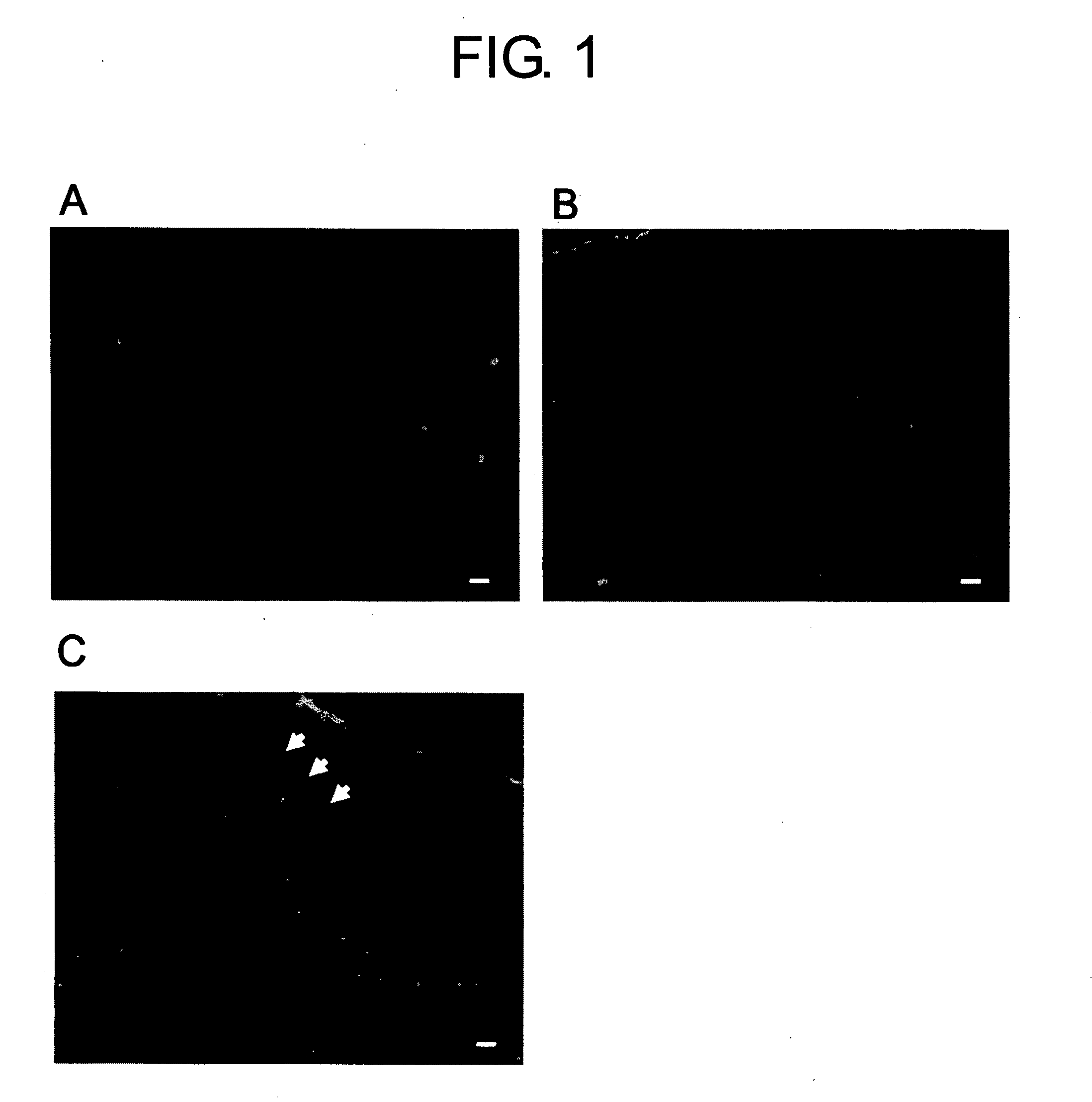
[0183] When bone marrow transplantations are performed, in many cases, host bone marrow cells are removed by radiation exposure. This method is suggested to partially disrupt the blood brain barrier or cause neurological deficits. To perform a bone marrow transplantation that was less invasive to the brain, host bone marrow cells were removed using a drug, 5-FU.

[0184] To confirm that the blood brain barrier was undamaged, Evans blue was injected into the tail vein of 5-FU-treated B6 mice, normal B6 mice, and B6 mice that were physically injured using an injection needle, and dye leakage through the blood brain barrier was examined (FIG. 1). As a result, dye leakage was not observed in 5-FU-treated mice, and damage to the blood brain barrier could not be confirmed.

example 2

Correlation between Foreign Cells Found in the Cerebral Parenchyma and those in the Bone Marrow on Day 7 after Transplantation

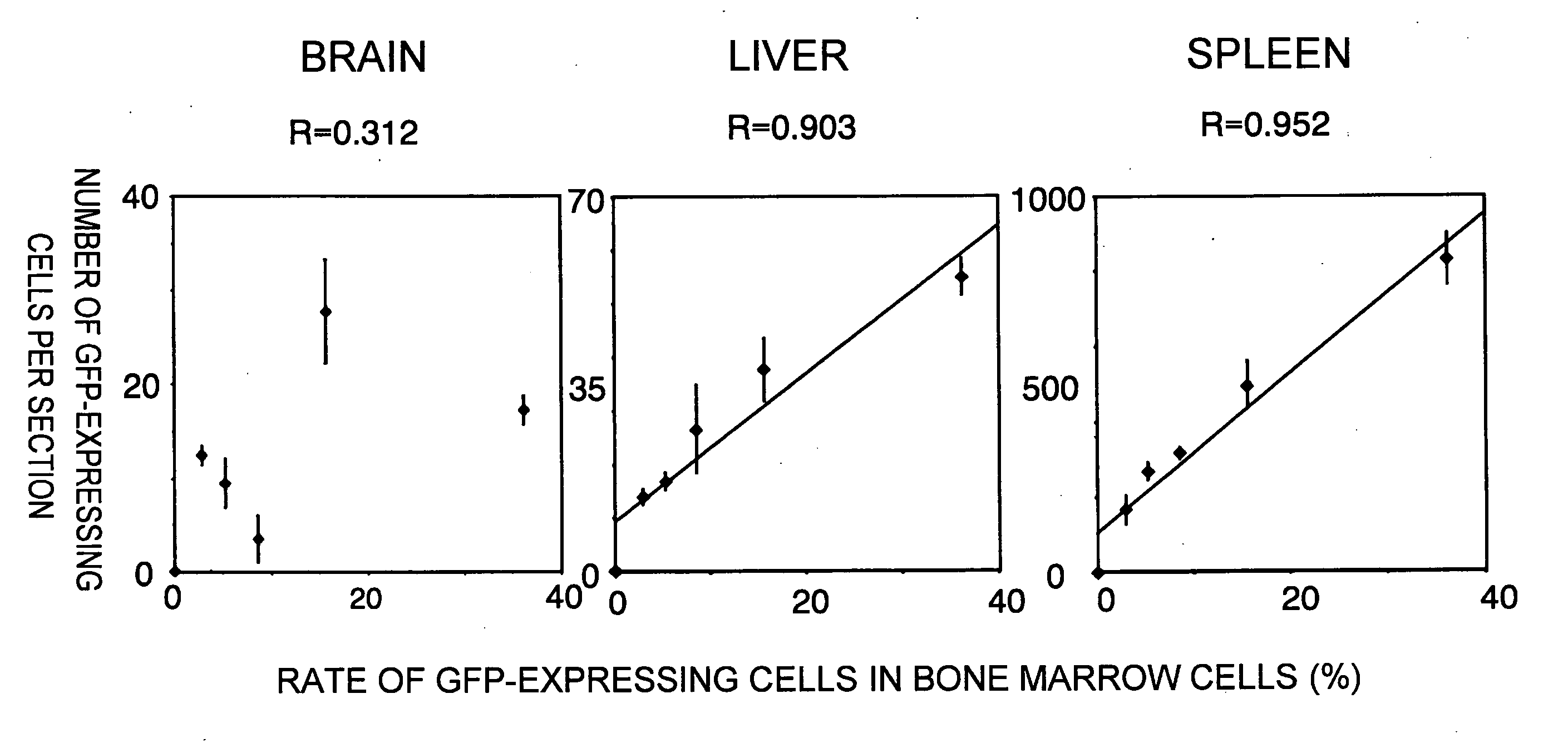
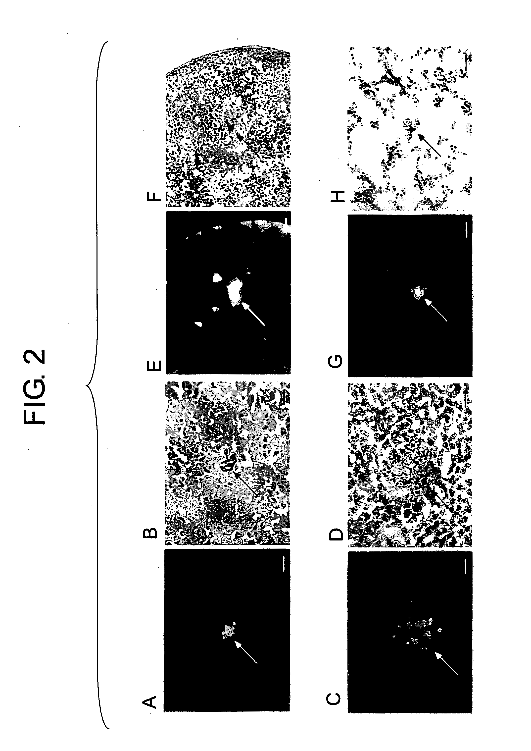
[0185] Bone marrow cells collected from GFP transgenic mice were transplanted via the tail vein of 5-FU-treated mice. GFP-expressing cells were confirmed in the brain, liver, spleen, and lung on day 7 after transplantation (FIG. 2). When correlations between GFP-expressing cells that translocated to the bone marrow and those that translocated to tissues were examined, a positive correlation was observed in peripheral tissues such as the liver or spleen, whereas no correlation was observed in the brain (FIG. 3).

example 3

Examination of the Stage of Brain-Localization of Cells

[0186] The distributions of GFP-expressing cells in the bone marrow, bloodstream, and tissues were examined over time (FIG. 4). In the bone marrow and blood, the proportion of GFP-expressing cells significantly increased on day 7 after transplantation, and GFP expression had completely disappeared in weeks 3 to 4. GFP-expressing cells that translocated to the liver were virtually undetectable in week 6 after transplantation, and expression had completely disappeared in week 18. In contrast, the GFP-expressing cells that translocated to the brain decreased in number after supply from the bloodstream was terminated; however, expression was still observed in week 18.

[0187] To investigate at what stage GFP-expressing cells translocate into the brain, the brains of transplanted mice were collected over time, and GFP expression was examined using RT-PCR methods (FIG. 5). GFP expression was observed in the brain on days 1 to 2 after ...


PUM

PropertyMeasurementUnit
fluorescent imagesaaaaaaaaaa
time-aaaaaaaaaa
timeaaaaaaaaaa

Abstract

The present inventors discovered that brain-localizing cells exist in the progenitor cell fraction of bone marrow cells. They also elucidated various characteristics of the brain-localizing cells. When these cells are infused into the bloodstream, they circulate in the blood and directly translocate into the cerebral parenchyma from the bloodstream. Furthermore, the present inventors succeeded in developing methods for efficiently preparing brain-localizing cells from bone marrow or bone marrow cells. These methods can be applied to less-invasive regenerative medicines targeting the brain and using autologous cells.

Description

TECHNICAL FIELD [0001] The present invention relates to cells with brain-localizing activity, and methods for preparing the same. BACKGROUND ART [0002] The transport of substances and cells to the brain, which is the center of higher functions, is restricted by a barrier structure called blood-brain barrier. Therefore, it has been difficult to effectively treat the brain, except by surgical operation. Even when white blood cells such as monocytes, which circulate through the bloodstream, are collected and transplanted to adult animals, it is known that the cells do not transfer to the cerebral parenchyma, except when the blood-brain barrier is disrupted due to external factors (see Non-patent Document 1). On the other hand, microglia are present in the brain as macrophage-like cells; however, when microglia isolated from a neonatal brain are injected into blood, they are known to show brain-specific infiltration (see Non-patent Document 2). However, microglia are cells in the cerebr...

Claims


Application Information

Patent Timeline
Patent Type & Authority Applications(United States)
IPC IPC(8): A61K35/28A61P43/00C12N15/10C12N5/06C12N5/0793
CPCA61K31/7088C12N2506/1353C12N5/0619A61K35/28A61P9/10A61P25/00A61P25/16A61P25/28A61P35/00A61P37/04A61P43/00
Inventor SAWADA, MAKOTOONO, KENJI
Owner ACTGEN
Why Eureka
Try Eureka