
Multi-lumen catheter and endoscopic method

a multi-lumen catheter and endoscope technology, applied in the field of multi-lumen catheters, can solve the problems of edema, swelling and blocking the normal flow, and permanent disruption of the flow through the sinus system, and achieve the effects of enlarge the pathway, and improve the effect of edema

Inactive Publication Date: 2008-04-17
QUEST MEDICAL
View PDF7 Cites 19 Cited by
  • Summary
  • Abstract
  • Description
  • Claims
  • Application Information

AI Technical Summary

Benefits of technology

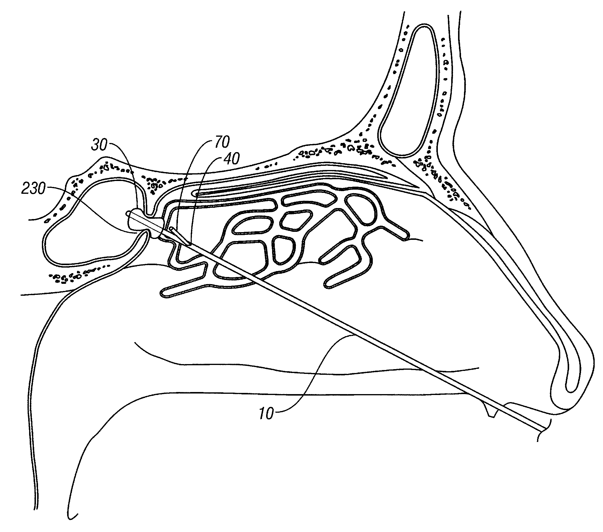
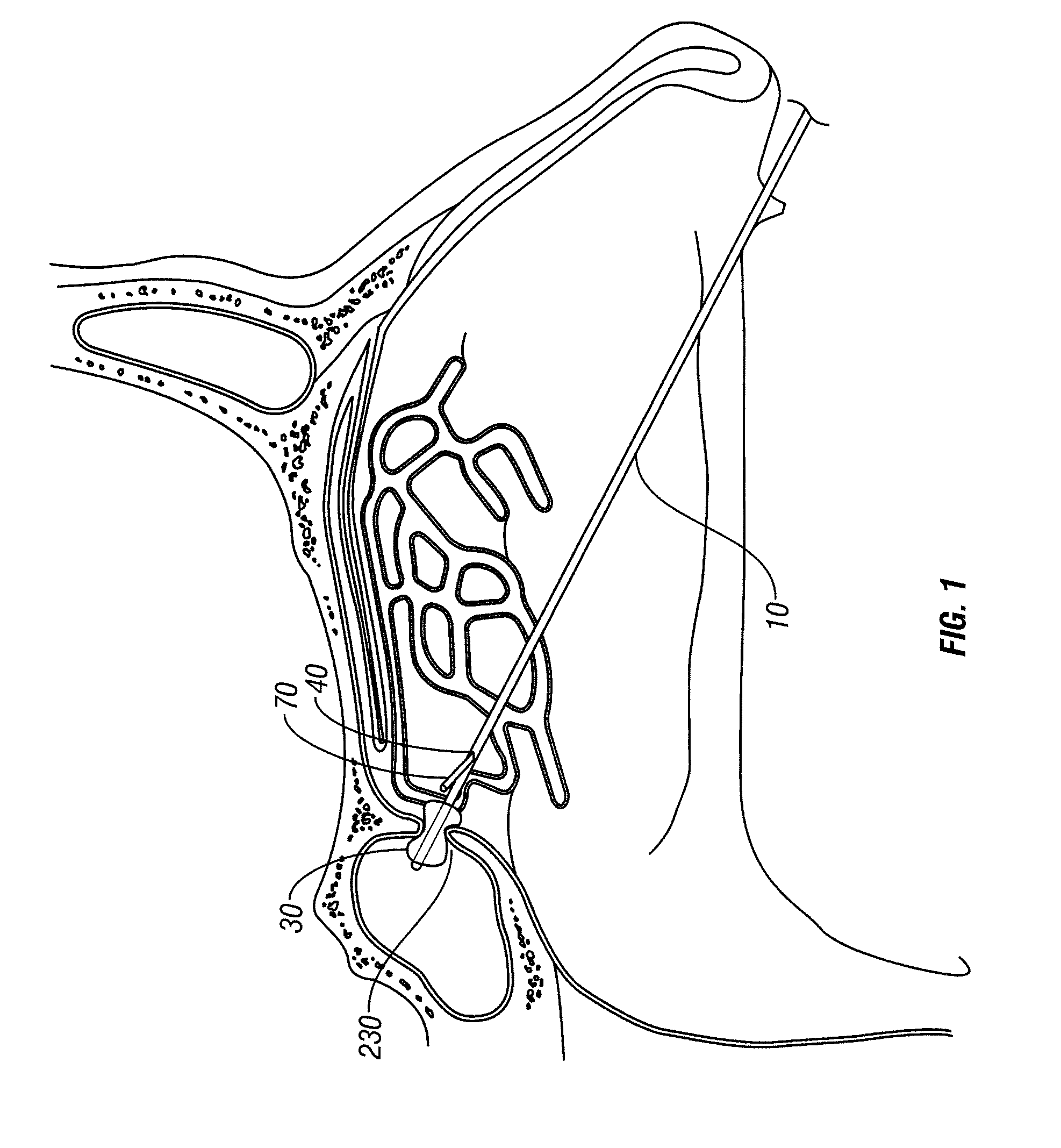
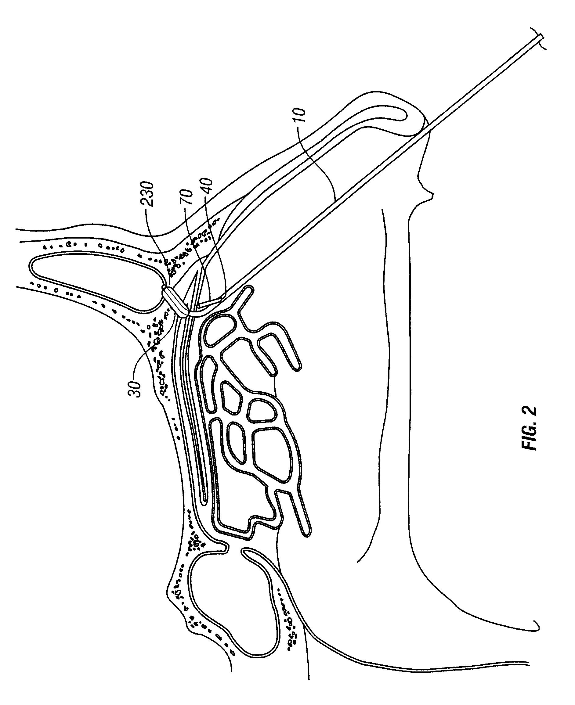
[0005]The first embodiment of the present invention is a four lumen catheter with an expandable device, such as a balloon, built directly into the tip of the catheter. Each of the four lumens is designed to be useful for different functions. One lumen is dedicated to inflating and deflating the balloon. A second lumen is used to insert a traditional balloon catheter into the obstructed pathway. A third lumen carries an endoscope or fiberscope, which allows the surgeon to guide the catheter to the correct location. The fourth lumen is designed to allow the surgeon to remove the fiberscope out of the third lumen and run it back through the fourth lumen. The fourth lumen, however, allows the fiberscope to escape from the catheter shaft before it gets to the tip, thereby giving the surgeon a better view of the balloon on the tip of the multi-lumen catheter inflating.
[0007]The second embodiment of the present invention is a three lumen catheter with an expandable device, such as a balloon, built directly into the tip of the catheter. Each of the three lumens is designed to be useful for different functions. The first lumen is dedicated to inflating and deflating the balloon. The second lumen carries an endoscope or fiberscope, which allows the surgeon to guide the multi-lumen catheter to the correct location. The second lumen in the second embodiment has a portion of the shaft that allows the surgeon to partially retract the fiberscope into the second lumen and make it travel outside the catheter shaft behind the balloon built into the tip of the multi-lumen catheter. The third lumen is used to insert a traditional balloon catheter into the obstructed pathway.
[0009]Both the devices and the methods of the present invention are novel over the prior art in several respects. The present invention allows the surgeon to use two balloons of different size to produce better results from the endoscopic surgery. The first balloon is a traditional balloon catheter that opens up the obstructed pathway to a certain degree. The balloon built into the tip of the multi-lumen catheter is larger in diameter, so it enlarges the pathway more than the traditional balloon catheter was able to. The present invention also eliminates the need for the surgeon to navigate the sinus system and direct a balloon catheter to the affected region twice. Instead, the larger balloon on the tip of the multi-lumen catheter is already very close to the affected region when the smaller balloon is removed and can be used immediately. The present invention also allows the surgeon to have a complete view of the entire process by allowing the endoscope to travel outside the catheter shaft directly behind the balloon.

Problems solved by technology

Inflammation can occur in the tissues that make up the ducts and ostia, causing them to swell and block the normal flow.
Over time there can be a pathologic increase in inflamed tissue causing permanent disruption in the flow through the sinus system.
Obstruction of the narrow ducts and ostia between the paranasal sinuses and nasal cavity develops, resulting in a vicious cycle of increased secretions, edema and ultimately complete blockage of the sinus pathways.
However, some patients become resistant to medical treatment and surgery becomes necessary.
Since space inside the nasal cavity is limited, use of standard 4 millimeter diameter endoscopes for direct visual placement assistance is prohibitive.

Method used


Image

  • Multi-lumen catheter and endoscopic method
  • Multi-lumen catheter and endoscopic method
  • Multi-lumen catheter and endoscopic method

Examples


Embodiment Construction

[0022]The present invention is directed towards a multi-lumen catheter and method for using it to perform endoscopic surgery.

[0023]Referring initially to FIG. 7, therein is depicted the first embodiment of the present invention. The first embodiment is a multi-lumen catheter 10 containing four lumens. It is understood that more lumens can be included in the catheter as needed. A lumen is a hollow, tubular portion of the multi-lumen catheter 10, approximately circular in cross section. The diameter of the lumens can vary from about 0.2 millimeters to about 1.3 millimeters. The overall diameter of the catheter shaft is approximately 4 millimeters. The multi-lumen catheter shaft is flexible and has a proximal end and a distal end. The proximal end is not depicted in because it is not important to the claimed invention. The distal end is the tip of the multi-lumen catheter and is inserted into the surgical patient's body. The distal end of the multi-lumen catheter incorporates an inflat...


PUM


Abstract

The present invention is directed towards a multi-lumen catheter with an expandable device, such as a balloon, incorporated into its surface. The multi-lumen catheter also has an opening in its shaft that allows an imaging instrument to exit the shaft and view the expandable device built into its surface. The present invention also covers an endoscopic surgical method utilizing the multi-lumen catheter.

Description

FIELD OF THE INVENTION[0001]This invention relates to multi-lumen catheters and methods for using such catheters for treating paranasal sinuses.BACKGROUND OF THE INVENTION[0002]In order to fully understand this invention, it is important to consider the anatomy of the sinus system. The sinus system consists of many different pathways, called ducts or ostia, which allow mucus, air and other substances to drain and flow through the system. Inflammation can occur in the tissues that make up the ducts and ostia, causing them to swell and block the normal flow. Inflammation may be caused by allergies, noxious agents, nasal polyps, and other factors. Over time there can be a pathologic increase in inflamed tissue causing permanent disruption in the flow through the sinus system. Obstruction of the narrow ducts and ostia between the paranasal sinuses and nasal cavity develops, resulting in a vicious cycle of increased secretions, edema and ultimately complete blockage of the sinus pathways...

Claims


Application Information

Patent Timeline
IPC IPC(8): A61B1/05
CPCA61B17/24A61B2017/003A61B2017/22051A61M2025/004A61B2019/5217A61M2025/0036A61B2017/22072A61B2090/3614
Inventor DOLLAR, MIKE
Owner QUEST MEDICAL
Learn More