Analysis of brain patterns using temporal measures

a technology of brain pattern and time series, applied in the field of neurologic analysis, can solve the problems of a huge and growing problem, no good tests of brain function available, and complicated research and treatment efforts, and achieve the effect of low cost and high throughpu

Active Publication Date: 2008-04-17
RGT UNIV OF MINNESOTA
View PDF31 Cites 62 Cited by
  • Summary
  • Abstract
  • Description
  • Claims
  • Application Information

AI Technical Summary

Benefits of technology

[0021] Advantageously, embodiments of the invention may enable obtaining the brain characterization, diagnosis, and other results from a single trial, or measurement session, which may last only a few minutes or less. This offers a relatively low cost and high throughput for the use of measurement equipment that collects the data from subjects. For subjects, the comfort of short, non-invasive data collection sessions such as using MEG instruments, is a clear advantage over having to make multiple trips to the clinic or having to endure lengthy testing. Embodiments of the invention may overcome the disadvantages of the qualitative and subjective evaluations of brain disorders that are commonplace today with current indirect diagnostic techniques based on observation and other clinical data by providing a quantitative, statistically correlated measure of multiple brain conditions. Another benefit offered by aspects of the invention is the ability to accept as input non-preprocessed data. Thus, standard measurement instrument output can be utilized without the need for special equipment upgrades, and a subject can be fully evaluated for a variety of conditions without any a priori anticipation, or preparation for screening of certain hypothesized or speculated conditions.

Problems solved by technology

However, unlike any other organ of the body, no good tests of brain function are available.
Neurological disease, including for example, cognitive impairment, is a huge and growing problem.
While the clinical characterization of AD and mild cognitive impairment is usually accurate, misdiagnoses do occur, complicating research and treatment efforts.
An objective test for AD, cognitive impairment, or other neurological conditions would be desirable, but the various approaches proposed to-date have significant drawbacks, limiting their potential for application as a sensitive, reliable, diagnostic or evaluative tool.
One drawback of ERP-based measurements is the evoked response to the stimulus causes certain brain regions to become very active while other brain regions remain relatively inactive.
Using this approach, a measurement representing overall brain activity, taking into account the activity of less active regions, is not possible.
This problem is exacerbated by the use of conventional EEG instrumentation, which tends to detect primarily electrical activity near the outer surface of the brain, with substantially reduced sensitivity at deeper brain regions.
However, the measurement and data analysis approaches disclosed involve mainly spectral analysis and are not capable of recognizing subtle characteristic indicia of certain diseases or conditions from among all of the measurements collected.
While this work produced some interesting insights into measurement techniques for taking MEG readings (such as the advantageous use of 1 kHz sampling, and preprocessing of the data to pre-whiten it), this work explored only individual sensor signal interactions, and did not take into account whole brain modeling in which large numbers of sensor groupings must be studied.
Indeed, for reasons that will become apparent from the following disclosure, the disclosure of Leuthold et al. does not enable analyzing brain activity for purposes of characterizing a brain condition of a subject or making a diagnosis of a brain condition.

Method used


Image

  • Analysis of brain patterns using temporal measures
  • Analysis of brain patterns using temporal measures
  • Analysis of brain patterns using temporal measures

Examples


example 1

[0043] Magnetoencephalography (MEG) can be used in elderly subjects and in subjects with MCI and AD.

[0044] In one example, a fixation task and MEG are used to assess the dynamic status or dynamic function of the brain in three groups of elderly subjects (77.1±1.5 y, mean ±SEM, N=1 1): normal (N=4, 76.5±2.1 y), subjects with MCI (N=4, 75.7±3.7 y), and subjects with AD (N=3, 79.7±0.3 y). Data is acquired from 248 axial gradiometers (Magnes 3600 WH, 4-D Neuroimaging) while subjects fixate on a spot for 45 s, and were preprocessed to remove cardiac artifacts or eye blink artifacts.

[0045] After prewhitening the time series by fitting an AutoRegressive Integrative Moving Average (ARIMA) model and taking the residuals, all pairwise, zero-lag, partial cross correlations are calculated, providing estimates of the strength and sign (positive, negative) of direct synchronous coupling between neuronal populations at 1 ms temporal resolution.

[0046] An analysis of covariance is performed in wh...

example 2

Methodology

[0056] The subject lies supine on a bed and is instructed to look at a spot in front of them for 1 minute. The subjects are asked to keep their eyes fixated on the spot and not to blink. Then they close their eyes for 3 more minutes, and this is the end of the test. MEG data from the fixation period are used for all analyses. In one embodiment, the data acquired during the eyes closed condition are useful for identifying and removing signal artifacts, such as the cardiac artifact.

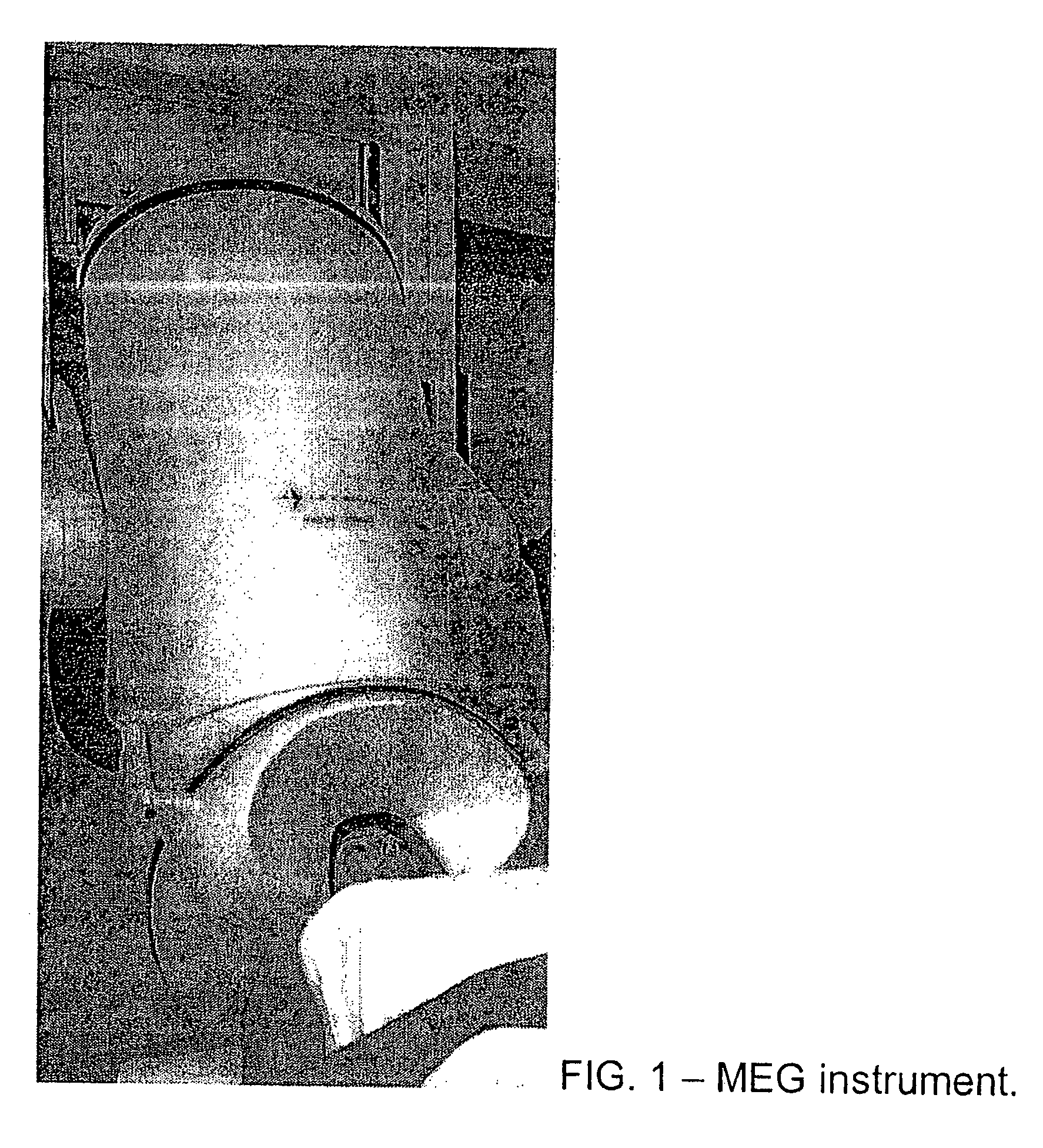
[0057] MEG Instrument Data is acquired using the MEG instrument. Subjects lie on a bed in the magnetically shielded room and MEG signals are acquired from 248 axial gradiometers (0.1-400 Hz, sampled @ 1017 Hz, Magnes 3600 WH, 4-D Neuroimaging, San Diego, Calif.) during the whole duration of the experiment (˜4 min).

Data Analysis

[0058] Data preprocessing. Cardiac artifacts may be removed using an event-synchronous subtraction method. Due to the very short duration of the eye-fixation period (1...

example 3

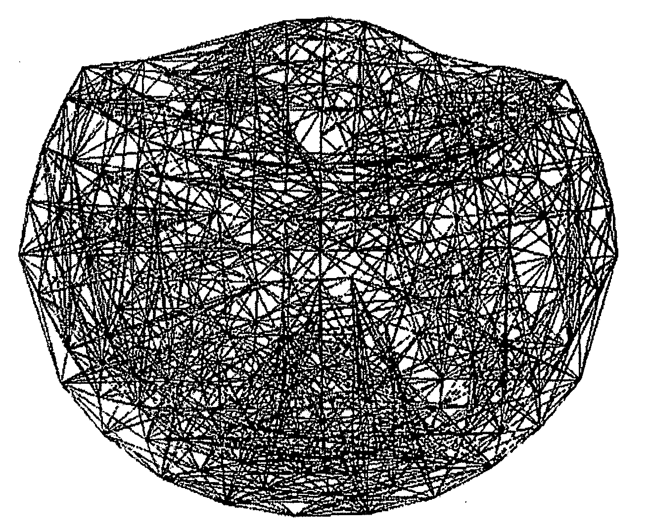
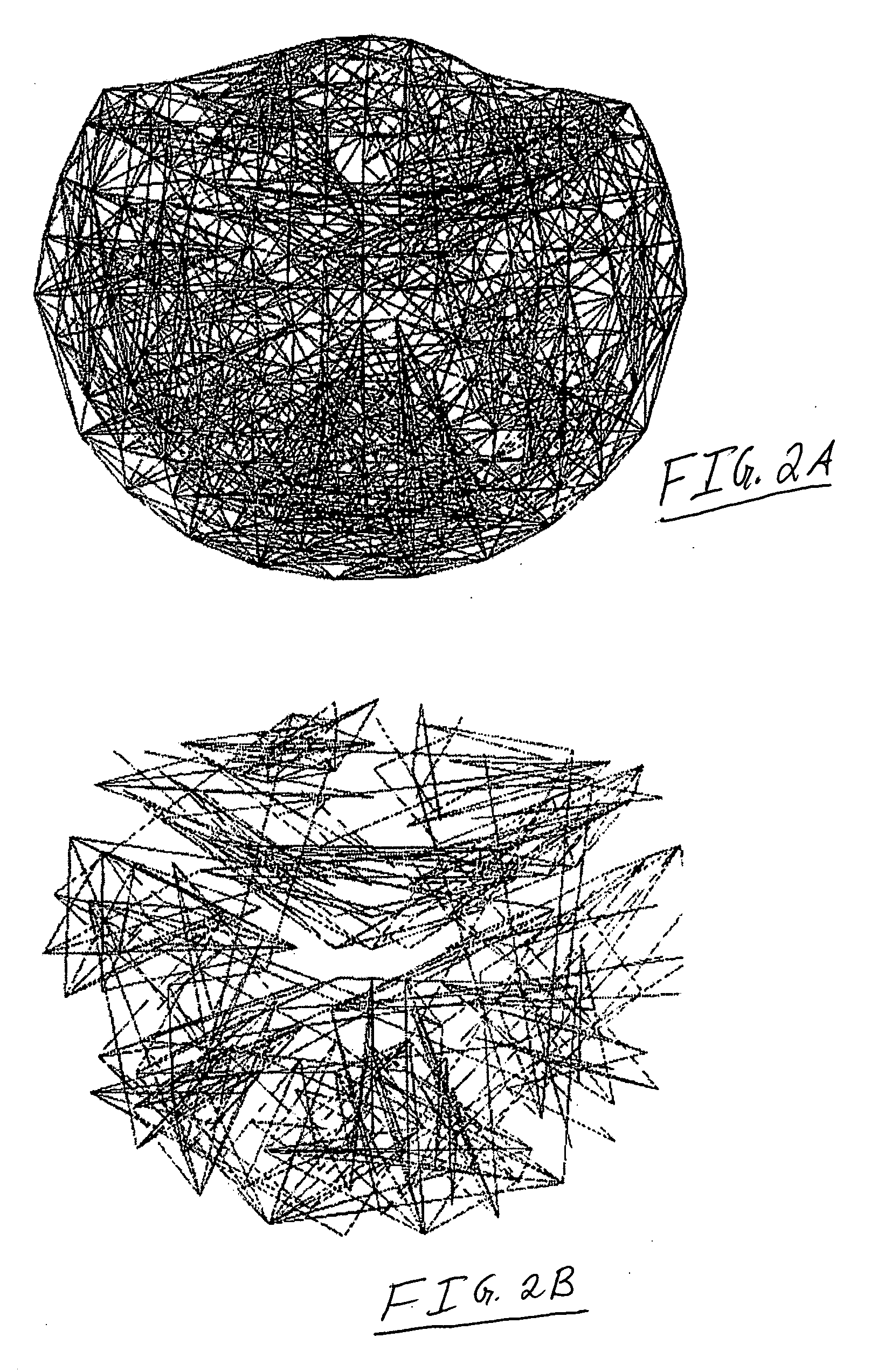
[0069] Synchronous dynamic brain networks are visualized by using prewhitened (stationary) magnetoencephalography signals. In one example, data is acquired from 248 axial gradiometers. After fitting an autoregressive integrative moving average model, and taking the residuals, all pairwise, zero-lag, partial cross correlations PCCIJ0 between the i and j sensors were calculated, providing estimates of the strength and sign (positive and negative) of direct synchronous coupling between neuronal populations at a 1-ms temporal resolution. In one example, 51.4% of PCCIJO were positive, and 48.6% were negative. Positive PCCIJO occurred more frequently at shorter intersensor distances and were 72% stronger than negative ones, on the average. On the basis of the estimated PCCIJO dynamic neural networks are constructed (one per subject) showing distinct features, including several local interactions. These features were robust across subjects and can serve as a blueprint for evaluating dynami...


PUM


Abstract

A set of brain data representing a time series of neurophysiologic activity acquired by spatially distributed sensors arranged to detect neural signaling of a brain (such as by the use of magnetoencephalography) is obtained. The set of brain data is processed to obtain a dynamic brain model based on a set of statistically-independent temporal measures, such as partial cross correlations, among groupings of different time series within the set of brain data. The dynamic brain model represents interactions between neural populations of the brain occurring close in time, such as with zero lag, for example. The dynamic brain model can be analyzed to obtain the neurophysiologic assessment of the brain. Data processing techniques may be used to assess structural or neurochemical brain pathologies.

Description

RELATED APPLICATIONS [0001] This application claims the benefit of U.S. Provisional Application No. 60 / 818,931, filed Jul. 6, 2006, and entitled “CLASSIFICATION AND QUANTITATIVE ASSESSMENT OF BRAIN PATTERNS USING MAGNETOECEPHALOGRAPHY;” and U.S. Provisional Application No. 60 / 851,599 Filed Oct. 13, 2006, and entitled “CLASSIFICATION AND QUANTITATIVE ASSESSMENT OF BRAIN PATTERNS USING MAGNETOECEPHALOGRAPHY,” both of which are incorporated by reference herein in their entirety.GOVERNMENT SUPPORT [0002] This invention was made with Government support under award DE-FG02-99ER62764 awarded by the Department of Energy to the MIND Institute and with support by the US Department of Veterans Affairs. The Government has certain rights in this invention.TECHNICAL FIELD [0003] This document pertains generally to neurophysiologic analysis, and more particularly, but not by way of limitation, to analysis of brain patterns using time series representation. BACKGROUND [0004] Like any other organ of...

Claims


Application Information

Patent Timeline
Patent Type & Authority Applications(United States)
IPC IPC(8): A61B5/0476A61B5/375
CPCA61B5/04008A61B5/0476A61B5/0482G06F19/3437A61B5/4064A61B5/4082A61B5/4088G16H50/50A61B5/245A61B5/7235A61B5/7264A61B5/369A61B5/7246A61B5/4076A61B5/0022A61B5/7203A61B5/388A61B5/375A61B5/372
Inventor GEORGOPOULOS, APOSTOLOS
Owner RGT UNIV OF MINNESOTA
Why Eureka
Try Eureka