Treatment for post partum hemorrhage

a technology for treating postpartum hemorrhage and uterine arteries, which is applied in the field of treatment of postpartum hemorrhage, can solve the problems of pharmaceutical agents producing side effects not just in the uterus but throughout the body, the uterine arteries cannot be palpated during a bimanual pelvic examination, and the uterine arteries cannot be analyzed. it can reduce or terminate the risk of further hemorrhage, reduce or eliminate blood flow, and reduce the risk of a

Inactive Publication Date: 2008-04-24
VASCULAR CONTROL SYST
View PDF99 Cites 5 Cited by
  • Summary
  • Abstract
  • Description
  • Claims
  • Application Information

AI Technical Summary

Benefits of technology

[0017] The invention is directed to instruments and the use of the instruments for accessing a blood vessel and occluding blood flow in the accessed blood vessel to reduce or eliminate hemorrhaging. The invention is particularly directed to the occluding of uterine arteries in the treatment of PPH following vaginal delivery.
[0019] One of the clamping elements of the clamping device is preferably larger than the other clamping element so that the smaller clamping element at least partially fits inside the larger clamping element when the clamping elements are brought together, i.e. when the clamping device is closed or partially closed. The larger clamping element may be longer or wider or both longer and wider than the smaller clamping element. At least one of the clamping members, preferably the outer and smaller clamping element, is provided with at least one blood vessel location sensor at or near the distal end of the clamping element. The sensors are mounted on the distal end of the clamping element to facilitate location of the target uterine artery when positioned adjacent thereto. The at least one sensor has a field of view which radiates distally from the distal end of the clamping element to which it is mounted to facilitate location and occlusion of the patient's uterine artery. One or more sensors may be mounted on the clamping elements having a field of view toward an opposing clamping element to aid in monitoring blood flow through an occluded or partially occluded artery compressed by one or both clamping elements.
[0020] The clamping device embodying features of the invention is configured to be inserted into a female patient's vaginal canal and advanced therein after a vaginal birth. The first or inner clamping element is disposed within the patient's uterine cervix and the second or outer clamping member disposed on the outside of the patient's uterine cervix. The inclination of the clamping elements with respect to the longitudinal axis of the handles helps with the proper placement of the clamping elements within the patient's post partum anatomy.
[0023] The invention provides a method of treating a patient suffering from PPH which includes locating the uterine artery with a location sensor on the distal end of a clamping element of the clamping device having features of the invention, and compressing a portion of the uterine artery by applying pressure to tissue with the clamping elements of the clamping device. To properly deploy the device, the inner clamping element is placed within a uterus of a patient and the outer clamping element is disposed about a portion of the uterine cervix near the adjacent vaginal fornix of a patient. The clamping device is pushed further into the patient's vaginal canal so the distal end of the outer clamping element distends the wall of the patient's vaginal fornix near to the patient's uterine artery so the outer clamping element folds the vaginal wall over the uterine artery. Closing of the clamping device compresses the uterine artery to effectively reduce or abolish blood flow in the uterine artery. Compression of the uterine artery may be maintained from several minutes up to several hours in order to reduce or terminate the post partum hemorrhage and to effect sufficient clotting so as to reduce the risk of further hemorrhage upon release of the clamping device from its closed or partially closed condition. Thus, compression may be maintained for a therapeutically effective amount of time, typically between about 5 minutes and about 7 hours, preferably about 10 minutes to about 5 hours.
[0024] The invention enables non-invasive identification and occlusion of blood vessels such as the uterine arteries to treat PPH. The devices and methods are simple and easy to use, enabling the rapid and effective occlusion of a female patient's uterine arteries after giving birth without surgical intervention.

Problems solved by technology

However, with the expansion of the uterus into the abdomen as the pregnancy term lengthens, the uterine arteries can no longer be palpated during a bimanual pelvic examination because they are too far away from the vaginal introitus to be reached by an examiner's fingers (typically about 19 cm).
Although uterine contractions following birth are generally sufficient to slow the velocity of blood flowing through the uterus to initiate blood clot formation throughout myometrium, in some women uterine contractions are inadequate.
However, these pharmaceutical agents produce side effects on the vasculature not just in the uterus but throughout the body.
If the uterine packing fails, more complex surgery is performed.
Though noninvasive, this method has no means for positively identifying whether or nor any artery has been compressed by the two sets of forceps.
As a result, the method has not been adopted.
However, because vascular surgery is not generally taught in obstetric residencies, the technique is not widely practiced.
Though effective, these techniques are not universally available and there is considerable time delay between the onset of postpartum hemorrhage and the transfer to an angiographic suite—commonly in different parts of the same hospital.
Furthermore, since angiography is performed by radiologists and babies are delivered by obstetricians, communication gaps arise between the two specialties.
Ligation of the uterine arteries is effective at stopping postpartum hemorrhage, but the procedure is invasive and time consuming.
However, because the angiographic methods are performed by different specialists at different locations, there is frequently insufficient time to perform them for effective treatment of PPH.
Moreover, the procedure leaves the mother infertile.
Accordingly, there is need for uncomplicated instruments that can be rapidly deployed to partially or completely terminate blood flow in blood vessels such as the uterine arteries, particularly in the case of PPH.

Method used


Image

  • Treatment for post partum hemorrhage
  • Treatment for post partum hemorrhage
  • Treatment for post partum hemorrhage

Examples


Embodiment Construction

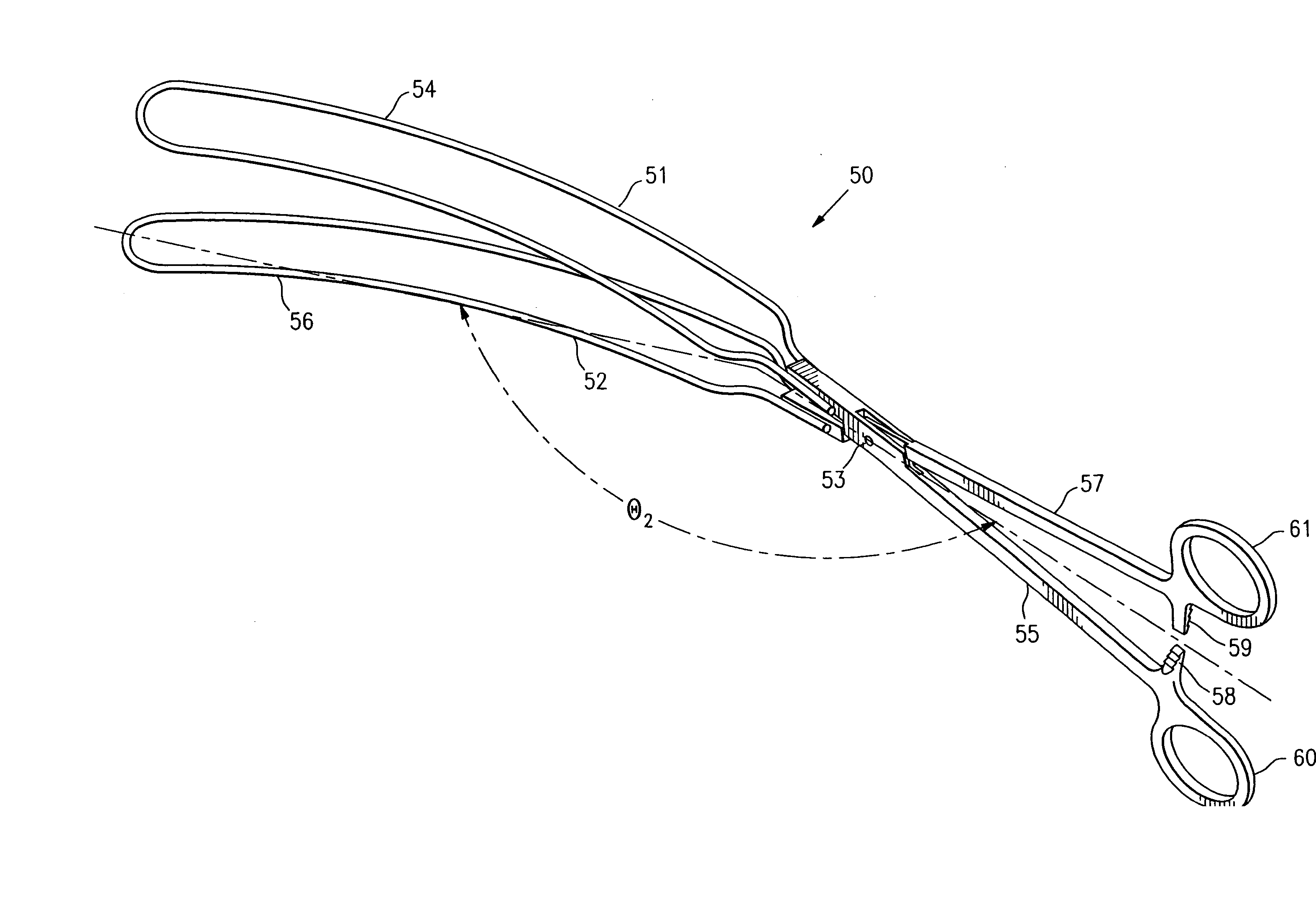
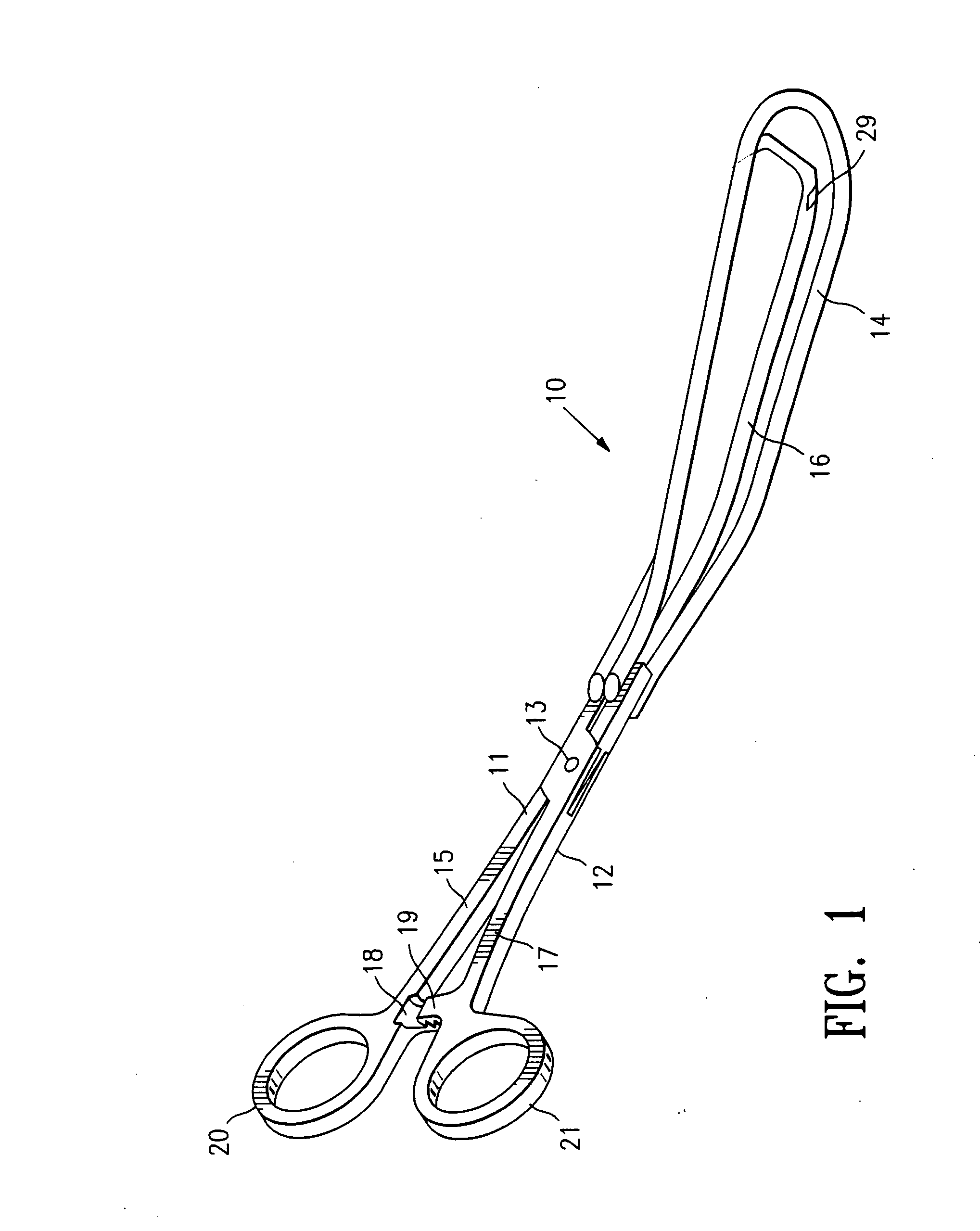
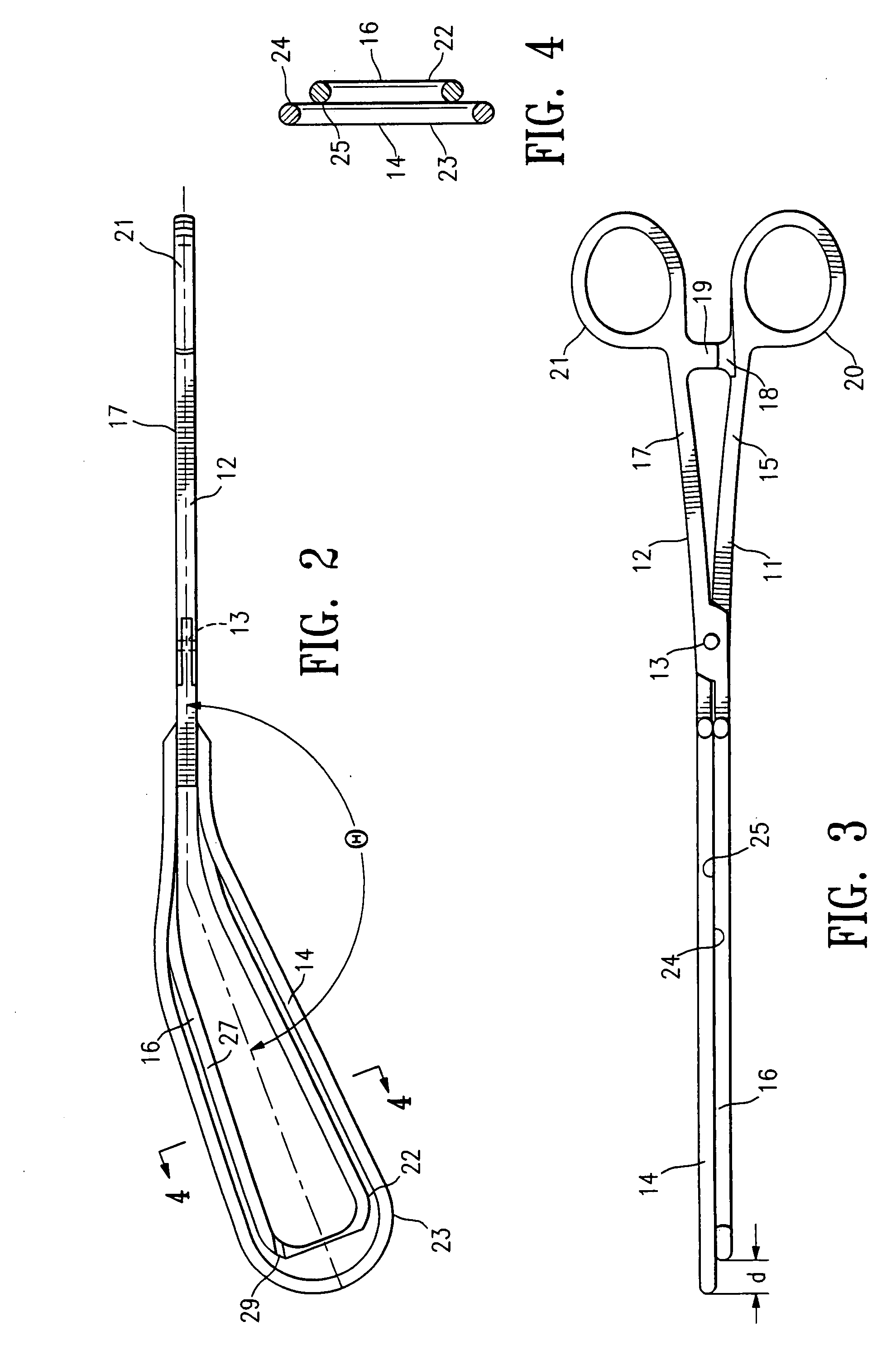
[0041]FIGS. 1-5 illustrate a clamping device 10 embodying features of the invention including first and second clamping members 11 and 12 which are pivotally connected at pivot point 13. First clamping member 11 has a first clamping element 14 secured to the distal end of handle 15. Second clamping member 12 has a second clamping element 16 secured to the distal end of handle 17. The handles 15 and 17 are provided with ratchet members 18 and 19 respectively to provide a releasable locking connection therebetween and finger grips 20 and 21 respectively for rotating the handles about the pivot point 13 to open and close the clamping elements 14 and 16. Each of the clamping elements 14 and 16 are at angles with respect to the longitudinal axis of the handles 15 and 17 to facilitate deployment within the patient's post partum vaginal canal and uterine cervix. The clamping elements 14 and 16 form an obtuse included angle θ with respect to the longitudinal axis of handles 15 and 17 so as ...


PUM


Abstract

The invention is directed to instruments and procedures using such instruments for temporarily reducing or terminating blood flow through a female patient's uterine artery to treat post partum hemorrhage (PPH). The uterine artery is occluded by a clamping device which includes a pair of pivotally connected clamping members, with each of the clamping members having a handle and a clamping element at the distal end of the handle. The clamping elements are inclined with respect to the longitudinal axes of the handles at an included obtuse angle between about 120° and about 170°, preferably between about 130° and 160°. An artery locating sensor is provided on the distal end of at least one of the clamping elements. Preferably, the artery locating sensor is a Doppler ultrasound blood flow sensor. After birth, the clamping device is inserted into the female patient's post partum vaginal canal and advanced therein until one of the clamping elements Is in the patient's uterine cervix and the other clamping element is on the exterior of the uterine cervix. The clamping element on the exterior of the patient's uterine cervix is pressed against the patient's vaginal fornix and the clamping device closed so as to occlude the uterine artery disposed within tissue grasped by the clamping device. The clamping device is locked in the closed configuration and maintained in the condition until the patient's uterus is sufficiently clotted up to ensure termination of the hemorrhaging, typically about 5 minutes to about 7 hours.

Description

[0001] This application is a continuation of application Ser. No. 10 / 355,809, filed Jan. 30, 2003 from which priority is claimed and which is incorporated herein in its entirety.FIELD OF THE INVENTION [0002] The invention relates generally to the field of devices and treatments of diseases and conditions by the regulation of blood flow in blood vessels. In particular, the invention is directed to the treatment of post partum hemorrhage by reducing or terminating blood flow to a female patient's uterus after giving birth. BACKGROUND OF THE INVENTION [0003] Some women bleed to death following childbirth. If the blood supply to the uterus can be controlled soon after birth, such deaths can be prevented. The blood supply to the uterus is predominantly from the right and left uterine arteries. In most women, the origin of each uterine artery is from the anterior division of the internal iliac artery. The uterine arteries reach the uterus traveling in the retroperitoneum and broad ligamen...

Claims


Application Information

Patent Timeline
Patent Type & Authority Applications(United States)
IPC IPC(8): A61B17/42A61B17/00A61B17/28A61B19/00
CPCA61B17/12A61B17/2812A61B17/42A61B2019/5276A61B2017/2837A61B2019/464A61B2017/00057A61B2090/064A61B2090/378
Inventor BURBANK, FRED H.JONES, MICHAEL L.UYENO, JILL
Owner VASCULAR CONTROL SYST
Why Eureka
Try Eureka