Method and Apparatus for Irradiating Body Tissue

a body tissue and irradiation technology, applied in biochemistry apparatus, biochemistry apparatus and processes, diagnostics, etc., can solve the problems of increasing the likelihood of long-lasting serious damage, increasing the likelihood of cancer, and molecular damage to the tissue, so as to increase the information content

Inactive Publication Date: 2008-05-22
GAVED MATTHEW +1
View PDF5 Cites 4 Cited by
  • Summary
  • Abstract
  • Description
  • Claims
  • Application Information

AI Technical Summary

Benefits of technology

[0006]It is a general aim of the present invention to provide approaches to irradiating biological tissue that can be used to increase the information content of measured data, whilst respecting dos

Problems solved by technology

However, as with any in vivo X-ray (or other penetrating, particularly ionising radiation) technique, the absorption of X-ray radiation by body tissue (the principle on which X-ray imaging relies) causes molecular damage to the tissue.
Over exposure (in one session or cumulatively over time) can significantly increase the likelihood of long-lasting serious damage and has been shown, for example, to increase the possibility of cancer.
Although necessary under the current approach, biopsie

Method used


Image

  • Method and Apparatus for Irradiating Body Tissue
  • Method and Apparatus for Irradiating Body Tissue
  • Method and Apparatus for Irradiating Body Tissue

Examples


Embodiment Construction

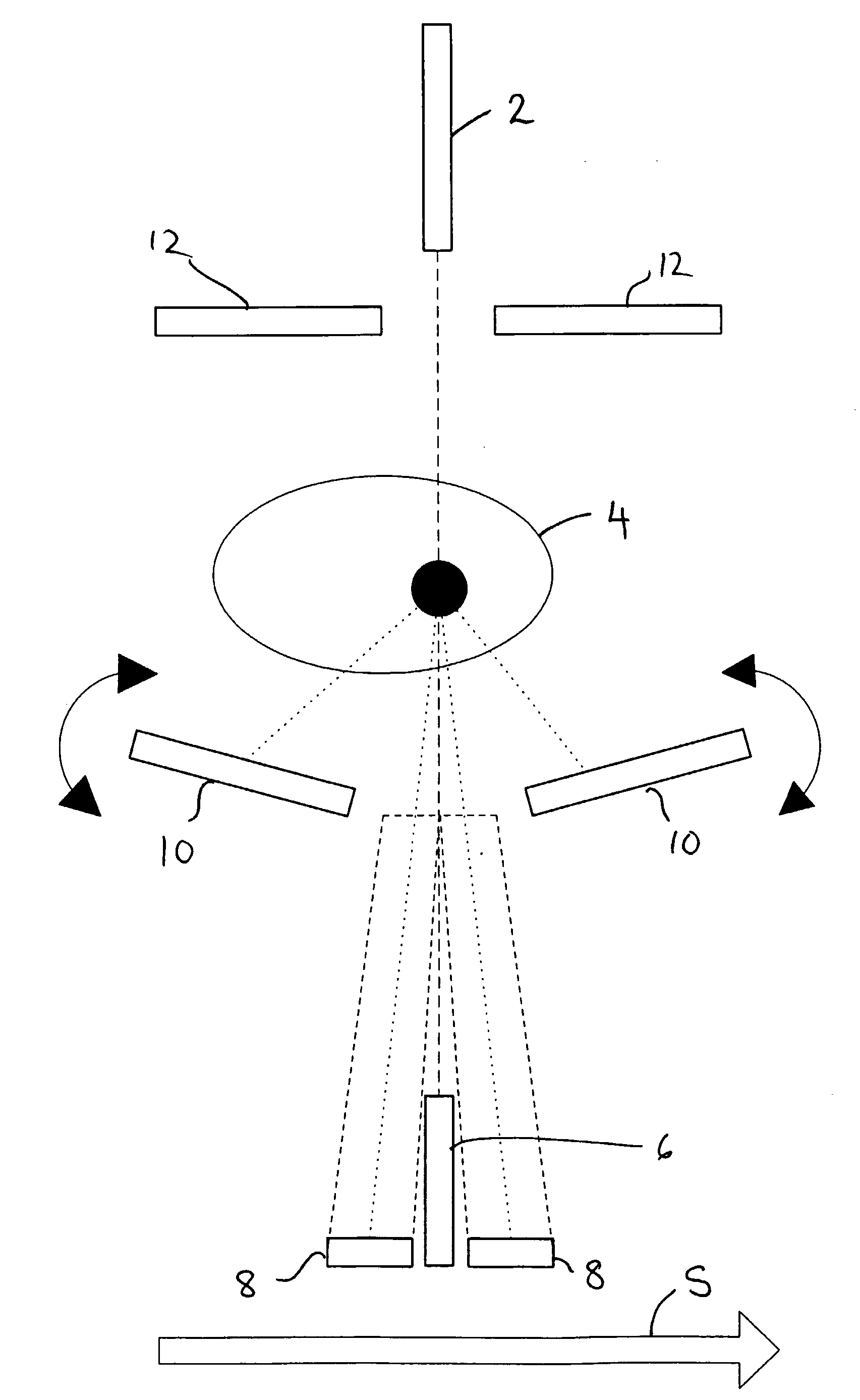
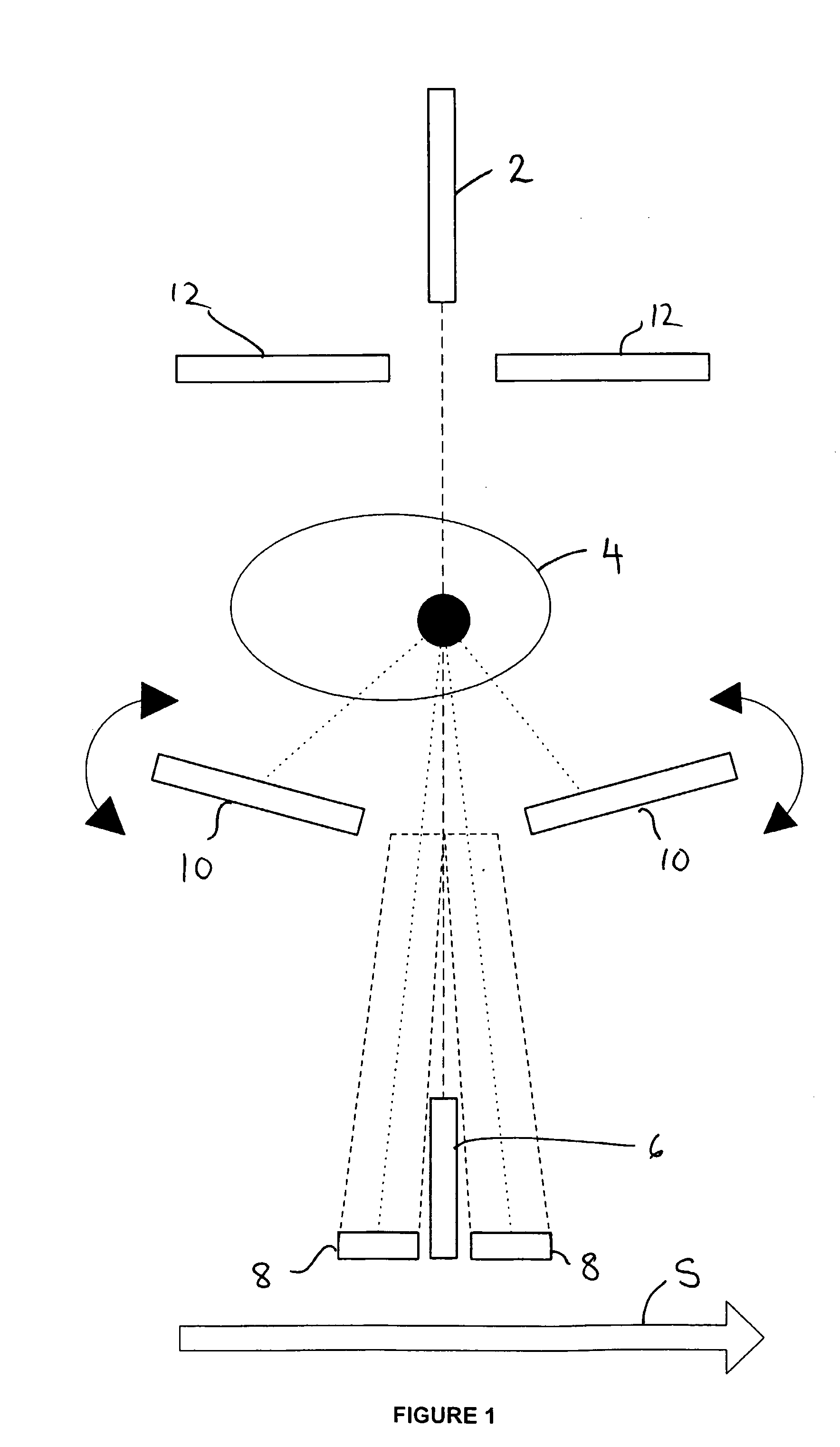
[0045]FIG. 1 illustrates and apparatus suitable for in vivo irradiation of a tissue sample (e.g. a breast). The apparatus comprises a penetrating radiation (in this example X-ray) beam source 2 that directs a beam of X-ray radiation onto the tissue sample 4 being examined. A series of detectors 6, 8, 10, 12 are arranged below and above the sample 4 to detect both transmitted and scattered X-ray radiation.

[0046]A more detailed explanation of the source and detector arrangement is given in our co-pending UK patent application filed on the same date as the present application with the title “Penetrating Radiation Measurements”.

[0047]In use, the source and detector arrangement is scanned across the full length of the tissue sample (e.g. breast), as indicated by arrow ‘S’, whilst the sample is held stationary. The scan is completed in step-wise fashion, with measurements being taken from the detectors at each step.

[0048]The incident beam can be a slit-form beam having a width (into the p...


PUM


Abstract

A method for irradiating a biological tissue sample is provided, the method comprising: irradiating a portion of a biological tissue sample with a penetrating radiation beam for a first exposure period; subsequently irradiating the same or an adjacent biological tissue portion with a penetrating radiation beam for a second exposure period; the radiation dose incident on the tissue sample during the second exposure period being higher than the dose during the first exposure period. Also provided is an apparatus operative in accordance with the method. The method and apparatus have particular application in the characterisation of body tissue by x-ray diffraction, both in vitro and in vivo.

Description

FIELD OF THE INVENTION[0001]The present invention relates to methods and systems for irradiating body tissue. The invention has particular, although not necessarily exclusive application in the characterisation of body tissue, for instance characterisation of tissue as normal (e.g. healthy) or abnormal (e.g. pathological) and has both in vitro and in vivo applications. It is useful in the diagnosis and management of cancer, including breast cancer.BACKGROUND[0002]Mammography is a conventional X-ray technique typically used in the early detection of breast tumours. However, as with any in vivo X-ray (or other penetrating, particularly ionising radiation) technique, the absorption of X-ray radiation by body tissue (the principle on which X-ray imaging relies) causes molecular damage to the tissue. The potential damage increases with absorbed dose (a measure of energy absorbed per unit area).[0003]Over exposure (in one session or cumulatively over time) can significantly increase the l...

Claims


Application Information

Patent Timeline
IPC IPC(8): A61B6/04C12N13/00C12M1/00A61B6/00A61B6/02A61B10/00
CPCA61B6/482A61B6/483A61B6/488A61B6/508A61B6/542A61B10/0041A61B6/502
Inventor GAVED, MATTHEWFARQUHARSON, MICHAEL
Owner GAVED MATTHEW
Why Eureka
Try Eureka