

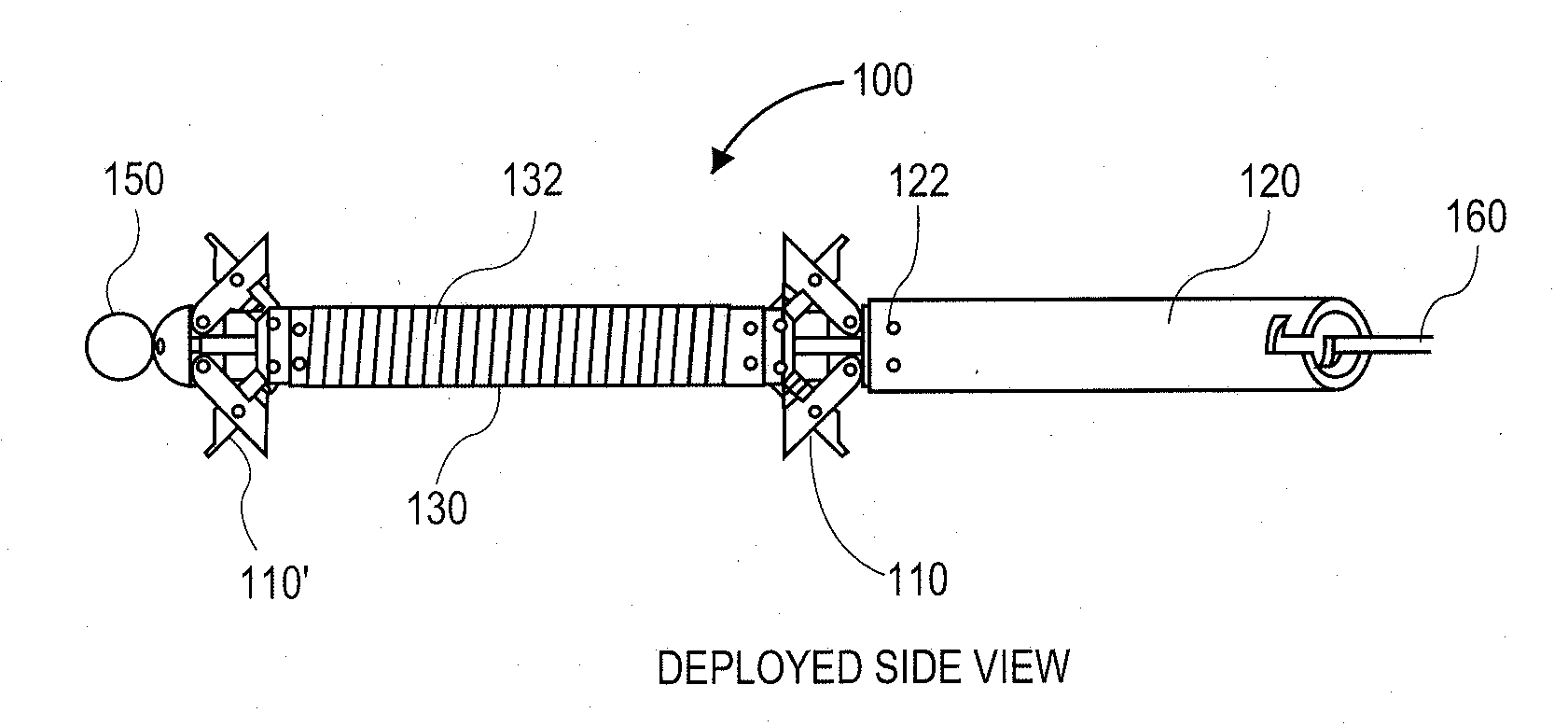
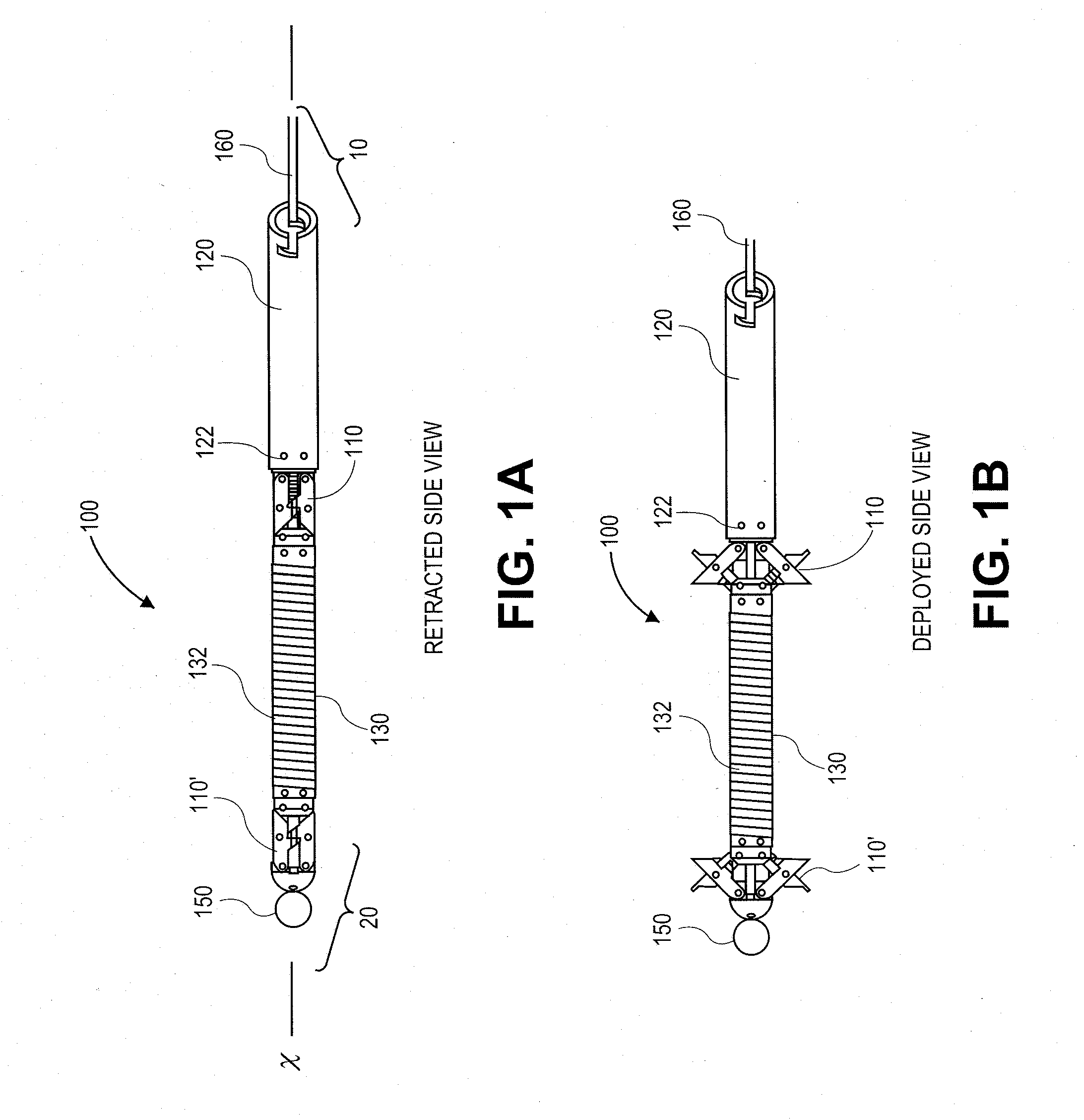
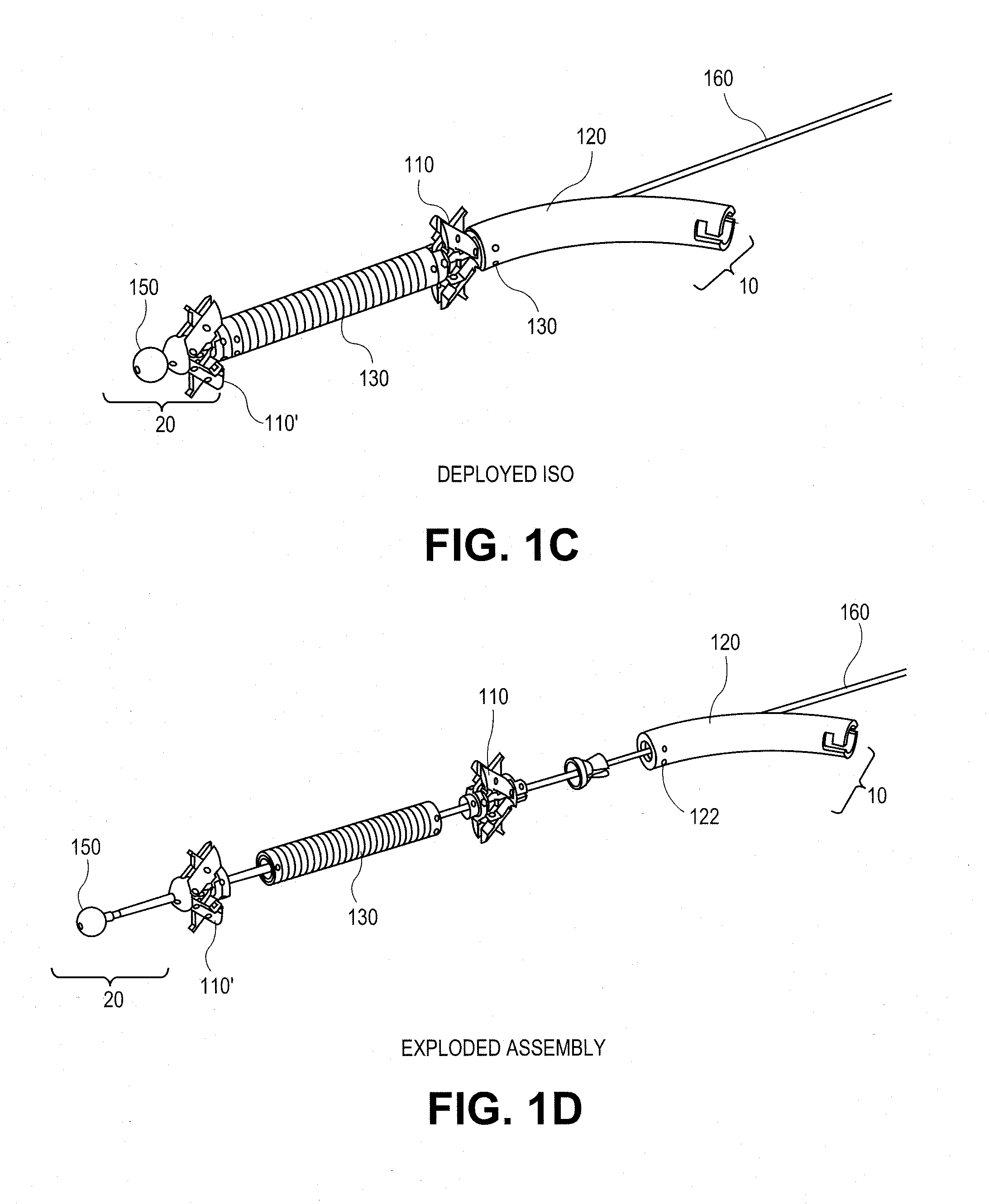
Fracture fixation device, tools and methods
a fixation device and bone technology, applied in the field of methods and systems for providing bone reinforcement, can solve the problems of pain or necrosis of bone, significant number of bone fractures, and take several months for bone to heal, and achieve the effect of convenient us
- Summary
- Abstract
- Description
- Claims
- Application Information
AI Technical Summary
Benefits of technology
Problems solved by technology
Method used
Image

Examples
Embodiment Construction
[0073]By way of background and to provide context for the invention, it may be useful to understand that bone is often described as a specialized connective tissue that serves three major functions anatomically. First, bone provides a mechanical function by providing structure and muscular attachment for movement. Second, bone provides a metabolic function by providing a reserve for calcium and phosphate. Finally, bone provides a protective function by enclosing bone marrow and vital organs. Bones can be categorized as long bones (e.g. radius, femur, tibia and humerus) and flat bones (e.g. skull, scapula and mandible). Each bone type has a different embryological template. Further each bone type contains cortical and trabecular bone in varying proportions. The devices of this invention can be adapted for use in any of the bones of the body as will be appreciated by those skilled in the art.
[0074]Cortical bone (compact) forms the shaft, or diaphysis, of long bones and the outer shell...
PUM
 Abstract
Description
Claims
Application Information