

Non-Evasive Method and Apparatus of Detection of Organism Tissues
a tissue and non-evasive technology, applied in the field of non-evasive methods and apparatus of organism tissues, can solve the problems of complex sample preparation techniques, long invasive sampling process, and inability to detect lesion before, and achieve the effect of simple, quick and accura
- Summary
- Abstract
- Description
- Claims
- Application Information
AI Technical Summary
Benefits of technology
Problems solved by technology
Method used
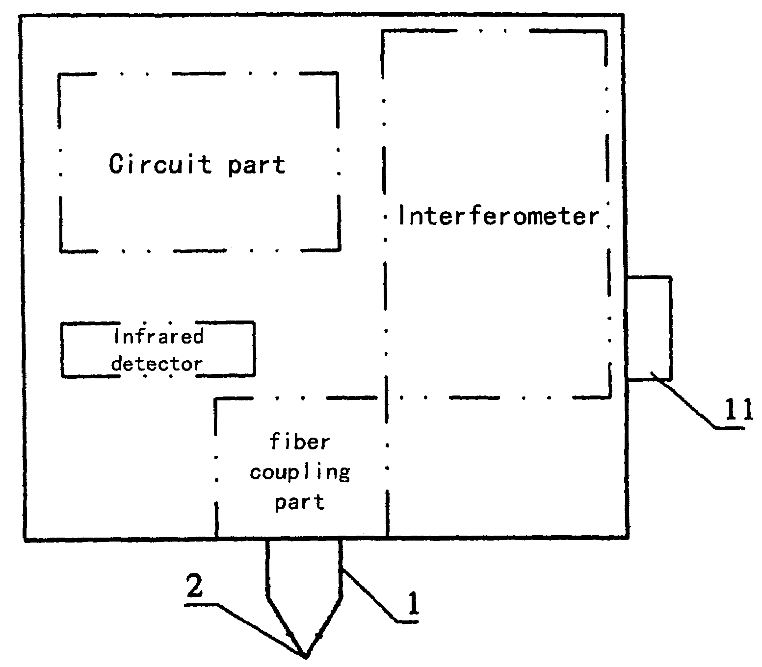
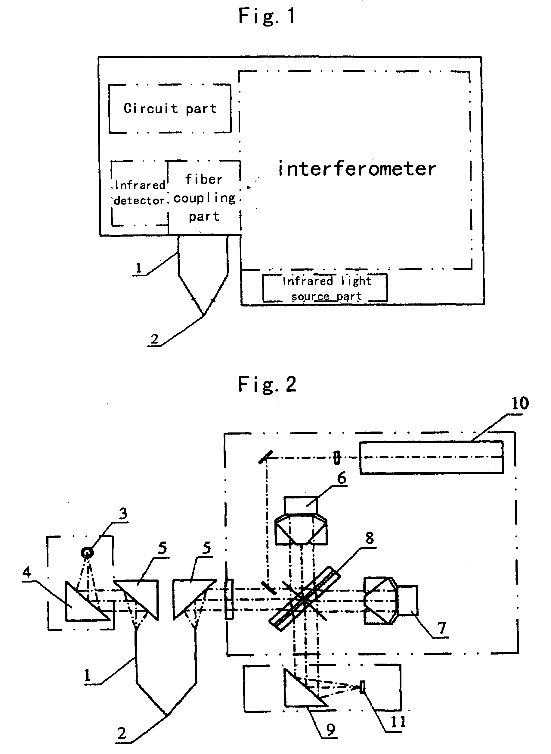
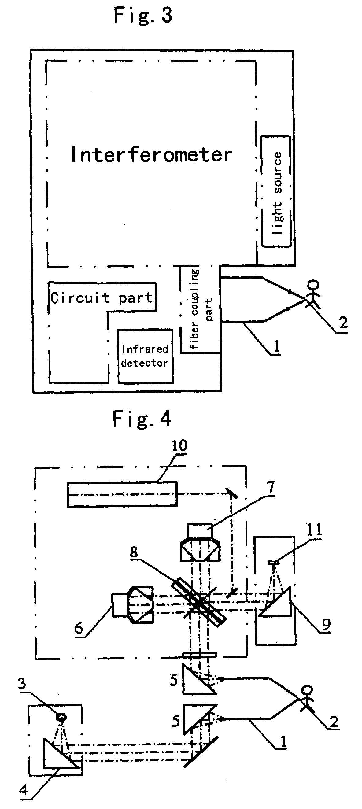
Image

Examples
embodiment 1
Diagnosis of Pathologic Changes in Mammary Glands
[0053]Apparatus being used: a dedicated sealed mid-infrared spectrometer, manufactured by Beijing No. 2 Optical Instruments Factory, provided with mid-infrared fibers, an ATR probe and a MCT (tellurium-cadmium-mercury) detector cooled by liquid Nitrogen.
[0054]Detection Method:
[0055]1. Switching on and adjusting to maximize the output energy of the fiber in which the energy coefficient at 2450 cm−1 is about 56 (Gain=4).
[0056]2. Cleaning the skin of the mammary gland region to be tested with alcohol or water, then placing the ATR probe on the skin of the region to be tested (for patients with mastalgia or touchable lumps, the ATR probe should be placed on the skin outside the pathologic region), scanning and detecting 32 times and based on variations (variations relative to characteristic peaks of normal mammary glands) in characteristic peaks (band of 1000 cm−1-1800 cm−1 and band of 2800 cm−1-3000 cm−1) detection of pathologic changes ...
embodiment 2
Diagnosis of Pathologic Changes in Thyroid Glands
[0059]In accordance with the method and detection conditions described above as Embodiment 1, the ATR probe is placed on the cleaned surface of the skin outside the thyroid gland on one side. Based on the general regularities of variations in healthy and pathologic spectra concluded from the detection results of a number of patients, and on the differences between spectra in the region of 2800-3800 cm−1 and 1000-1800 cm−1, it is possible to diagnose the property and the degree of the pathologic change of the thyroid gland from a spectrum. For example, variations (relative to characteristic peaks of normal thyroid glands) in peak shape and intensity in 2800-3000 cm−1, 1400 cm−1-1500 cm−1 (mainly observing the band of 1200-1580 cm−1) can provide relevant information, which can be inferred by comparison with the standard spectrum data.
embodiment 3
Diagnosis of Pathologic Changes in Parotid Glands
[0060]In accordance with the method and detection conditions described above as Embodiment 1, the ATR probe is placed on the cleaned surface skin outside the parotid glands on both sides (wherein, usually one side is the side with suspected cancerous lumps and the detection results of the other side are used for comparison). As result of comparison, it can be found that there is asymmetry between the spectra on the two sides of a patient with lumps in parotid gland. Based on the general regularities of variations in normal and pathologic spectra concluded from the detection results of a number of patients of different ages and genders, the sensitivity of response in the spectrum is high in the region of 1000-1800 cm−1. It is possible to observe whether the parotid tumor is benign or malignant and the degree of deterioration particularly in the range of 1300-1650 cm−1. The variations (relative to characteristic peaks of normal parotid ...
PUM
 Abstract
Description
Claims
Application Information