

Method for inspecting quality of core material for electrophotographic ferrite carrier
a core material and electrophotographic ferrite technology, applied in chemical methods analysis, resistance/reactance/impedence, instruments, etc., can solve the problems of not being able to judge the level of a surface property of the whole particle constituting a powder as the core material of the electrophotographic ferrite carrier, and the accuracy of the bet method for inspecting the particle is not sufficien
- Summary
- Abstract
- Description
- Claims
- Application Information
AI Technical Summary
Benefits of technology
Problems solved by technology
Method used
Image

Examples
example 1
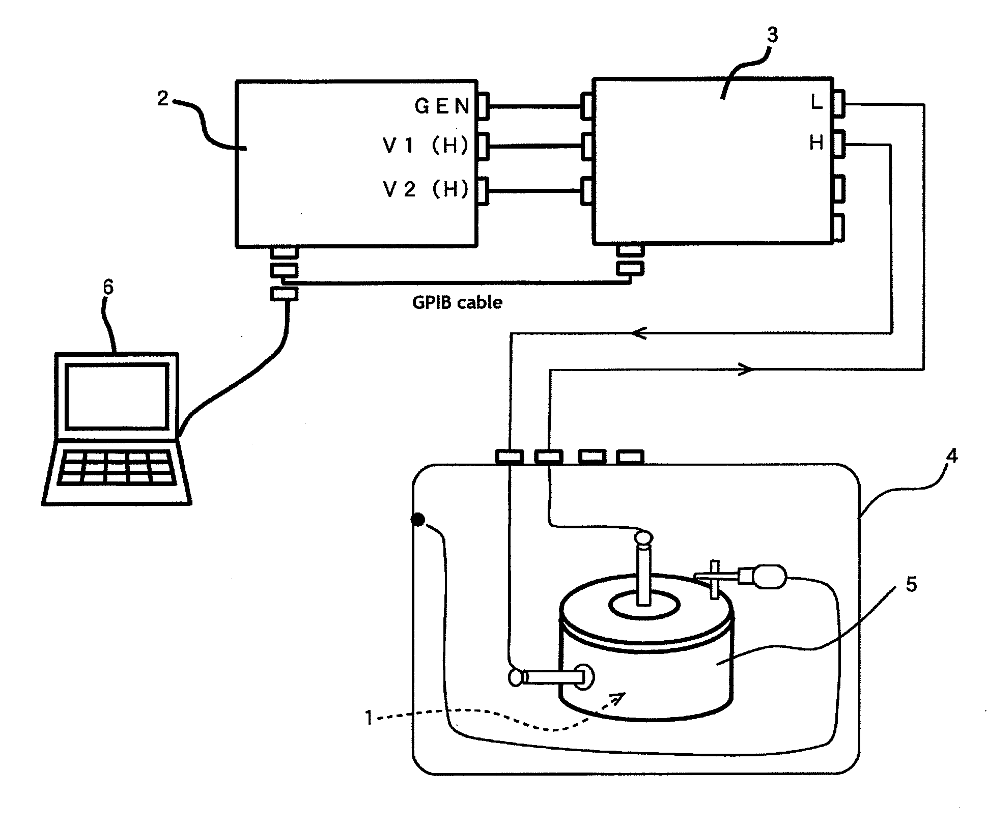
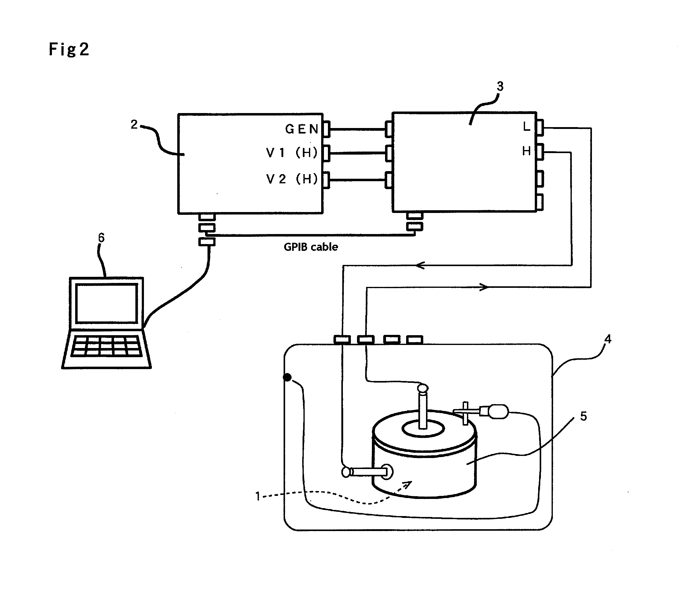
[0041]Example 1 show whether there is a correlation or not between a value obtained in AC-resistance measurement and a specific surface area measured by a BET method which is a conventional measurement. The AC-resistance was measured by using a type 1260 frequency response analyzer (FRA) 2 made by Solartron Analytical (U.K.) having a basic construction illustrated in FIG. 2 connected with a type 1296 dielectric interface 3. As is shown in FIG. 2, the terminals of the type 1296 dielectric interface 3 were wired to each electrode of an input electrode, an output electrode and a guard electrode in a sample cell 5 in a shielded box 4 of the AC-resistance measuring system. In the construction, a sample 1 of a core material for an electrophotographic ferrite carrier was put in the sample cell 5 (suggested by a broken arrow in FIG. 2). The core material for the electrophotographic ferrite carrier used was the Mn—Li ferrite carrier core material which contains a certain amount of manganese ...
example 2
[0049]In the example 2, it will be described that the value obtained by AC-resistance measurement can show a state of each particle of the core material in an electrophotographic ferrite carrier, which never be shown by the value of a specific surface area measured by a BET method of a conventional measurement.
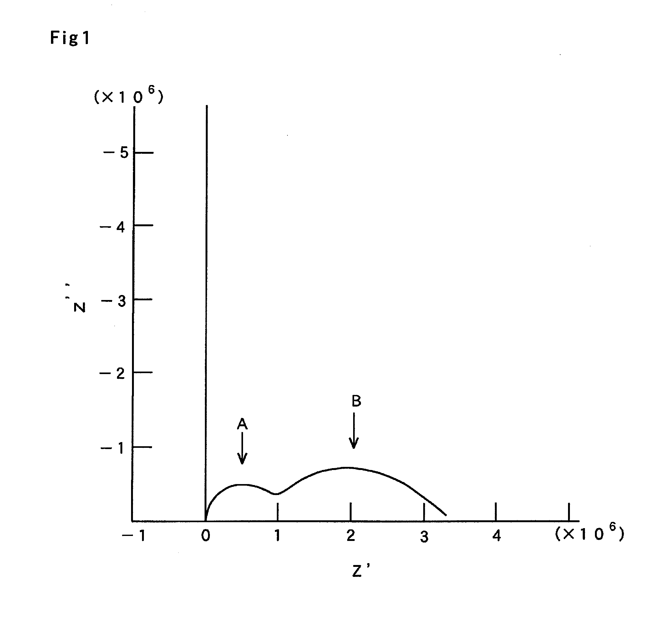
[0050]The sample (A) which had the volume median diameter (D50) of 35 μm and the specific surface area of 0.08 m2 / g measured by a BET method was used as a control sample. FIG. 5 shows an observation image (with magnification of 450 times) of the sample (A), which was obtained by using a scanning electron microscope. The sample (A) showed the value of 0.89 for CPE-P1.
[0051]In contrast, the sample (B) was prepared, which had the same volume median diameter (D50) of 35 μm, but had a completely different specific surface area (0.28 m2 / g) measured by a BET method. FIG. 6 shows an observation image (with magnification of 450 times) of the sample (B), which was obtained by using the ...
PUM
 Abstract
Description
Claims
Application Information