
Apparatus and method for image alignment for combined positron emission tomography (PET) and magnetic resonance imaging (MRI) scanner

a combined positron emission tomography and magnetic resonance imaging technology, applied in tomography, instruments, applications, etc., can solve the problem of limited ability to fully integrate pet and mri systems

Active Publication Date: 2008-10-30
SIEMENS MEDICAL SOLUTIONS USA INC
View PDF5 Cites 23 Cited by
  • Summary
  • Abstract
  • Description
  • Claims
  • Application Information

AI Technical Summary

Benefits of technology

[0007]Thus, there is a clear need for an improved method and apparatus for providing image alignment for combined positron emission tomography (PET) and magnetic resonance imaging (MRI) scanning.
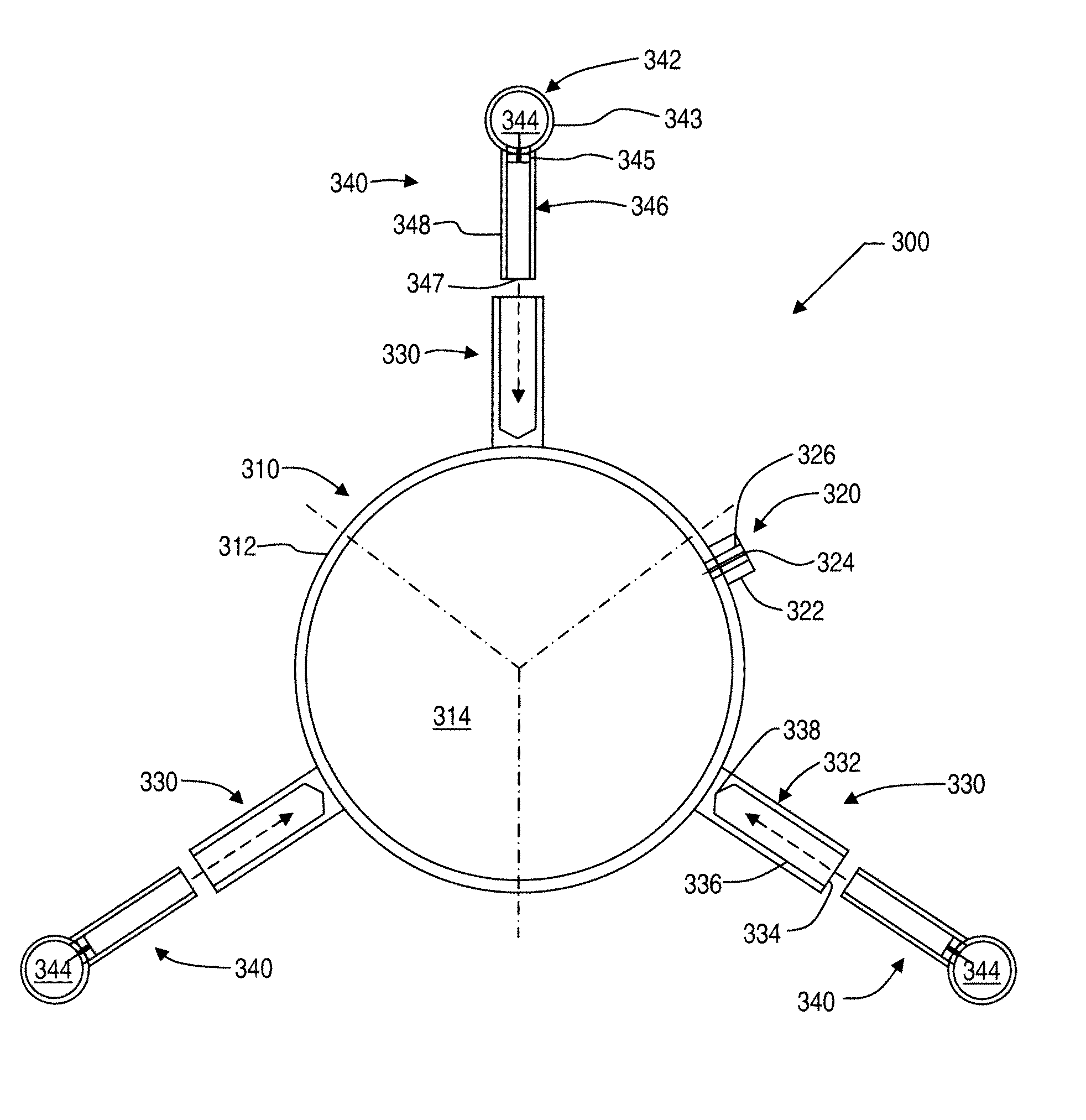
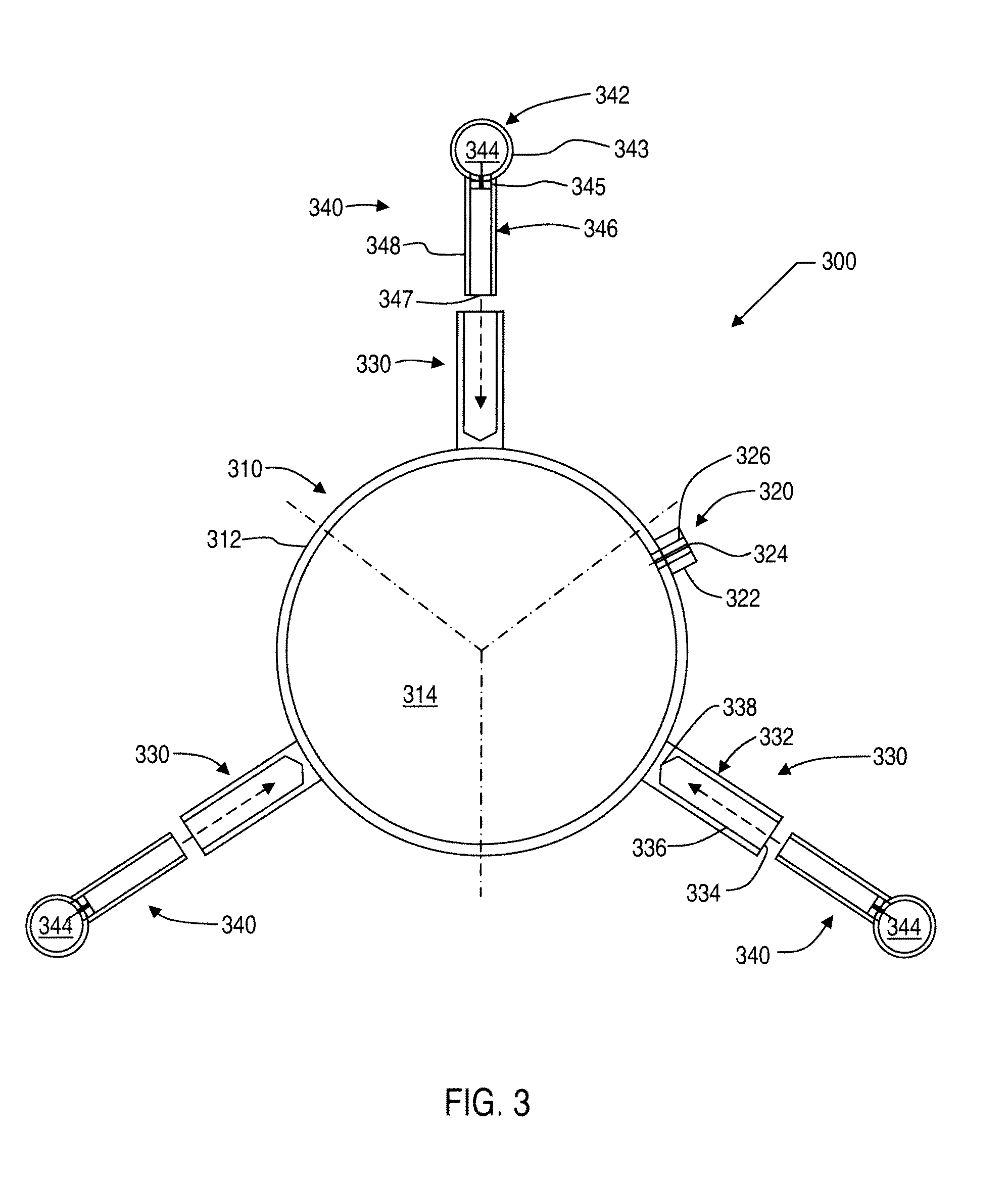
[0008]The present disclosure advantageously provides a phantom that can be used, for example, for co-registering a magnetic resonance image and a nuclear medical image, where an embodiment of the phantom includes a first housing defining a first chamber configured to receive a magnetic resonance material upon which magnetic resonance imaging can be

Problems solved by technology

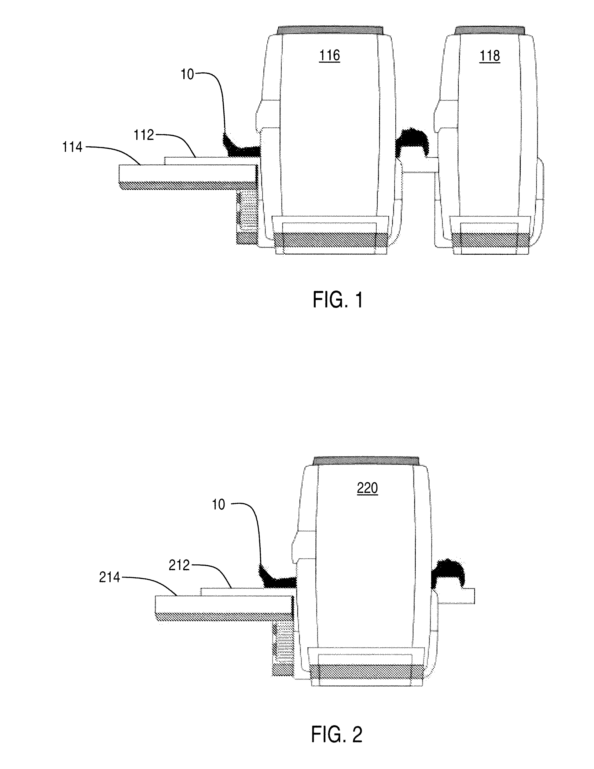
However, numerous obstacles have been present that have limited the ability to fully integrate PET and MRI systems into a combined scanner that produces accurate co-registration of the PET and MRI images.

Method used


Image

  • Apparatus and method for image alignment for combined positron emission tomography (PET) and magnetic resonance imaging (MRI) scanner
  • Apparatus and method for image alignment for combined positron emission tomography (PET) and magnetic resonance imaging (MRI) scanner
  • Apparatus and method for image alignment for combined positron emission tomography (PET) and magnetic resonance imaging (MRI) scanner

Examples


Embodiment Construction

[0016]A method and apparatus for providing image alignment for combined positron emission tomography (PET) and magnetic resonance imaging (MRI) are described. In the following description, for the purposes of explanation, numerous specific details are set forth in order to provide a thorough understanding of the embodiments of the invention. It is apparent, however, to one skilled in the art that the embodiments of the invention may be practiced without these specific details or with an equivalent arrangement. In other instances, well-known structures and devices are shown in block diagram form in order to avoid unnecessarily obscuring the embodiments of the invention.

[0017]Co-registration of anatomical information can greatly improve the diagnostic value of functional imaging. For example, the combination of PET and MRI images can offer numerous advantages, such as higher soft tissue contrast in the MRI anatomical images, real simultaneous acquisition, and minimum radiation exposur...


PUM


Abstract

A phantom and method are provided for co-registering a magnetic resonance image and a nuclear medical image. The phantom includes a first housing defining a first chamber configured to receive a magnetic resonance material upon which magnetic resonance imaging can be performed in order to produce the magnetic resonance image. The phantom also includes three or more second housings configured to be attached to the first housing, where the second housings each define a second chamber configured to receive a radioactive material upon which nuclear imaging can be performed in order to produce the nuclear medical image and upon which the magnetic imaging can be performed in order to produce the magnetic resonance image. The first chamber has a volumetric capacity that is larger than a volumetric capacity of each second chamber.

Description

PRIORITY CLAIM TO RELATED APPLICATION[0001]This application claims priority from U.S. Provisional Patent Application Ser. No. 60 / 914,403, filed Apr. 27, 2007, the disclosure of which is incorporated by reference in its entirety herein.FIELD[0002]The present invention, according to certain embodiments, relates to combined nuclear medicine imaging, such as positron emission tomography (PET), and magnetic resonance imaging (MRI).BACKGROUND[0003]Magnetic resonance imaging (MRI) is primarily used in medical imaging to visualize anatomical structure of a patient's body. MRI technology can provide detailed images of the body in any plane. MRI has the ability to show soft tissue contrasts, which makes MRI scans especially useful in neurological, musculoskeletal, cardiovascular, and oncological imaging. MRI scans use a powerful magnetic field to align the magnetization of hydrogen atoms in the body. Radio waves are used to systematically alter the alignment of such magnetization, thereby cau...

Claims


Application Information

Patent Timeline
IPC IPC(8): A61B5/055A61B6/00
CPCG01R33/481G01R33/58A61B6/583A61B6/037G01T1/1603
Inventor PAUL, RAINERSCHMAND, MATTHIAS J.HAYDEN, CHARLES H.CORBEIL, JAMESMICHEL, CHRISTIAN J.BURBAR, ZIADMATSCHL, VOLKER
Owner SIEMENS MEDICAL SOLUTIONS USA INC
Learn More