Implantable Biocompatible Immunoisolatory Vehicle for Delivery of Gdnf

a biocompatible, immunoisolation technology, applied in the direction of peptide/protein ingredients, drug compositions, genetically modified cells, etc., can solve the problems of reducing the stability of the blood brain barrier, and reducing the risk of zoonosis and adverse immune reactions. , the effect of reducing the risk of “units”

Inactive Publication Date: 2008-11-20
NSGENE AS
View PDF24 Cites 2 Cited by
  • Summary
  • Abstract
  • Description
  • Claims
  • Application Information

AI Technical Summary

Benefits of technology

[0012]By using human cells the risk of zoonosis and adverse immune reactions is reduced.
[0014]In a particularly preferred embodiment, the cells are capable of phagocytising. In this way debris from dying cells within the device may be removed and the environment within the device may be kept “clean”.

Problems solved by technology

Because GDNF does not readily cross the blood brain barrier, its administration into the central nervous system requires the use of invasive procedures, which may compromise the integrity of the blood brain barrier.
Every occasion in which the pump reservoir must be replaced or the injection syringe reinserted through the cannulae represents another opportunity that contaminants might be introduced into the brain, which is especially susceptible to infection.
Even with the careful use of sterile procedures, there is risk of infection.
In addition to the risk of infection, there seems to be some risk associated with the infusion procedure.
However, gene therapy requires the use of virus vectors which use is inherently associated with risks of insertional mutagenesis and tumorigenesis as well as the inability to stop the GDNF secretion should untoward effects occur.
However, naked cells integrate into the tissue into which they have been transplanted making a termination of the treatment almost impossible.
Furthermore, transplanted naked cells may migrate once inside the brain and establish undesirable populations of GDNF secreting cells in the brain.
Although for both BHK and C2C12 cells proof of concept in treatment of PD was provided in rats and baboon disease models, these cells have not been used in clinical studies.
The major reason for this is that both BHK and C2C12 cells even if encapsulated are not useful for therapy of human beings, because of the potential risk of triggering an immune-response or zoonosis from the use of xenogeneic cells and the poor long-term viability BHK and C2C12 cells show after encapsulation possibly due to continued proliferation.

Method used


Image

  • Implantable Biocompatible Immunoisolatory Vehicle for Delivery of Gdnf
  • Implantable Biocompatible Immunoisolatory Vehicle for Delivery of Gdnf
  • Implantable Biocompatible Immunoisolatory Vehicle for Delivery of Gdnf

Examples


example 1

Cloning and Expression of GDNF

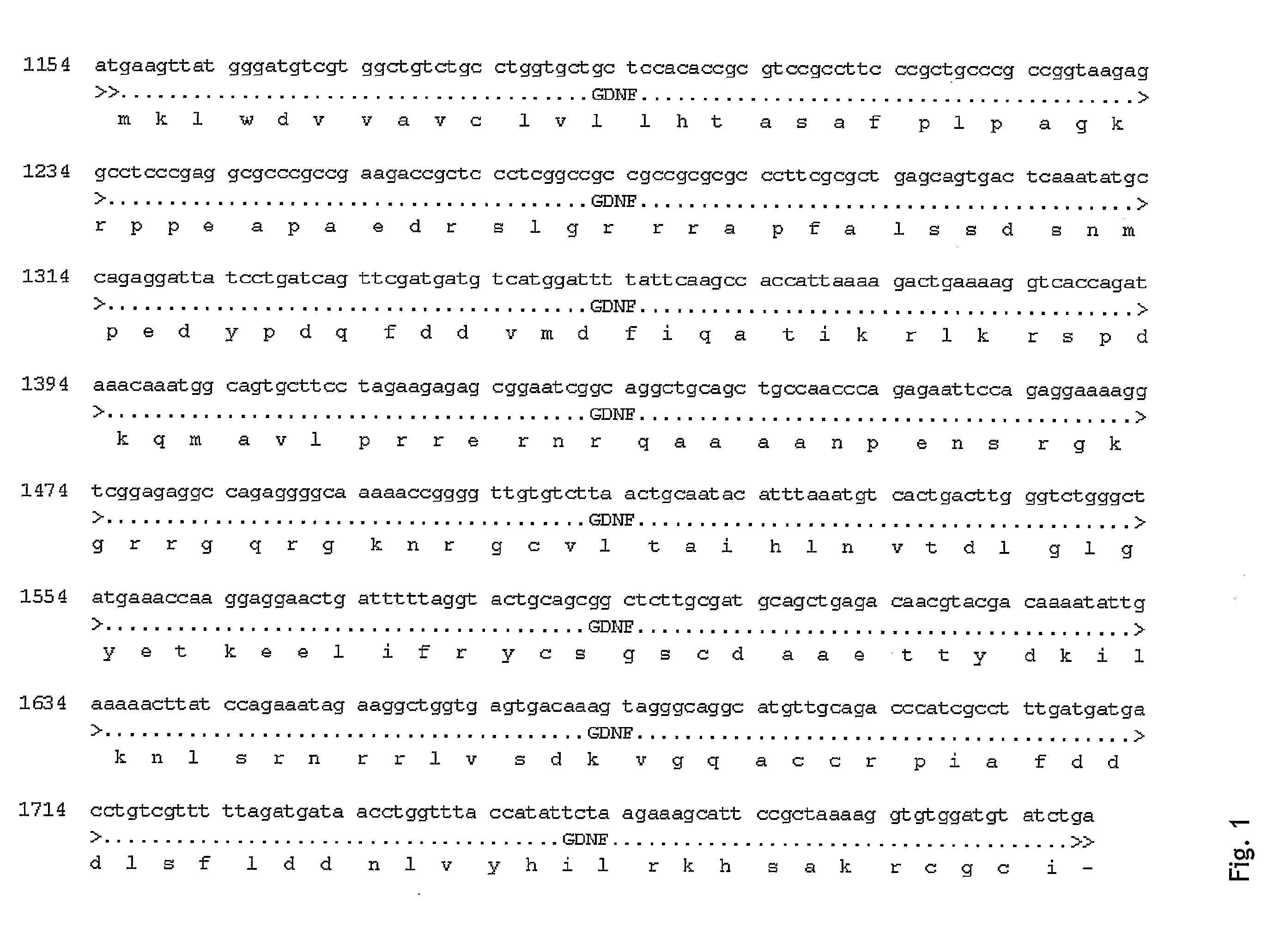
[0189]Cloning of GDNF from caudate nucleus polyA mRNA

[0190]cDNA was synthesized from Caudate nucleus polyA mRNA (Clontech, Becton Dickinson, catalog number 636132) as described previously (Current Protocols in Molecular Biology). A fragment encoding long isoform of pre-pro- was amplified from cDNA by PCR using the primers rGDNFs (5′-GGTCTACGGAGACCGGATCCGAGGTGC-3′; SEQ ID No 15) and rGDNFas (5′-TCTCTGGAGCCAGGGTCAGATACATC-3′; SEQ ID No 16) essentially as described in Schaar et al. 1994 (Exp. Neurol. 130, 387-393). The resulting PCR product was purified and cloned into the SrfI site of pCRScript vector (Stratagene). Subsequently, the GDNF cDNA fragment was subcloned into the BamHI / XhoI sites in pNS1n and pCln.hNGF to yield pNS1n.hGDNF and pCln.hG, respectively. pNS1n is a customised vector derived from pcDNA3 (Invitrogen) with expression under control of the cytomegalovirus promoter carrying the neo resistance marker instead of zeo (Jensen et al 2002, J Bi...

example 2

Evaluation of GDNF Release from Confluent Stable Clones

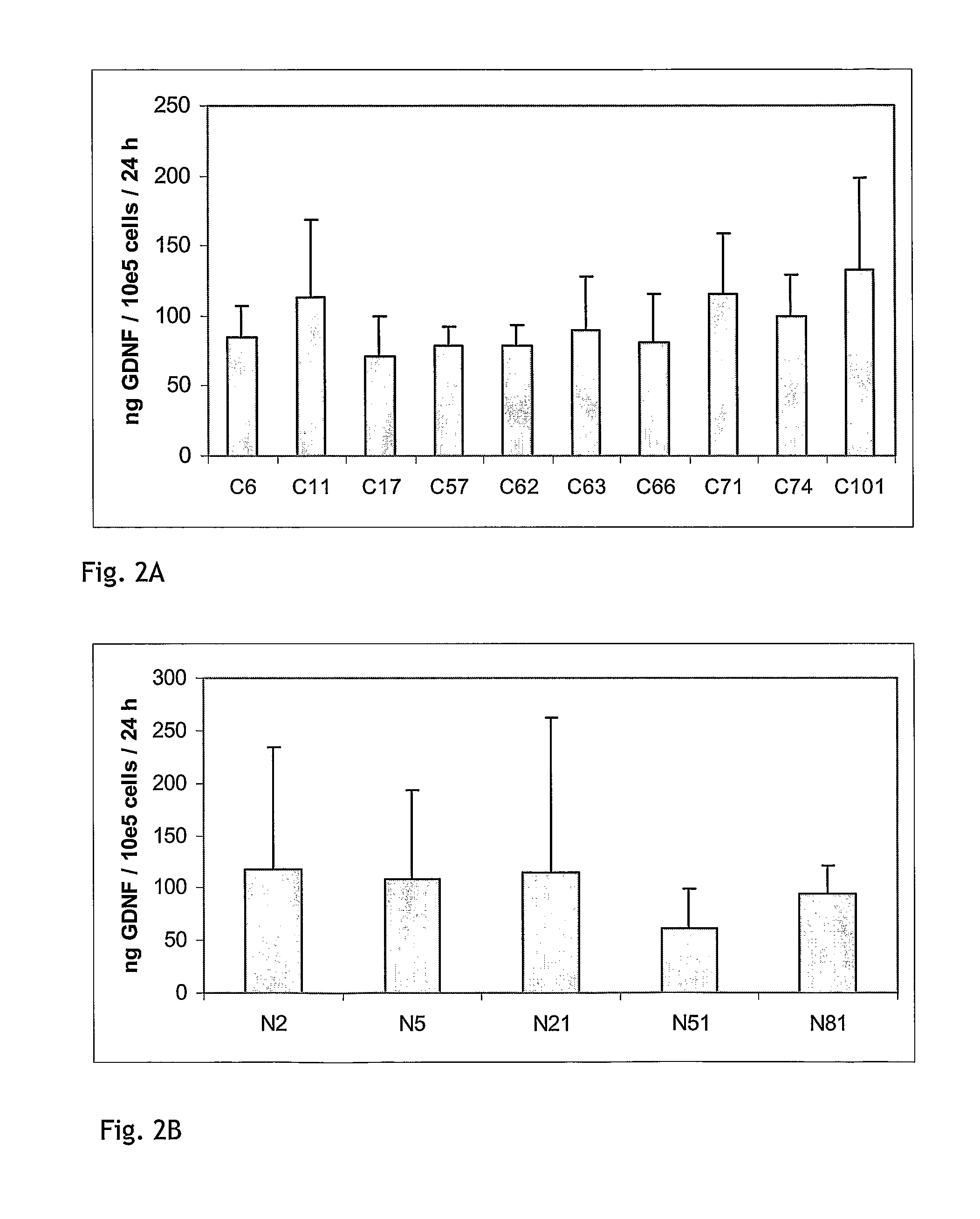
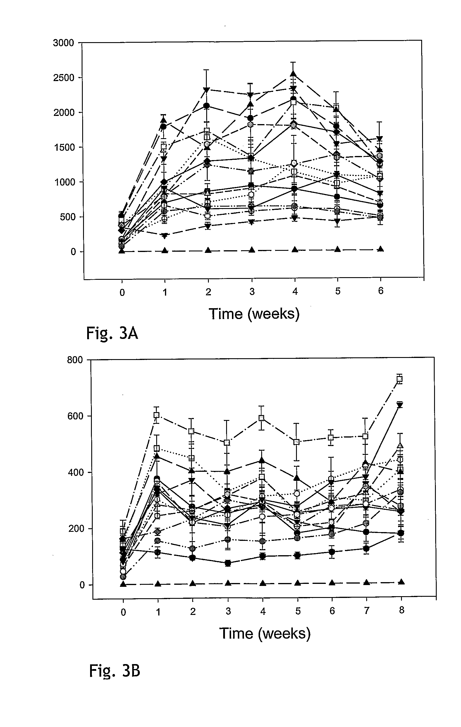
[0195]To evaluate the stability of GDNF release in confluent cultures of stable clones, a fixed number of cells were seeded in growth medium. The next day, medium was replaced with Human Endothelial Serum-free Medium (HE-SFM) from Invitrogen (cat#11111-044) or growth medium. After 4 h incubation, media were removed and subjected to GDNF ELISA analysis using the DuoSet human GDNF ELISA kit (REF RED systems) according to the manufacturer's recommendations. ELISA values were calculated as ng GDNF / ml / 24 h. Cells were allowed to grow to confluency in HE-SFM or growth medium and were maintained in culture for up to 8 weeks. GDNF release in 4 h media was determined every week. The GDNF levels for representative clones transfected with the constructs pCln.hG and pNS1n.hGDNF are shown in FIGS. 3A and 3B. At the end of the experiment, cells were trypsinated and counted. FIG. 4A shows GDNF release for week 4 in HE-SFM calculated as ng GDNF...

example 3

Processing and Glycosylation of GDNF Secreted from ARPE-19 Cells

[0196]The purpose of this experiment is to analyse GDNF secreted from transfected ARPE-19 clones in Western blot analysis to confirm glycosylation and correct processing of the secreted GDNF.

[0197]Briefly, conditioned media from GDNF-producing ARPE-19 cells were diluted in deglycosylation reaction buffer (Prozyme Enzymatic Deglycosylation kit #GK80110) to a concentration of 0.2 ng / μl according to ELISA results. Recombinant mammalian produced hGDNF (R&D systems #212GD) was diluted to the same concentration and used as reference. Deglycosylations with and without denaturing step were performed according to protocol 3.2 and 3.3 provided by manufacturer. Samples were etectrophoresed on 8-18% gradient SDS gets. E. coli produced hGDNF (Alomone Labs #G-240) was used as a reference for non-glycosylated GDNF. Proteins were transferred to PVDF membrane. The blocked membrane was incubated with anti-GDNF antibody (R&D Systems, No A...


PUM

PropertyMeasurementUnit
volumeaaaaaaaaaa
volumeaaaaaaaaaa
diameteraaaaaaaaaa

Abstract

The present invention relates to devices comprising a composition of human cells secreting a therapeutically effective amount of GDNF (Glial cell-line-derived neurotrophic factor) encapsulated in a device comprising a core and a semipermeable membrane allowing for the diffusion of GDNF protein. The human cells are from one cell line.

Description

[0001]The present application relates to devices containing GDNF secreting cells, which devices may be used for the treatment of Parkinson's Disease, Amyotrophic Lateral Sclerosis, Huntington's Disease and retinopathies. All references cited herein are incorporated by reference.PRIOR ART[0002]GDNF is a neurotrophic factor capable of promoting the survival of dopaminergic neurons. In Parkinson's disease, the dopaminergic neurons degenerate. There are ample reports in literature showing that that long-term delivery of GDNF is a feasible way to treat Parkinson's disease. Because GDNF does not readily cross the blood brain barrier, its administration into the central nervous system requires the use of invasive procedures, which may compromise the integrity of the blood brain barrier.[0003]Long-term delivery of GDNF to the central nervous system behind the blood-brain barrier can be accomplished in different ways: continuous infusion using implanted pumps or cannulae, in vivo gene therap...

Claims


Application Information

Patent Timeline
Patent Type & Authority Applications(United States)
IPC IPC(8): A61K48/00A61P25/16A61P27/02A61K35/12
CPCA61K38/185A61K48/0008A61K48/005A61K2035/126C12N5/0621C12N2501/13C12N2510/00A61P9/00A61P25/00A61P25/14A61P25/16A61P25/28A61P27/02A61P27/06
Inventor TORNOE, JENSWAHLBERG, LARS U.
Owner NSGENE AS
Why Eureka
Try Eureka