Methods for pre-stressing and capping bioprosthetic tissue

a bioprosthetic tissue and pre-stressing technology, applied in the field of methods, can solve the problems of undesirable stiffening or degradation of the bioprosthetic, inability to address the changes within the tissue, and inability to achieve the desired effect of reducing in vivo calcification

Active Publication Date: 2008-12-11
EDWARDS LIFESCIENCES CORP
View PDF101 Cites 66 Cited by
  • Summary
  • Abstract
  • Description
  • Claims
  • Application Information

AI Technical Summary

Benefits of technology

[0019]Another aspect of the invention is a method of treating bioprosthetic implant tissue to reduce in vivo calcification, comprising cyclically stressing the bioprosthetic implant tissue until the rate of acid production in the bioprosthetic implant tissue decreases by about 10%, and then applying a calcification mitigant to the stressed tissue. Desirably, the bioprosthetic implant tissue comprises a bioprosthetic heart valve, and the step of stressing comprises subjecting the heart valve to pulsed fluid flow therethrough. The step of stressing may be done until the rate of increase of the acid level of the bioprosthetic implant tissue levels off. The calcification mitigant may be a capping agent solution having at least one constituent that can bind to calcium or phosphate, and/or immunogenic binding sites. Alternatively, the calcification mitigant comprises a linking agent solution including a long elastic molecule.
[0020]Another aspect of the invention is method of treating a bioprosthetic heart valve to reduce in vivo calcification, comprising mounting a biopro...

Problems solved by technology

One problem associated with the implantation of many bioprosthetic materials is that the connective tissue proteins (i.e., collagen and elastin) within these materials can become calcified following implantation within ...

Method used


Image

  • Methods for pre-stressing and capping bioprosthetic tissue
  • Methods for pre-stressing and capping bioprosthetic tissue
  • Methods for pre-stressing and capping bioprosthetic tissue

Examples


Embodiment Construction

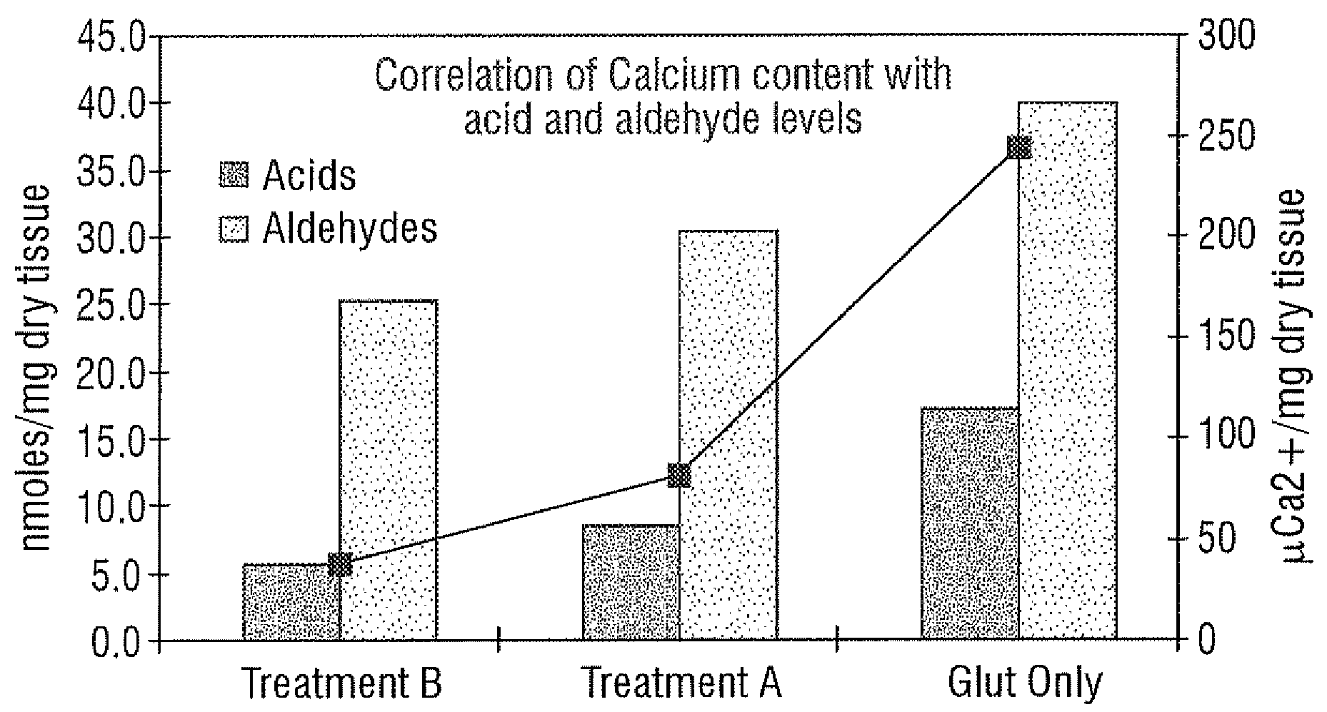
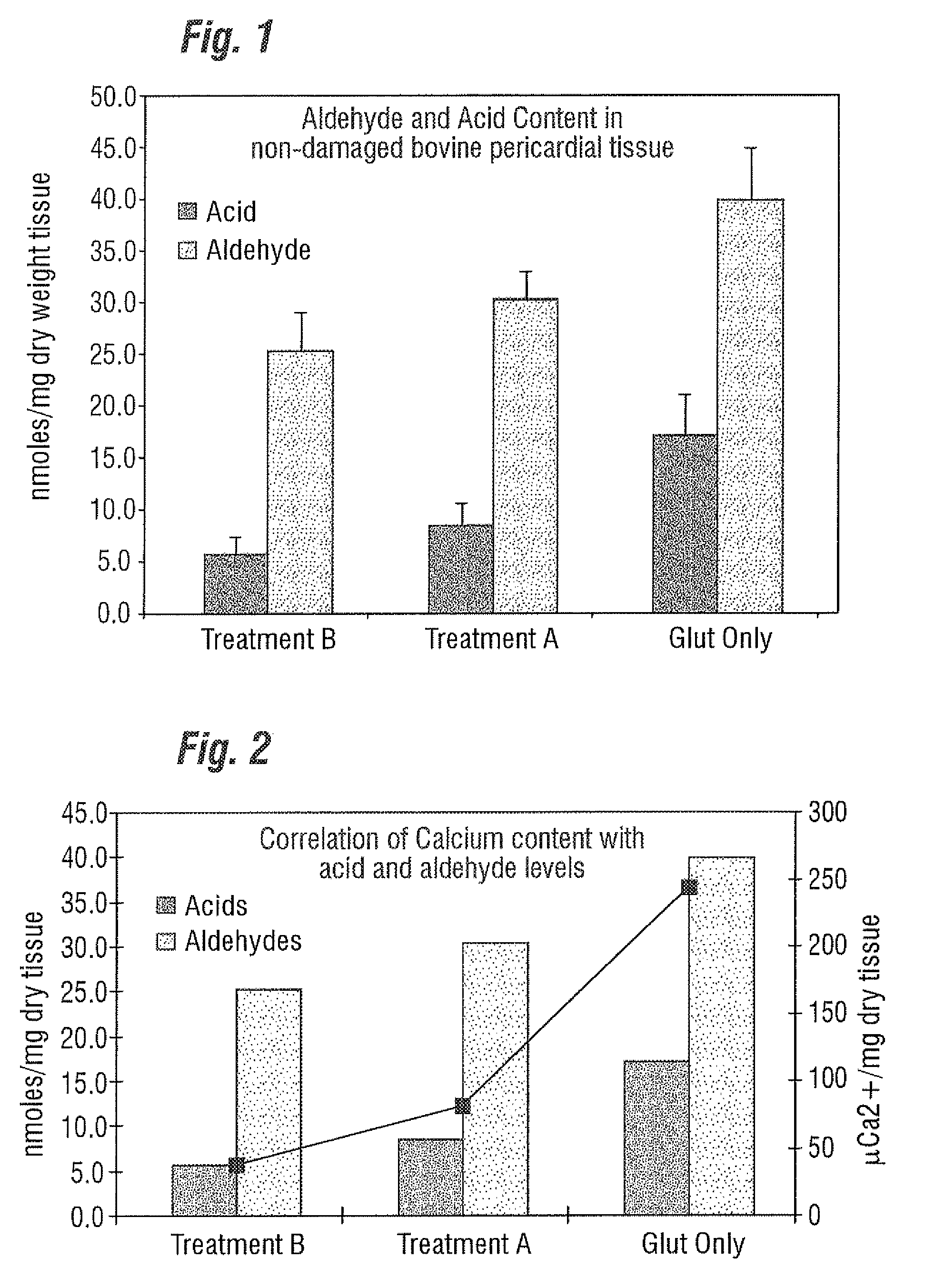
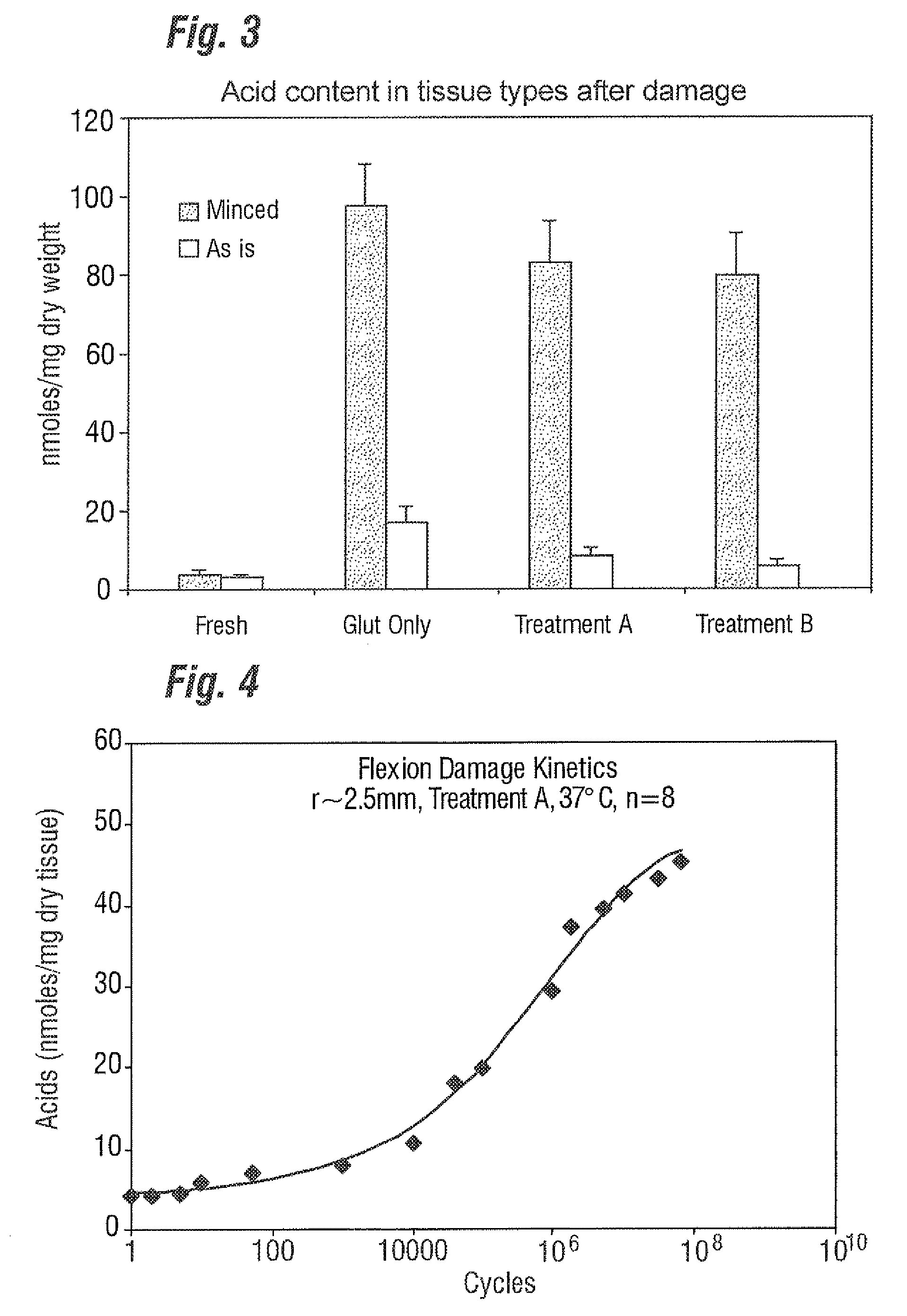
[0029]Unlike the prior art that focuses only on “as-processed tissue” and without any consideration of how the tissue calcification propensity can change from imposed service stresses, the present invention provides an improved bioprosthetic tissue treatment process that is believed to greatly reduce the potential for calcification after implantation. “Bioprosthetic tissue” in this sense means at least bovine pericardium and whole porcine valves which are commonly used in bioprosthetic heart valves that must endure years and even decades without calcifying. Other tissues that may be improved by the treatment include blood vessels, skin, dura mater, pericardium, small intestinal submucosa (“SS tissue”), ligaments and tendons. “Implants” in the present application refers not only to heart valves but also to vascular prostheses and grafts, tissue grafts, bone grafts, and orbital implant wraps, among others.

[0030]A “bioprosthetic heart valve” in the present application refers to a fully...


PUM

PropertyMeasurementUnit
Temperatureaaaaaaaaaa
Fractionaaaaaaaaaa
Frequencyaaaaaaaaaa

Abstract

A treatment for bioprosthetic tissue used in implants or for assembled bioprosthetic heart valves to reduce in vivo calcification is disclosed. The method includes preconditioning, pre-stressing, or pre-damaging fixed bioprosthetic tissue in a manner that mimics the damage associated with post-implant use, while, and/or subsequently applying a calcification mitigant such as a capping agent or a linking agent to the damaged tissue. The capping agent suppresses the formation of binding sites in the tissue that are exposed or generated by the damage process (service stress) and otherwise would, upon implant, attract calcium, phosphate, immunogenic factors, or other precursors to calcification. The linking agent will act as an elastic reinforcement or shock-absorbing spring element in the tissue structure at the site of damage from the pre-stressing. In one method, tissue leaflets in assembled bioprosthetic heart valves are preconditioned by simulating actual flow conditions for a predetermined number of cycles, during or after which the valve is exposed to the capping agent.

Description

[0001]The present Application for Patent claims priority to U.S. Provisional Application No. 60 / 943,118 entitled, “METHODS FOR PRE-STRESSING AND CAPPING BIOPROSTHETIC TISSUE,” filed Jun. 11, 2007, and assigned to the assignee hereof ad hereby expressly incorporated by reference herein.FIELD OF THE INVENTIONBackground of the Invention[0002]Heart valve replacement may be indicated when there is a narrowing of the native heart valve, commonly referred to as stenosis, or when the native valve leaks or regurgitates, such as when the leaflets are calcified. In one therapeutic solution, the native valve may be excised and replaced with either a biologic or a mechanical valve.[0003]Tissue-type or “bioprosthetic” valves have flexible leaflets supported by a base structure that project into the flow stream and function much like those of a natural human heart valve by coapting against each other to ensure one-way blood flow. In tissue-type valves, a whole xenograft valve (e.g., porcine) or a ...

Claims


Application Information

Patent Timeline
IPC IPC(8): A61L27/00A61L27/28A61L27/50
CPCA61L27/3625A61L27/3683A61L27/3691A61L27/507A61L2400/02A61L2430/40A61F2210/0004A61F2210/0076A61L27/3687A61F2/2412A61L2430/20
Inventor DAVIDSON, JIMDOVE, JEFFDOBLER, DARIN
Owner EDWARDS LIFESCIENCES CORP
Why Eureka
Try Eureka