Agents for prophylaxis or treatment of neurological related diseases and conditions

a technology for applied in the field of agents for prophylaxis or treatment of neurological related diseases and conditions, can solve problems such as difficult studies and achieve the effect of reducing binding

Inactive Publication Date: 2008-12-11
CHILDRENS MEDICAL RES INST +2
View PDF1 Cites 9 Cited by
  • Summary
  • Abstract
  • Description
  • Claims
  • Application Information

AI Technical Summary

Benefits of technology

[0031]FIG. 1: Phosphorylation-dependent interaction of syndapin I and endophilin with dynamin I in vitro (a) Dynamin consists of four distinct domains: the GTP hydrolysis domain (GTPase), a pleckstrin homology (PH) domain, an assembly domain (AD) and a proline-rich domain (PRD) and is phosphorylated by Cdk5 at Ser-774 and Ser-778 in vivo in the DynI “phospho-box” (769PAGRRSPTSSPTPQRRAPAVPPARPGSRGP798) (SEQ ID No. 6). Point mutations were made in phospho-box residues at the indicated positions (arrows). (b) GST-DynI-PRD either WT, dmA or dmE coupled to GSH-sepharose were used in pull-downs from brain lysates. Bound proteins were separated by SDS-PAGE and stained with Coomassie Blue. (c) MALDI-MS peptide mass spectrum of a 52 kDa band identified as syndapin I. (d) MALDI-MS of the 52 kDa band revealed 42% sequence coverage of syndapin I (SEQ ID No. 7). (e) Effect of individual DynI-PRD mutants on syndapin binding in pull-down experiments (upper panels). The amount of syndapin (f) or endophilin (g) bound to GST-DynI-PRD mutants was quantified by densitometry analysis of Western blots (n=5). The data is expressed as a percent of DynI-PRD-WT ±S.E.M.
[0032]FIG. 2: Phosphorylation-dependent interaction with syndapin I in vivo. (a) Amphiphysin SH3 domain binds all synaptosomal dynamin. (b) Effect of in vivo dynamin phosphorylation on binding SH3 domains. The samples were probed with phospho-Ser-774 and phospho-Ser-778 antibodies (bottom four panels). (c) Effect of in vivo dynamin I phosphorylation on binding full length proteins. (d) Phospho-dynamin I showed reduced binding to full-length syndapin I but not full-length endophilin.

Problems solved by technology

The former peptide included Ser-778, but no PxxP motif, making it difficult to reconcile the studies.

Method used


Image

  • Agents for prophylaxis or treatment of neurological related diseases and conditions
  • Agents for prophylaxis or treatment of neurological related diseases and conditions
  • Agents for prophylaxis or treatment of neurological related diseases and conditions

Examples


example 1

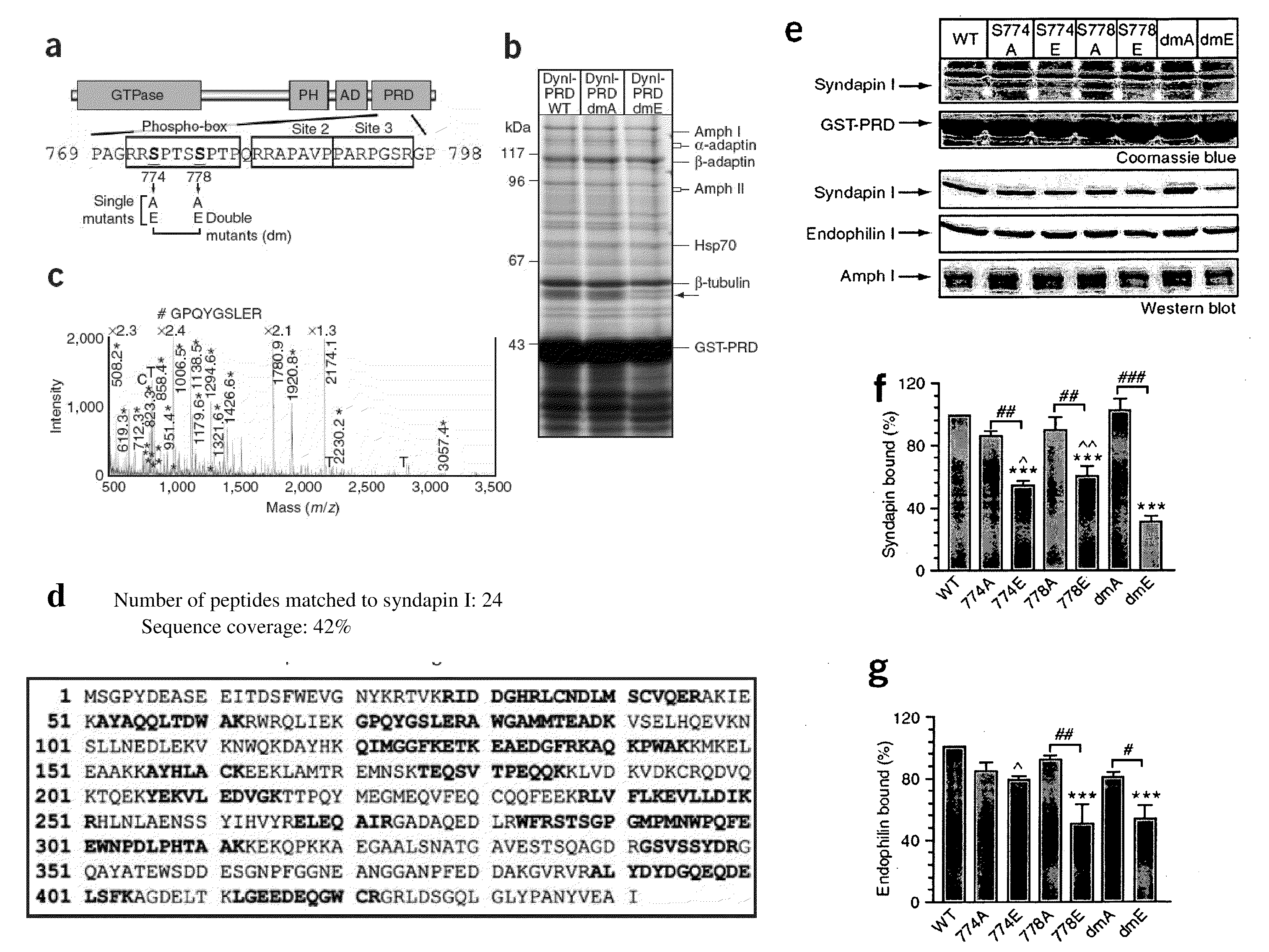
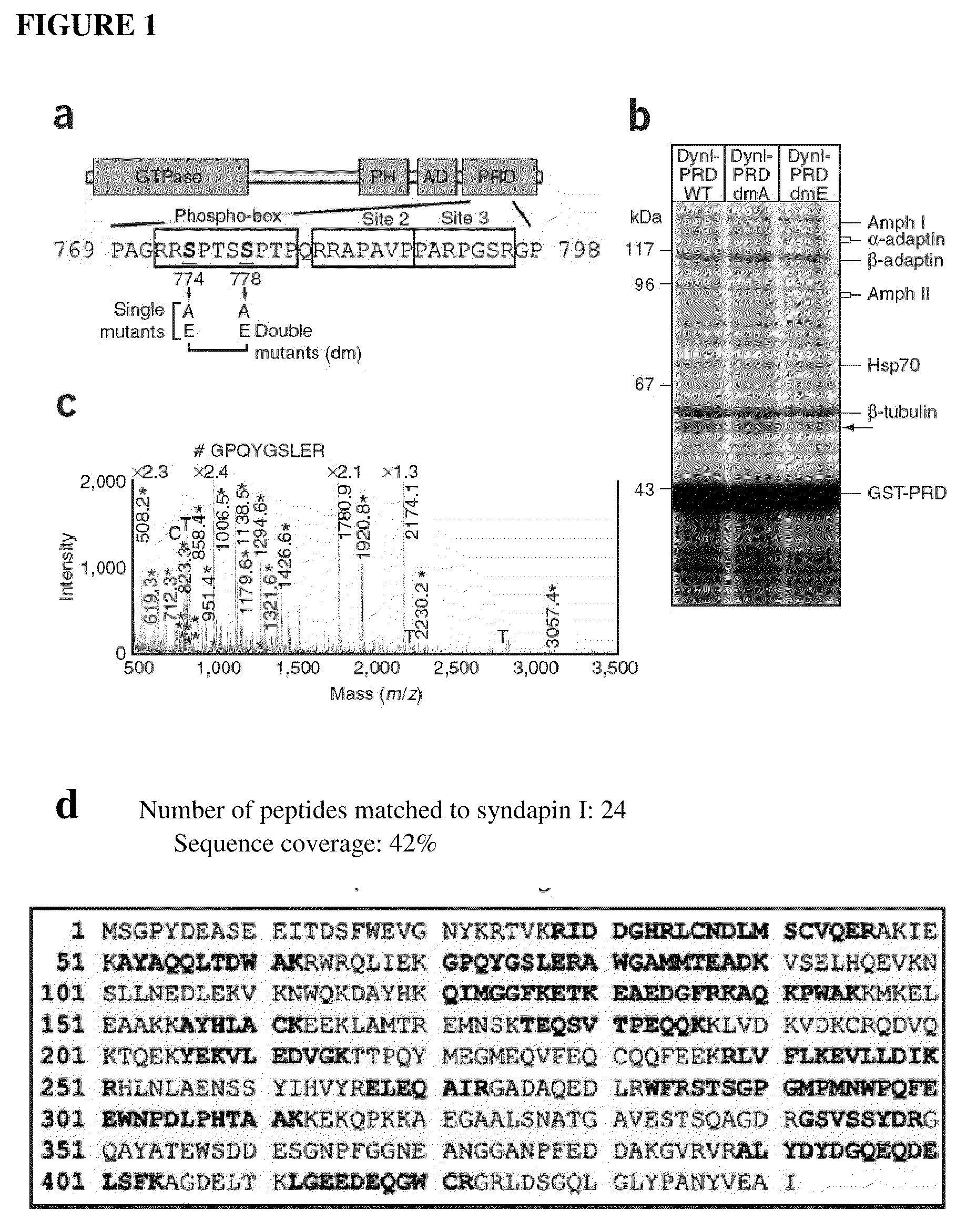
Identification of Syndapin I Binding to DynI

1. Materials and Methods

1.1.1 DNA Constructs

[0080]Dynamin I-Green fluorescent protein (rat sequence, Iaa isoform) in pEGFP-N1 was provided by Dr Mark A. McNiven (Mayo Clinic, Minnesota) (Cao et al. 1998). The sequence encoding the dynamin Iaa-PRD (rat, amino acids 746-864) was amplified from this GFP-tagged dynamin Iaa with the oligonucleotides 5′-CGGCGAATTC-AACACGACCACCGTCAGCACGCCC-3′ (SEQ ID No. 14) and 5′-CTGCAGAATT-GCGGCCGCTTAGAGGTCGAAGGGG-3′ (SEQ ID No. 15) and then subcloned into pGEX4T-1 vector (Amersham Biosciences) as previously described (Anggono et al. 2006). Underlining indicates unique restriction sites used for subcloning the amplified cDNA. Dynamin I point mutants were generated using the QuickChange site-directed mutagenesis kit (Stratagene) and were confirmed by DNA sequencing. All GST-fusion proteins were expressed in Escherichia coli and purified using glutathione (GSH)-sepharose beads (Amersham Biosciences) according to...

example 2

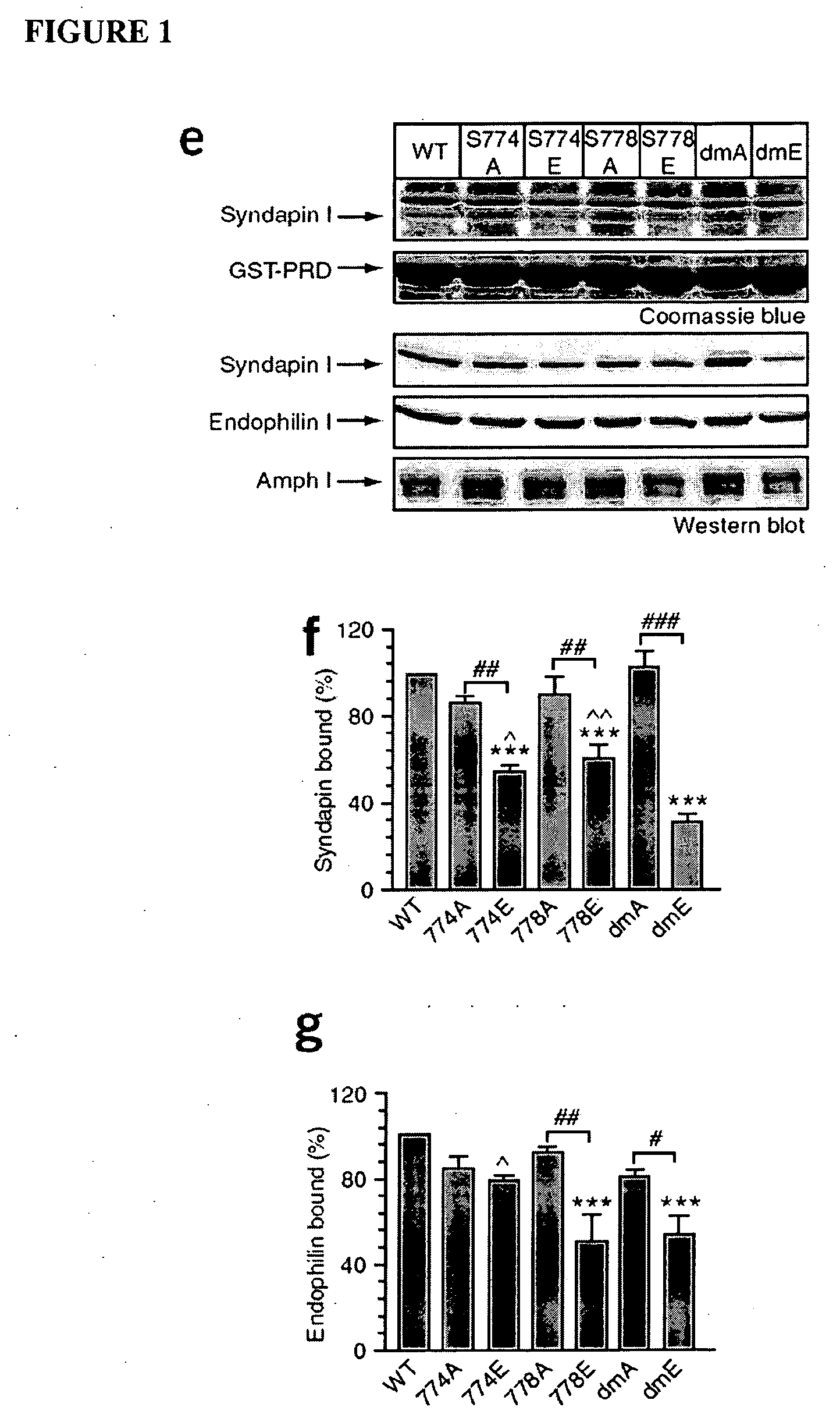
Identification of the DynI Binding Sites for Syndapin I and Endophilin I

2.1 Materials and Methods

[0109]Peptides were synthesised by Mimotopes (Clayton, Australia). FM4-64 was purchased from Molecular Probes (Eugene, Oreg.). Tissue culture plastics were from Falcon (Franklin Lakes, N.J.). Penicillin / streptomycin, phosphate buffered salts, foetal calf serum and Minimal Essential Medium (MEM) were obtained from Invitrogen (Carlsbad, Calif.). All other general laboratory chemicals were from Sigma (St. Louis, Mo.) unless otherwise stated.

2.1.1 Antibodies and Western Blots

[0110]Anti-syndapin I, anti-endophilin I and anti-p85 antibodies were obtained from Santa Cruz Biotechnology (Santa Cruz, Calif.). Anti-Grb2 was purchased from Transduction Laboratory (Lexington, Ky.). Anti-amphiphysin monoclonal antibody was from Pietro De Camilli (Yale, New Haven, Conn.). Anti-dynamin I antibody was as previously described (Tan et al. 2003). Protein samples were separated by SDS-PAGE on 10% or 12% acry...


PUM

PropertyMeasurementUnit
pHaaaaaaaaaa
lengthaaaaaaaaaa
synaptic transmissionaaaaaaaaaa

Abstract

Inhibitors of syndapin I binding to dynamin I (DynI) are provided. Examples include mimetics of a region of DynI including the serine residues S774 and S778 or phosphorylatable amino acids in homologous positions. Typically, the mimetics exclude or do not imitate at least one phosphorylation site provided by the serine residues or phosphorylatable amino acids. Peptide fragment inhibitors comprising or consisting of this region of DynI are also described. The inhibitors have application in the prophylaxis or treatment of neurological diseases or conditions. The inhibitors can also be used to inhibit neuronal cell vesicle trafficking and synaptic signal transmission.

Description

RELATED DISEASES AND CONDITIONS[0001]This application claims priority to U.S. Provisional Patent Application Ser. No. 60 / 915,025, filed Apr. 30, 2007, which is incorporated by reference herein in its entirety.FIELD OF THE INVENTION[0002]The invention relates to methods of screening for inhibitors of the interaction of syndapin I with dynamin I (DynI). The invention further relates to mimetics of a region of dynamin I (DynI), and methods for the prophylaxis or treatment of neurological related diseases or conditions.BACKGROUND OF THE INVENTION[0003]Neurons communicate via the release of neurotransmitter by exocytosis from nerve terminals. After exocytosis, synaptic vesicles (SV) are retrieved by endocytosis to accommodate multiple cycles of synaptic transmission. Synaptic vesicle endocytosis (SVE) is triggered by a coordinated calcineurin-dependent dephosphorylation of a group of at least eight proteins called the dephosphins, namely DynI, amphiphysin I / II, synaptojanin, epsin, eps15...

Claims


Application Information

Patent Timeline
Patent Type & Authority Applications(United States)
IPC IPC(8): A61K38/10C07K7/08A61P25/00
CPCA61K38/46C07K14/00C07K14/4722G01N33/5058G01N2800/28G01N2800/2821G01N2800/2835C12Y306/05005A61P25/00
Inventor MCCLUSKEY, ADAMROBINSON, PHILLIPCOUSIN, MICHAEL
Owner CHILDRENS MEDICAL RES INST
Why Eureka
Try Eureka