
Apparatus and Method for Endoscopic Surgical Procedures

a surgical procedure and endoscopic technology, applied in the field of endoscopic subxiphoid surgical procedures, can solve the problems of inability to perform thoracotomy, too invasive, and all procedures are quite invasive, and achieve the effect of facilitating the dissection of the extrapericardial tract using the endoscopic subxiphoid cannula

Inactive Publication Date: 2008-12-11
MAQUET CARDIOVASCULAR LLC
View PDF99 Cites 81 Cited by
  • Summary
  • Abstract
  • Description
  • Claims
  • Application Information

AI Technical Summary

Benefits of technology

[0029]In accordance with the present invention, apparatus and methods for using the apparatus provide safe and minimally invasive access to mediastinal structures including the pericardium that surrounds the heart. More specifically, the apparatus and methods access the pericardium via a subxiphoid approach, access the heart within the pericardium, and facilitate performing cardiac procedures thereon.

Problems solved by technology

These procedures, however, are all quite invasive, requiring large incisions or open heart surgery.
Thoracotomy is additionally invasive as it requires the deflation of one or both lungs, since the approach is via the pleural cavity.
Such a procedure, however, is too invasive, and thus not desirable.
These procedures, however, have been performed under direct vision, and thus still require a fairly large incision to assist the surgeon in observing the field of surgery.
This method, however, does not enable the surgeon to access all regions of the heart.
However, none of these minimally invasive methods allow access to all regions of the heart.
As the heart typically underlies the pericardium contiguously, the surgeon is presented with the difficult task of incising the pericardium without accidentally cutting the heart.
However, this technique requires multiple incisions in the patient and requires the advancement of multiple instruments in separate passageways to the pericardium.
In addition to requiring several incisions, the conventional techniques also typically require the incision in the pericardium to be lengthy.
With endoscopic post-access surgery, a long pericardial incision is difficult to close, due to the complexity of endoscopic suturing.
Another problem arising in conventional cardiac procedures is the dissection of a working tunnel from the initial incision to the pericardium.
Mechanical probing of heart tissue may cause severe or dangerous cardiac arrhythmias such as ventricular fibrillation.
However, the instruments currently available to perform cardiac procedures are typically large, and therefore a larger cavity must be dissected to allow these instruments to pass through to the pericardium.
Although using a larger dilator may create the necessary space, a larger dilator may cause damage to the heart by causing cardiac arrhythmias as discussed above.
If a small dilator is used to minimize this potential trauma, the working cavity may not be large enough to allow the larger instruments required in the procedure to be advanced to the pericardium.
A further problem with conventional dilators such as balloon dissectors is that such tools exert shear force on the surrounding tissue as they are advanced in the body.
Shear force has a tendency of causing vessel avulsion and tissue abrasion.
None of these systems permit reliable, safe entry under direct endoscopic visualization.
This concept suffers from unreliability, as it is difficult to ensure that the needle will pierce between two layers of pericardium that are compressed by the jaws of the device, without an active technique of holding the two opposed layers of pericardium apart.
Also, axial advancement of the needle carries the potential of myocardial puncture.
These techniques are unreliable, because there is generally a layer of fatty tissue adherent to the pericardial surface, and suction may pull fat into the tube instead of pericardium.
The multiple steps of pericardial grasping, pericardial puncture, guidewire advancement, and catheter insertion render this technique less practical.
In such cases, the heart may enlarge to such an extent that the adverse consequences of heart enlargement continue following recovery from the initial affliction with debilitating effect.
In dilated cardiomyopathy, the dilation is typically of the left ventricle with resultant failure of the heart as a pump.
In time, the heart becomes so enlarged that the heart cannot adequately supply blood.
An afflicted patient is fatigued, unable to perform even simple exerting tasks and experiences pain and discomfort.
Further, as the heart enlarges, the internal heart valves cannot adequately close.
This impairs the function of the valves and further reduces the heart's ability to supply blood.
With each type of cardiac dilation, there are associated problems ranging from arrhythmias resulting from increased stretching of myocardial cells, to leakage of the cardiac valves due to enlargement of the valvular annulus.
On the other hand, some drugs, for example, beta-blocking drugs, decrease the contractility of the heart and thus increase the likelihood of dilation.
Many of these drugs, however, have side effects which make them undesirable for long-term use.
However, none of these patents disclose a sheath to facilitate endoscopic introduction of the apparatus, or guide elements for positioning the cardiac restraint apparatus around the heart, and none of these patents disclose hollow guide tubes that permit an instrument to be advanced through such tubes to engage the mouth of the jacket and secure the mouth of the jacket to the pericardium.
Furthermore, none of these patents disclose introducing a cardiac restraint apparatus via a single subxiphoid incision.
Movement of the beating heart further complicates needle placement because of erratic movement of the coronary vessels as needle insertion is attempted.
The physical space occupied by two sets of endoscopic equipment is cumbersome in the operating room, and the expense is prohibitive to hospitals.
However, the jacket is difficult or impossible to place on the heart without opening the chest via a sternotomy or thoracotomy.
This repeated motion may tire the hand of a surgeon performing the procedure, and, further, stepwise dilation may result in formation of an uneven tunnel, with an irregular inner contour.

Method used


Image

  • Apparatus and Method for Endoscopic Surgical Procedures
  • Apparatus and Method for Endoscopic Surgical Procedures
  • Apparatus and Method for Endoscopic Surgical Procedures

Examples


Embodiment Construction

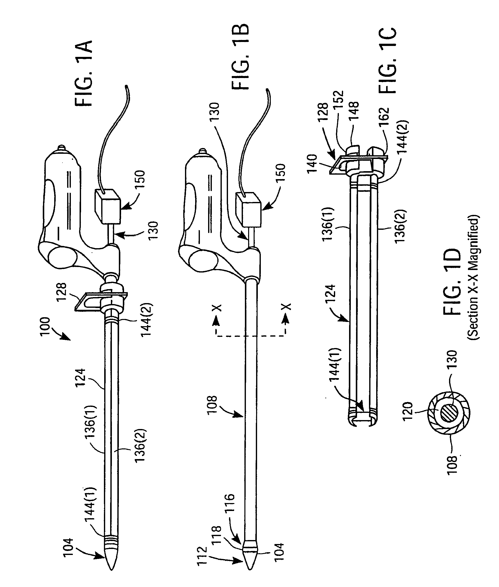
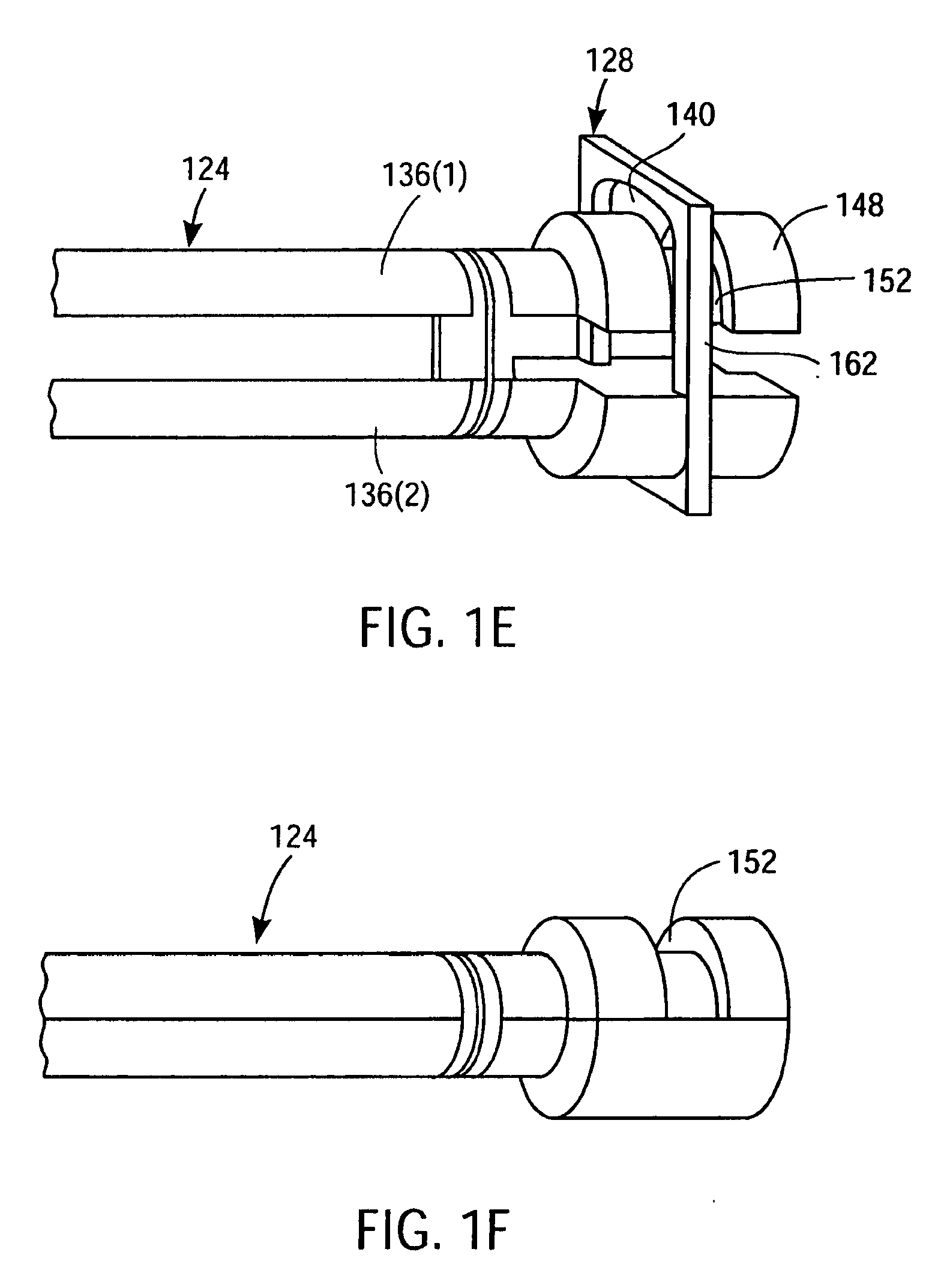
[0148]FIGS. 1A-D illustrate a preferred embodiment of a dilation tool 100 which embodies an aspect of the invention. Dilation tool 100 includes an inner cannula 108 having lumen 120 as shown in FIG. 1D, and an expandable sheath 124 comprised of shells 136(1) and 136(2) as shown in FIG. 1C. Preferably, the inner cannula is formed of a sufficiently rigid material, such as metal or plastic, that would allow tip 104 to be used to bluntly dissect a cavity from an incision point to the pericardium or other surgical site of interest. Lumen 120 is provided to allow the insertion of an endoscope 130 fitted with video camera 150 in the dilation tool 100, and tip 104 is transparent to allow endoscopic visualization during the surgical procedure. In a preferred embodiment, tip 104 has a long distal taper 112 as shown in FIG. 1B, which allows tip 104 to bluntly dissect away tissue encountered along the cavity to the pericardium. Conically-tapered tip 104 also provides a less distorted field of v...


PUM

PropertyMeasurementUnit
diameteraaaaaaaaaa
diameteraaaaaaaaaa
diameteraaaaaaaaaa

Abstract

Apparatus and method for performing surgical procedures within the mediastinum and within the pericardium include an endoscopic cannula having a transparent tip, and an endoscope for introduction into the mediastinum and optionally into the pericardium via a single subxiphoid incision. A cavity may be initially dilated for advancing the endoscopic cannula using a dilating tool that exerts a lateral-expansive force against surrounding tissue for evaluating the endoscopic cannula to be introduced into the mediastinum. Other surgical instruments are positioned through the endoscopic cannula to cut a flap of the pericardium as an opening through which other surgical apparatus may be introduced. The endoscopic cannula may be swept around selected regions of the heart through an aperture near the apex of the heart to facilitate placement of epicardial tacks about regions of the heart.

Description

CROSS-REFERENCES TO RELATED APPLICATIONS[0001]This is a divisional application of pending application Ser. No. 10 / 347,212, entitled “Apparatus and Methods for Endoscopic Surgical Procedures,” filed on Jan. 17, 2003, which in turn is a continuation-in-part application of pending application Ser. No. 10 / 174,454, entitled “Releasable Guide And Method For Endoscopic Cardiac Lead Placement”, filed on Jun. 17, 2002, which is a continuation-in-part of pending application Ser. No. 10 / 140,309 entitled “Methods And Apparatus For Endoscopic Cardiac Surgery”, filed on May 6, 2002, which is a continuation of pending application Ser. No. 09 / 635,721 entitled “Apparatus For Endoscopic Access”, filed on Aug. 9, 2000, which claims the benefit of provisional applications Ser. No. 60 / 148,130 filed on Aug. 10, 1999 and Ser. No. 60 / 150,737 filed on Aug. 25, 1999. This application is also a continuation-in-part application of the U.S. Pat. No. 6,569,082, entitled “Apparatus And Methods For Cardiac Restrai...

Claims


Application Information

Patent Timeline
Patent Type & Authority Applications(United States)
IPC IPC(8): A61B1/018A61B17/04A61B18/18A61B18/20A61H1/00A61B1/00A61B1/01A61B17/00A61B17/06A61B17/22A61B17/30A61B17/32A61B17/34A61B18/00A61B18/14A61B19/00A61K39/21A61N1/05C07K1/00
CPCA61B17/00008A61B17/06109A61B17/320068A61B17/3403A61B17/3417A61B17/3421A61B17/3478A61B18/1482A61B19/201A61B19/5202A61B19/54A61B2017/00243A61B2017/00247A61B2017/061A61B2017/22077A61B2017/306A61B2017/308A61B2017/320044A61B2017/3445A61B2017/3488A61B2018/00291A61B2018/00392A61B2018/00982A61B2019/306A61B2019/462A61N1/0587A61N2001/0578A61B2017/00323A61B1/00094A61B1/00154A61B1/00195A61B90/30A61B90/11A61B2090/036A61B2090/062A61B90/39A61B2017/320069A61B2017/32007
Inventor CHIN, ALBERT K.
Owner MAQUET CARDIOVASCULAR LLC
Learn More