Method for segmenting a myocardial wall and device for detecting a coronary artery with pathological changes

a technology of pathological changes and myocardial walls, which is applied in the field of myocardial wall segmentation and the detection of coronary arteries with pathological changes, can solve the problems of weakened arteries, negative effect of pump functions, and weakening cardiac output, and achieve the effect of better visualization of the pump function of the hear

Inactive Publication Date: 2009-01-01
SIEMENS HEALTHCARE GMBH
View PDF4 Cites 14 Cited by
  • Summary
  • Abstract
  • Description
  • Claims
  • Application Information

AI Technical Summary

Benefits of technology

[0010]The object of the present invention is therefore to ac

Problems solved by technology

Stenosed or calcified coronary arteries, which are no longer able to supply the myocardium with sufficient oxygen-rich blood, have the greatest negative effect on this pump function.
As a result the muscle of the region supplied by the corresponding coronary artery is weakened and in time ceases to play an active role in the contracting movement, thereby weakening cardiac output.
This standard protocol allows a distinction between contrast agent and tissue but makes a tissue classification between the myocardium and surrounding tissue, which is of interest for cardiological applications, virtually impossible.
Firstly this method is very time-consuming, since every individual branch of the coronary

Method used


Image

  • Method for segmenting a myocardial wall and device for detecting a coronary artery with pathological changes
  • Method for segmenting a myocardial wall and device for detecting a coronary artery with pathological changes
  • Method for segmenting a myocardial wall and device for detecting a coronary artery with pathological changes

Examples


Embodiment Construction

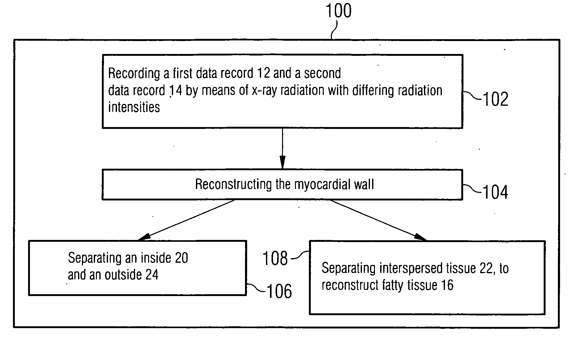
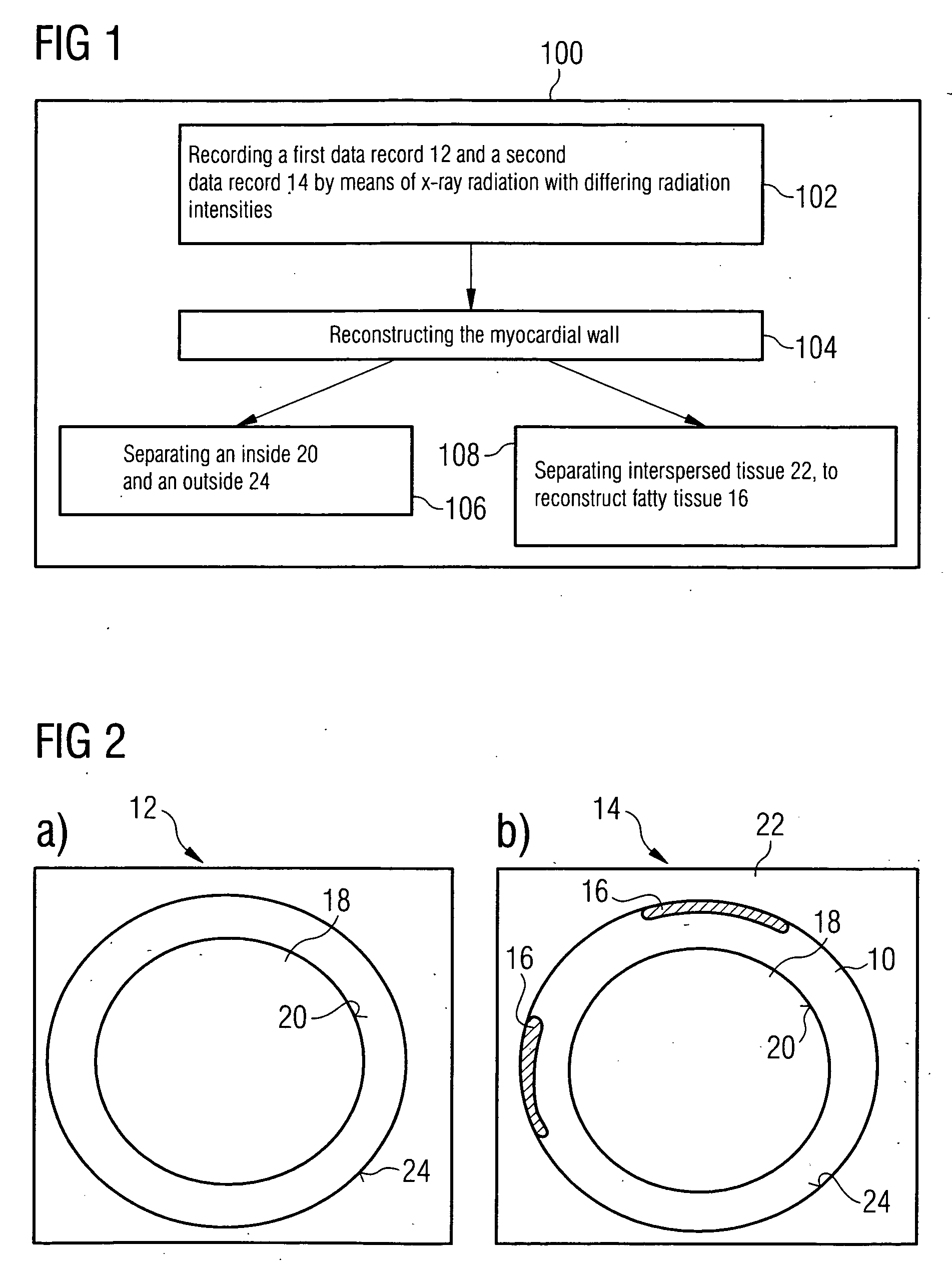
[0032]FIG. 1 shows an inventive method 100 for segmenting a myocardial wall 10. A first data record 12, which includes the myocardial wall 10, is recorded using x-ray radiation in a first method step 102. A second data record 12 of the myocardial wall 10 is recorded in the same cardiac phase. The two data records 12, 14 are recorded by an x-ray radiation source with differing radiation intensities as a detector transilluminates a person. Recording with ECG control allows the data records 12, 14 to be recorded in the same cardiac phase. Alternatively the second data record 14 is recorded at the same time as the first data record 12 using a second x-ray radiation source and assigned detector.

[0033]The data records 12, 14 essentially capture the radiation intensity of x-ray radiation remaining after transillumination and are used in a further method step 104 to reconstruct the myocardial wall 10. A reconstruction generated from the first data record 12 is shown in FIG. 2a. The radiatio...


PUM


Abstract

The present invention relates to a method for segmenting a myocardial wall with the following steps: recording a first data record and a second data record in the same cardiac phase by means of x-ray radiation with differing radiation intensities, reconstructing the myocardial wall from the first and second data record. To generate a detailed and complete reconstruction of the myocardial wall, it is proposed that the method be supplemented by the following steps: separating an inside and an outside of the myocardial wall from the data record recorded with higher radiation intensity, and separating tissue interspersing the myocardial wall between the inside and the outside, in particular fatty tissue, from the data record recorded with lower radiation intensity.

Description

CROSS REFERENCE TO RELATED APPLICATIONS[0001]This application claims priority of German application No. 10 2007 029 886.4 filed Jun. 28, 2007, which is incorporated by reference herein in its entirety.FIELD OF THE INVENTION[0002]The present invention relates to a method for segmenting a myocardial wall and a device for detecting a coronary artery with pathological changes.BACKGROUND OF THE INVENTION[0003]A generic method, in particular using an x-ray tomograph with two x-ray beam sources is known from the publication “First performance evaluation of a dual-source CT (DSCT) system” by Flohr et al., which appeared in Eur. Radiol. 2006, 16, pages 256 to 268.[0004]By recording a first data record and a second data record of the myocardial wall in the same cardiac phase by means of x-ray radiation with differing radiation intensities, the myocardial wall is segmented by the first data record and the second data record. However a corresponding image comprises only an external outline of t...

Claims


Application Information

Patent Timeline
IPC IPC(8): A61B6/00
CPCA61B5/02007A61B6/481A61B6/482A61B6/504G06T2207/30048A61B6/541G06T5/50G06T7/0081G06T2207/10116A61B6/5217G06T7/11G16H50/30
Inventor LAUTENSCHLAGER, STEFAN
Owner SIEMENS HEALTHCARE GMBH
Why Eureka
Try Eureka