Methods for isolation and analysis of sialylated and phosphorylated peptides

a technology of phosphorylated peptides and isolating methods, which is applied in the field of isolating and analyzing sialylated and phosphorylated peptides, can solve the problems of buffers that are not suited for mass spectrometry studies, expensive reagents, and inability to perform mass spectrometry studies

Active Publication Date: 2009-01-29
SYDDANSK UNIV
View PDF2 Cites 8 Cited by
  • Summary
  • Abstract
  • Description
  • Claims
  • Application Information

AI Technical Summary

Benefits of technology

[0012]In one preferred embodiment, proteins from control and patient samples are subject to enzymatic hydrolysis with trypsin in a buffer comprising unlabelled water. The control and patient samples are then subject to re-hydrolysis with trypsin, wherein ordinary samples are re-hydrolysed in a buffer comprising unlabelled water, while patient samples are re-hydrolysed in a buffer comprising O18-labelled water. The sialylated tryptic peptides from control and patient samples can then be isolated according to the methods of the present invention and comp

Problems solved by technology

These techniques generally require expensive reagents or relatively cumbersome

Method used


Image

  • Methods for isolation and analysis of sialylated and phosphorylated peptides
  • Methods for isolation and analysis of sialylated and phosphorylated peptides
  • Methods for isolation and analysis of sialylated and phosphorylated peptides

Examples


examples

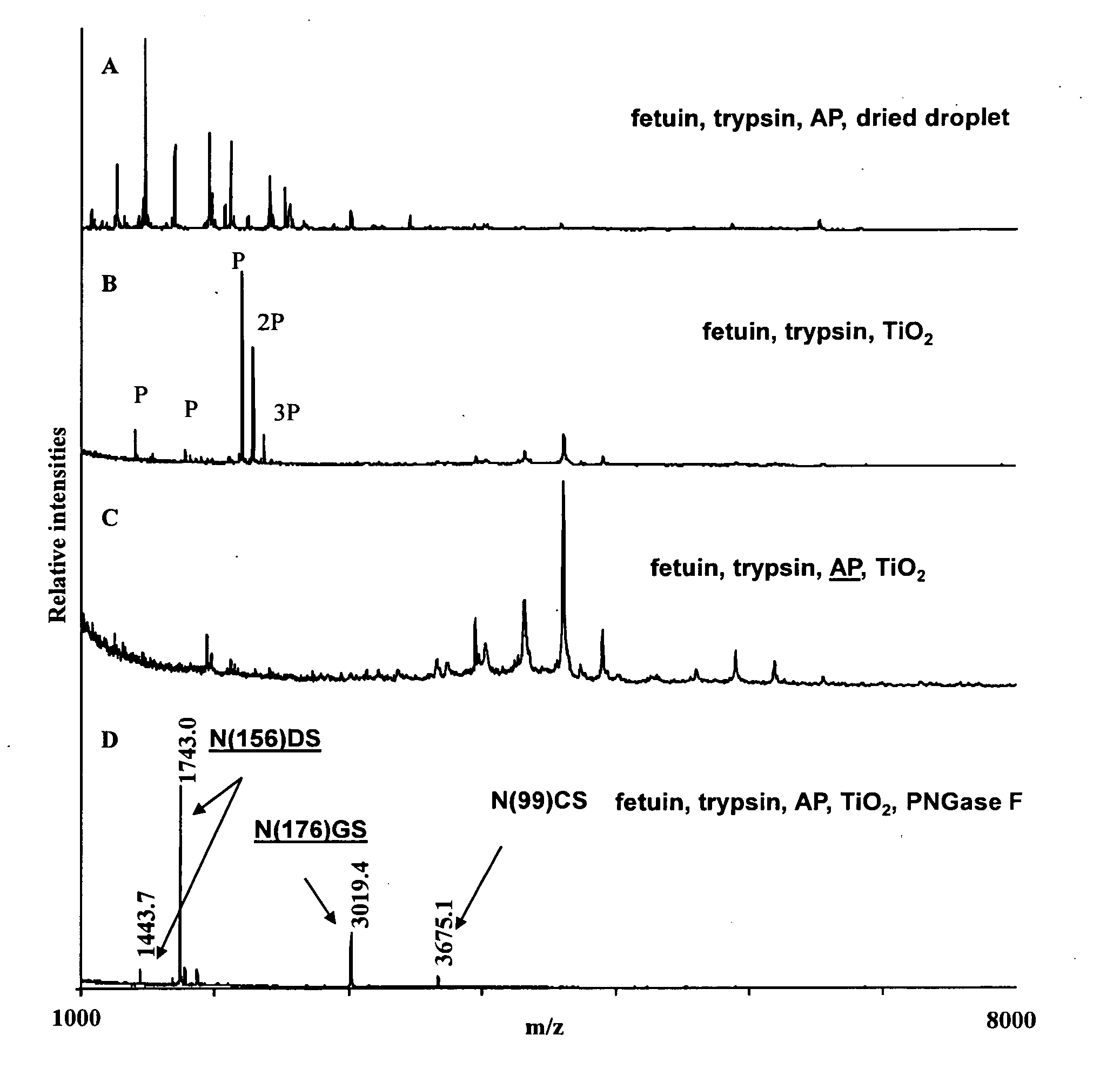
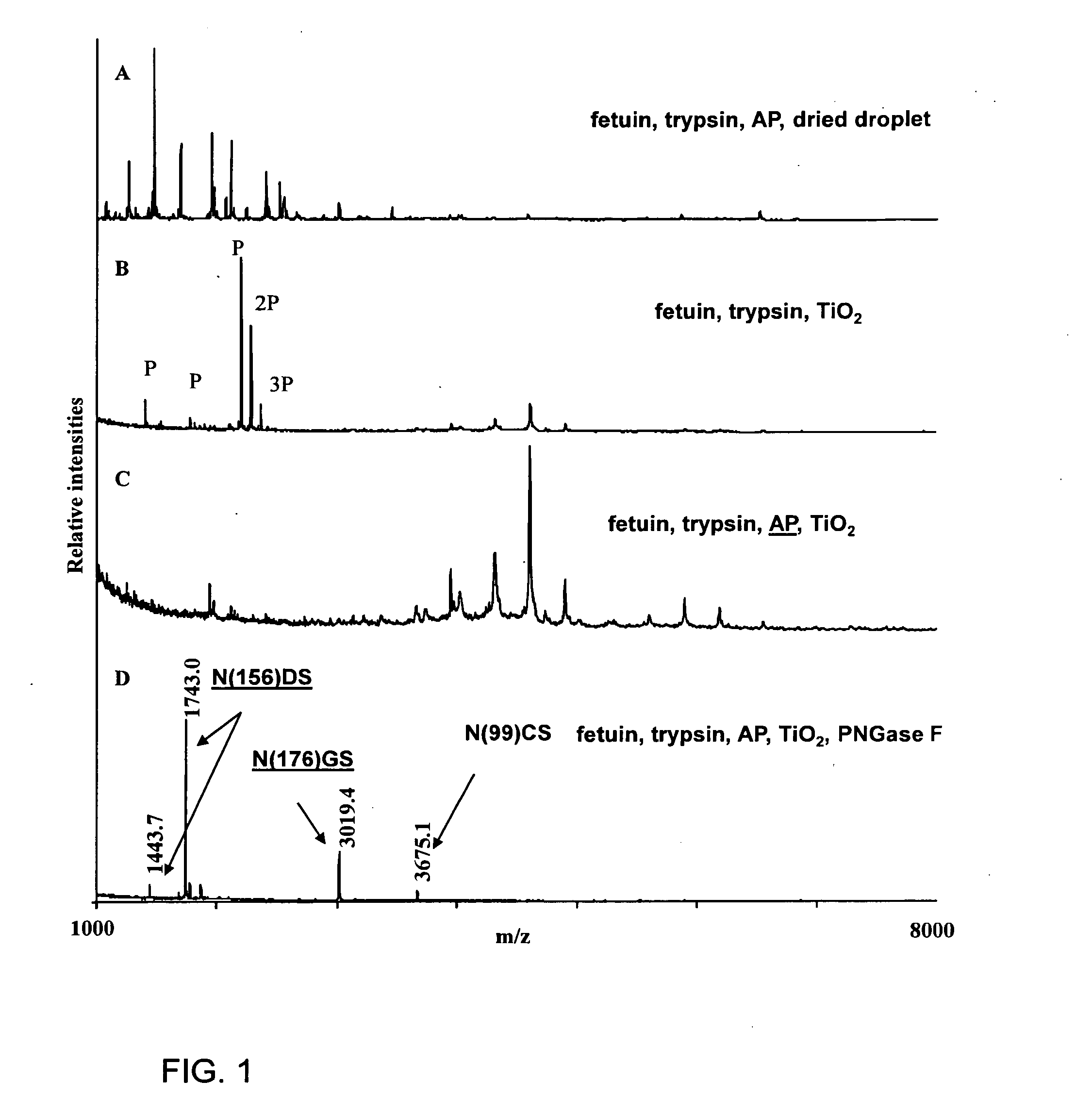
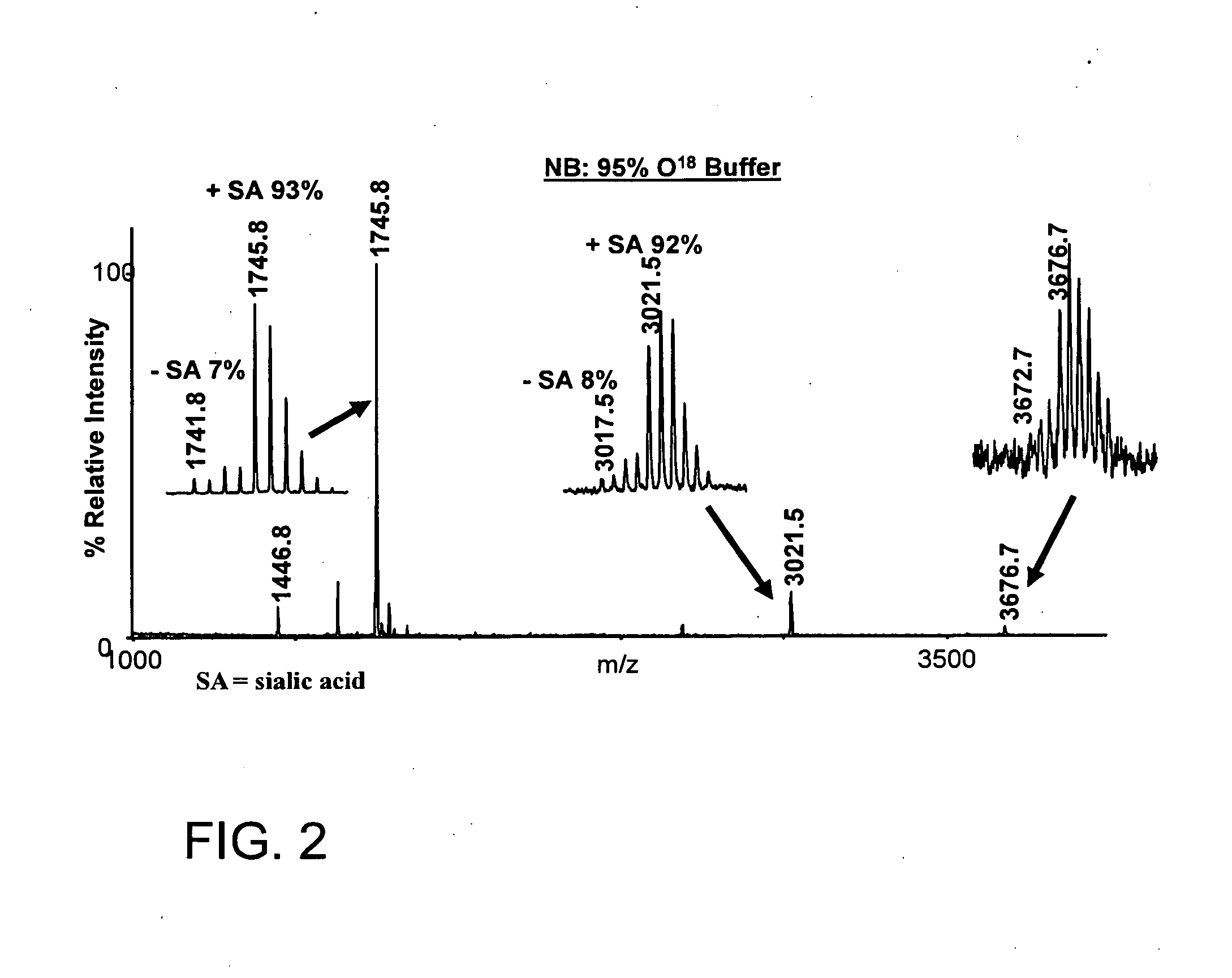
1. Isolation of Sialylated Tryptic Fragments of Fetuin with Titanium Dioxide Stationary Phase

Material

[0111]Modified trypsin was from Promega (Madison, Wis., USA). Fetuin was from Sigma. Poros R2 and Poros Oligo R3 reversed phase material were from PerSeptive Biosystems (Framingham, Mass.). GELoader tips were from Eppendorff (Eppendorf, Hamburg, Germany). 2,5-Dihydroxybenzoic acid (DHB) was from Fluka (St. Louis, Mo.). 3M Empore C8 disk was from 3M Bioanalytical Technologies (St. Paul, Minn., USA). Syringe for HPLC loading were from SGE (Victoria, Australia). The water was from a Milli-Q system (Millipore, Bedford, Mass.). Titanium dioxide beads were obtained from a disassembled TiO2 column (1350L250WO46 Titansphere, 5 micron, 250×4.6 mm) purchased from GL sciences Inc, Japan.

Fetuin

[0112]Bovine fetuin was dissolved in 50 mM NH4HCO3, pH 8, 10 mM DTT and incubated at 56° C. for 30 min. Iodoacetamide was added to the solution (final concentration 20 mM) and it was incubated at room temp...


PUM

PropertyMeasurementUnit
Fractionaaaaaaaaaa
Molar densityaaaaaaaaaa

Abstract

Changes in sialylation of cell surface or plasma proteins are often associated with various cancers and other disease conditions Provided are methods of detecting biomarkers of conditions associated with a change of sialylation status. Sialylated peptides are first isolated from biological and other samples by loading onto titanium dioxide (TiO2) or zirconium dioxide (ZrO2) stationary phase under acidic conditions in a solution comprising at least 20% organic phase and at least about 6.5 mM of substituted aromatic carboxylic acid, or, alternatively, at least about 1 mM short chain, non-aromatic, hydroxylated carboxylic acid. Sialic acid containing proteins can then be eluted from loaded stationary phase material by exposure to an alkaline solution having pH of 9.0 or greater, preferably at least 10.5. Sialylated peptides isolated by the methods provided can then be analysed by mass spectrometry to identify patterns of sialylation across a sialiome (the entire complement of sialic acid containing peptides in a biological sample) and/or to identify proteins in a sample that are sialylated or that show changes in sialylation status between two or more different samples. In preferred embodiments, sialylated peptides from control and patient samples can be differentially isotopically labelled and compared in a single mass sprectrometry experiment. Also provided are specific biomarkers of bladder cancer. The methods for isolating and analysing sialylated proteins can also by applied to phosphorylated proteins.

Description

REFERENCE TO RELATED APPLICATIONS[0001]The present application claims benefit of priority to U.S. Provisional applications Nos. U.S. 60 / 795,158, filed on Apr. 27, 2006, and 60 / 802,509, filed on May 23, 2006, the entire contents of which are incorporated by reference herein.FIELD OF THE INVENTION[0002]The present invention relates in general to methods for isolating and analyzing sialylated and phosphorylated peptides. More particularly, the invention relates to methods of identifying biomarkers of conditions associated with a change of protein sialylation status and to methods for derivatizing phosphorylated peptides while loaded onto stationary phase.BACKGROUND OF THE INVENTION[0003]Abnormal glycosylation is a well known attribute of cancer cells. (1-4) A number of specific glycan structures serve as biomarkers of tumor growth. Of particular significance are the sialic acids, of which more than 40 naturally occurring derivatives have been identified. Sialic acids are nine-carbon ca...

Claims


Application Information

Patent Timeline
IPC IPC(8): C12Q1/00C07K1/14C12P21/06C07K7/08
CPCC07K14/473C07K14/4732G01N2400/00G01N33/57407C07K14/77
Inventor LARSEN, MARTIN ROSSEL
Owner SYDDANSK UNIV
Why Eureka
Try Eureka