Methods And Compounds For Targeting Tissues

a tissue and target technology, applied in the field of molecular medicine, drug and gene delivery, imaging, etc., can solve the problems of low tissue penetration, high molecular weight, antibody-targeted liposomes, etc., and achieve the effect of increasing avidity

Active Publication Date: 2009-04-09
RGT UNIV OF CALIFORNIA
View PDF4 Cites 12 Cited by
  • Summary
  • Abstract
  • Description
  • Claims
  • Application Information

AI Technical Summary

Benefits of technology

[0032]FIGS. 5A-C. Time activity curves (TACs) and Logan Plot from dynamic PET analysis of various LPP liposomes. (a)-(b) TACs for heart muscle, (c) difference between CRPPR-3 and CRPPR-1 liposomes in TAC from heart muscle. Subtraction of non-binding CRPPR-1 compositions removes the effect of the blood pool, showing accumulation of compositions within heart muscle. (d) results of Logan analysis for 4 injection

Problems solved by technology

Among various drug-gene delivery particles, targeted phospholipid-based liposomes have been widely studied but have had limited clinical impact.
However, a major drawback of antibody targeted liposomes is low tissue pe

Method used


Image

  • Methods And Compounds For Targeting Tissues
  • Methods And Compounds For Targeting Tissues
  • Methods And Compounds For Targeting Tissues

Examples


example 1

Liposome Diameter, Zeta Potential, Lipo-PEG-Peptide (LPP) Incorporation, and in Vitro Binding

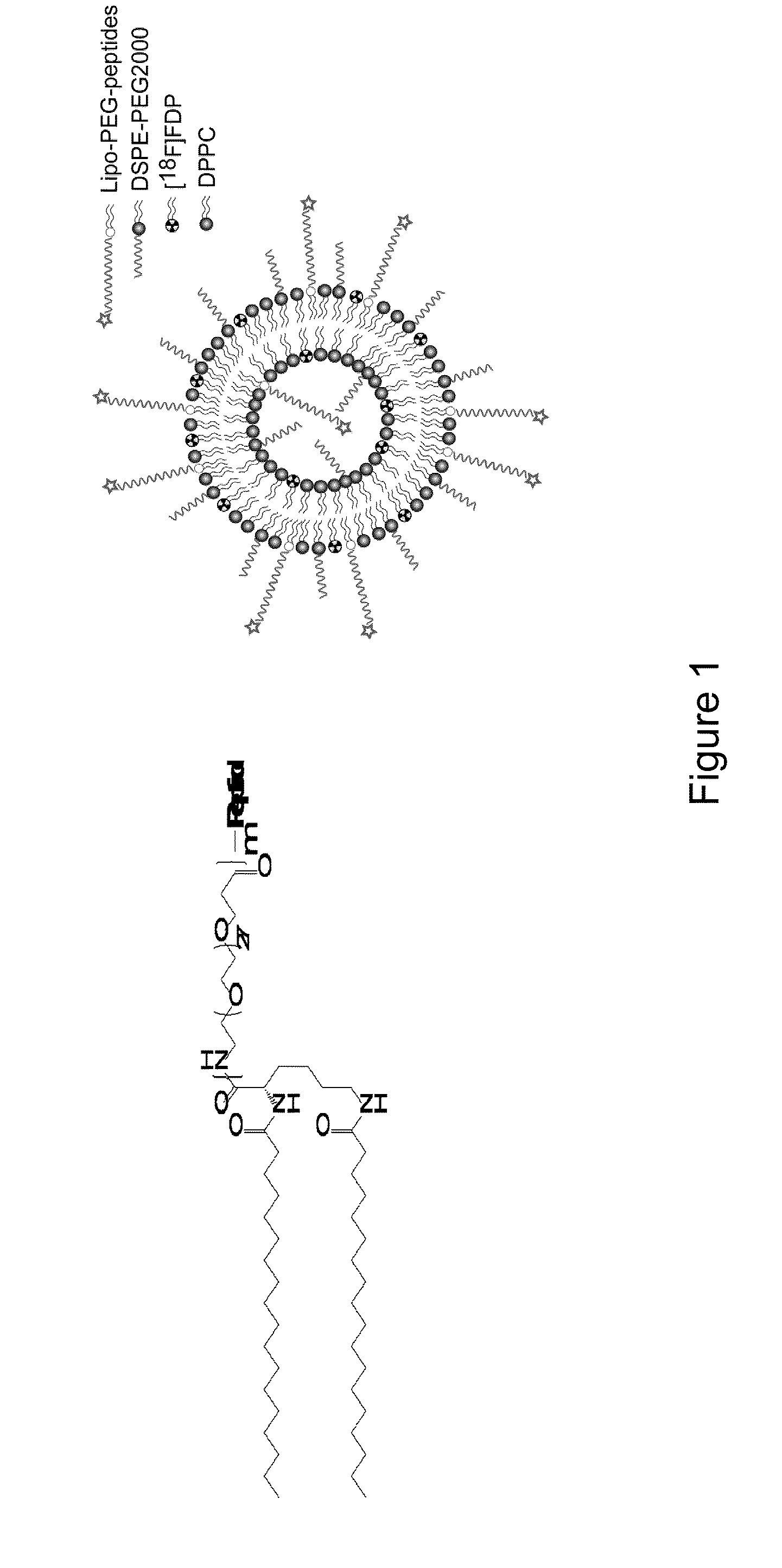
[0162]The LPP (structure shown in FIG. 1a), radiolabeled lipid ([18F]FDP), phospholipid (DPPC), and pegylated phospholipid (DSPE-PEG2000) were combined to produce a composition (FIG. 1b, with formulations and notation detailed in FIG. 1 caption), with a polymer brush layer of 2000 molecular weight (MW) and a ligand that was either exposed (m=3 in FIG. 1a) or buried (m=1). Initial in vitro studies indicated that the formulation of CRPPR-3:DSPE-PEG2000:DPPC=6%:6%:88% (mol / mol) produced effective targeting, therefore this formulation was employed in in vitro studies and as the baseline formulation in in vivo studies. In vivo results with the CRPPR-3:DSPE-PEG2000:DPPC=6%:6%:88% were compared with matched total PEG concentration (12%), LPP concentration (6%), DSPE-PEG2000 concentration (6%), or LPP:DSPE-PEG2000 ratio (1:1), while other parameters were varied (Table 1). For abbreviations in Table ...

example 2

PET Images

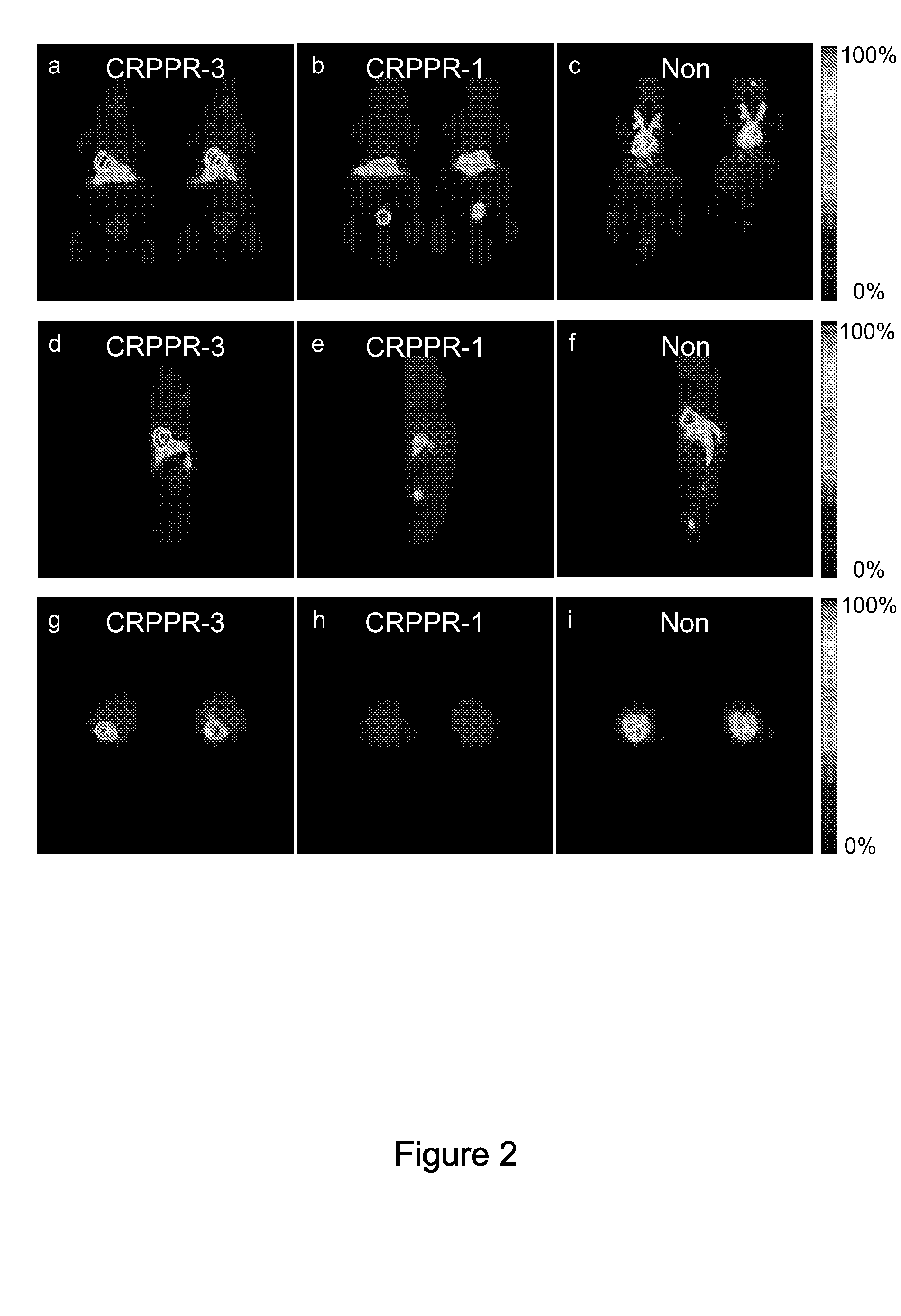
[0164]Ninety-minute accumulative PET images (FIG. 2a-i) acquired with the [18F]FDP and CRPPR-3 incorporated into the liposomal vectors (FIG. 2a, d, g) demonstrate the high level of the radiotracer within the heart, a lower density within the liver, and a low concentration within the spleen and bladder. Images acquired with the shorter PEG length LPP (CRPPR-1) loaded onto an identical vehicle demonstrate that radioactivity has accumulated within the liver and bladder at 90 minutes and a low level of activity is present in the heart (FIG. 2b, e, h). Clearance of the radiotracer occurs through the bladder, after metabolism in the liver separates the fatty acid chains (each with a molecular weight of 256) from the head group containing the isotope (with a molecular weight of 94). By comparison, images acquired with the identical vehicle, without a peptide attached to the liposome, demonstrate that the radioisotope is primarily circulating within the blood volume throughout the...

example 3

Effect of Peptide and Surface Architecture on Biodistribution at 90 Minutes

[0166]Well counts obtained from harvested tissues at the ninety-minute time point quantify the differences in biodistribution produced by the peptide (FIGS. 4a and b), the length of the PEG spacer between the fatty acid and CRPPR peptide portions of the LPP (FIG. 4c), and the molar fraction of the CRPPR-3 incorporated within the vehicle (FIG. 4d). When injected on a vehicle containing CRPPR-3, the concentration of the tracer was significantly higher within the heart than other organs (pb) at levels of 39±13, 26±13, 17±5% ID / g for CPPRR-3, CRRRR-3, and CRRPP-3, respectively, with target to skeletal muscle ratios of 32, 23 and 19. Injection of vehicles with the cyclized RGD peptide and an otherwise identical liposome surface architecture did not produce radioactivity above the baseline (no-peptide) case. The cyclized RGD and no-peptide controls also resulted in significantly lower activity levels within the liv...


PUM

PropertyMeasurementUnit
Fractionaaaaaaaaaa
Fractionaaaaaaaaaa
Fractionaaaaaaaaaa

Abstract

The present invention relates to peptides which home to cells, e.g. heart cells, with high selectivity and which can be useful in the form of compositions. Such compositions can be used, e.g., for selectively targeting a systemically administered therapeutic agent or imaging agent to a cell or tissue in a subject. The present invention further relates to methods of using the compositions for imaging, e.g. PET imaging, and targeting cells, e.g. for delivering a therapeutic agent to one or more target cells in a subject.

Description

CROSS REFERENCE TO RELATED APPLICATIONS[0001]This application claims the benefit of U.S. Provisional Application No. 60 / 970,923, filed Sep. 7, 2007, the entire disclosure of which is hereby incorporated by reference in its entirety for all purposes.STATEMENT REGARDING FEDERALLY SPONSORED RESEARCH OR DEVELOPMENT[0002]The U.S. Government has certain rights in this invention pursuant to Grant Nos. CA103828 and CA R24 110804 awarded by the National Institutes of Health.BACKGROUND OF THE INVENTION[0003]1. Field of the Invention[0004]The present invention relates generally to the fields of molecular medicine, drug and gene delivery, imaging, and, more specifically, to novel compositions and methods for treating and / or diagnosing diseases such as cardiovascular diseases and brain diseases.[0005]2. Description of the Related Art[0006]The vascular endothelium has been recognized as an important target for therapeutic interventions which use liposomes or other particles to carry drugs and / or ...

Claims


Application Information

Patent Timeline
IPC IPC(8): A61K51/08A61K47/42A61K31/7048A61K9/127
CPCA61K9/0019A61K9/1271A61K31/7048A61K51/1234A61K49/0002A61K49/0084A61K47/48815A61K47/6911
Inventor FERRARA, KATHERINEZHANG, HUAKUSUNOSE, JIRO
Owner RGT UNIV OF CALIFORNIA
Why Eureka
Try Eureka