

Reagents and Methods for miRNA Expression Analysis and Identification of Cancer Biomarkers
a cancer biomarker and expression technology, applied in the field of methods and reagents for amplifying and detecting micrornas (mirnas), can solve the problems of low yield of mirna and the number of cells obtained, and achieve the effect of assisting in the elucidation of the molecular basis of malignancy and/or disease pathology
- Summary
- Abstract
- Description
- Claims
- Application Information
AI Technical Summary
Benefits of technology
Problems solved by technology
Method used
Image

Examples
example 1
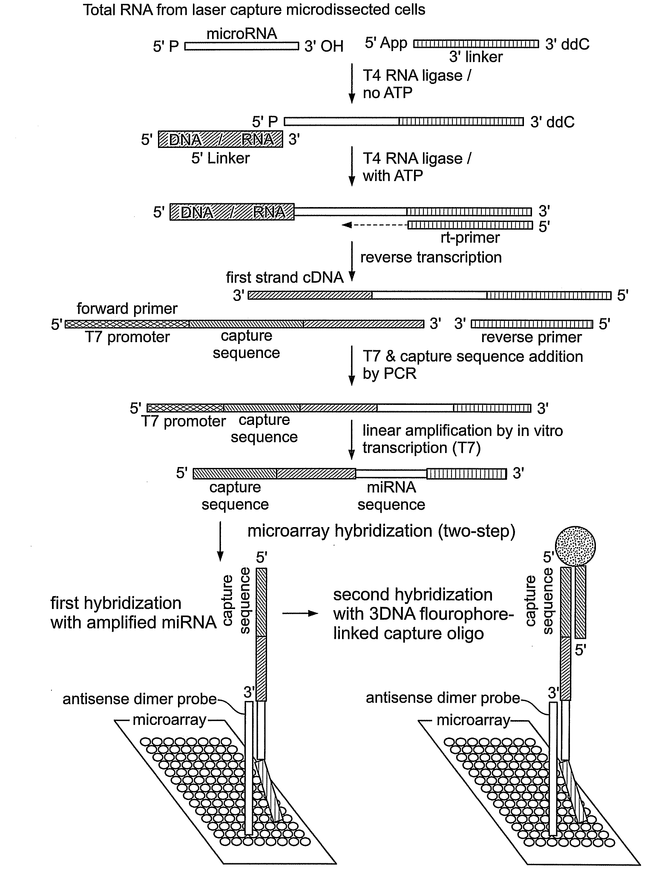
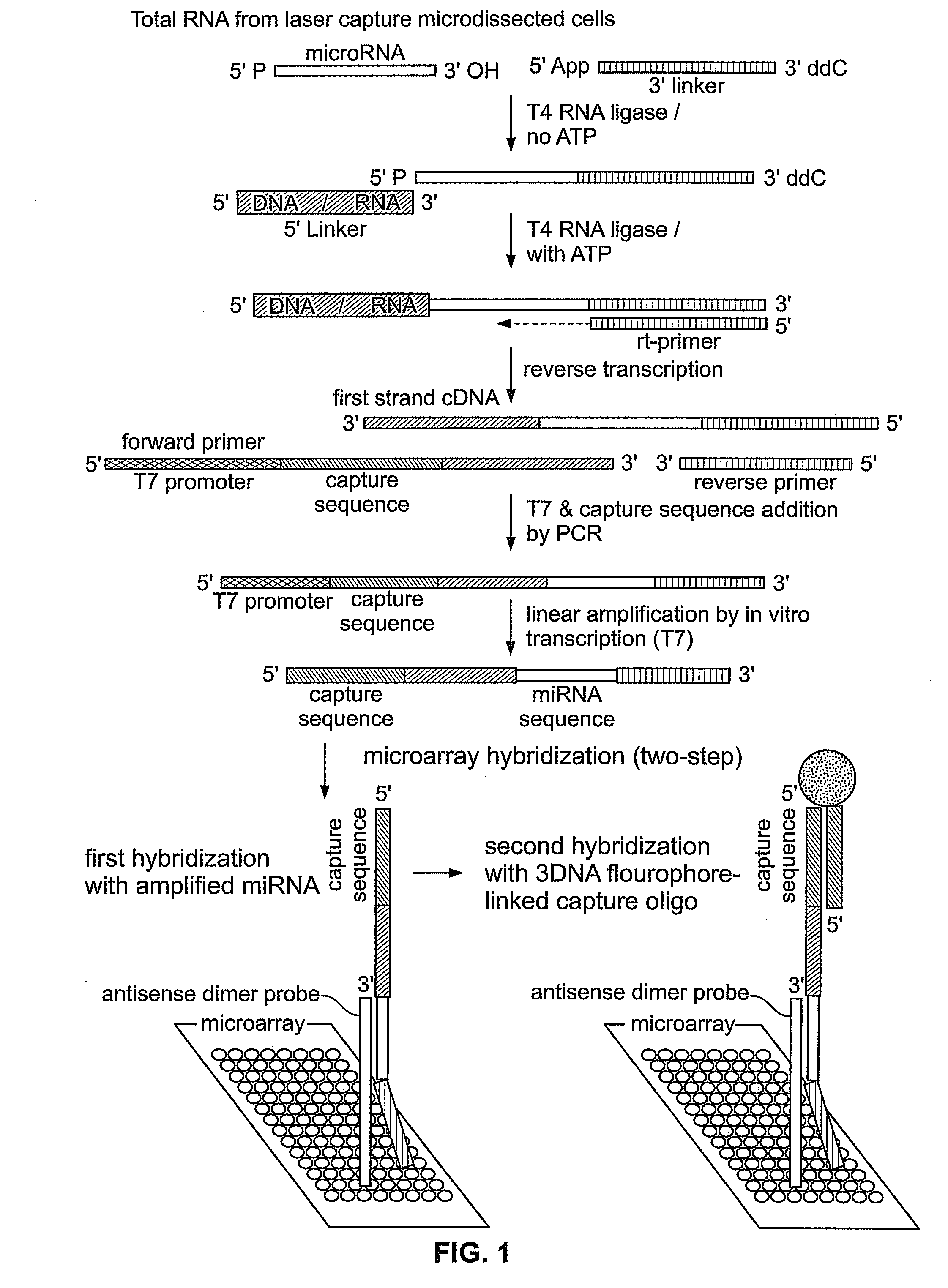
miRNA Isolation and Amplification
[0059]The methods described in this Example were developed to overcome deficiencies in the art associated with detection and differential expression analysis of miRNAs isolated from limited cell or tissue samples.
[0060]Total cellular RNA was isolated from tissue samples including nasopharyngeal carcinoma (NPC) tissue samples. Collection and processing of such samples, including histopathology, laser capture microdissection, and RNA extraction have been described in detail previously (Sengupta et al., 2006, Cancer Res. 66: 7999-8006), the disclosure of which is incorporated by reference herein. Here, a total of thirty-one NPC samples and ten normal nasopharyngeal tissue samples (including six normal tissue samples from non-NPC or biopsy-negative cases and four samples from tumor free nasopharyngeal area of NPC patients) were used. miRNA was amplified from total RNA isolated from laser microdissected / whole tissue sections without any size selection fol...
example 2
Microarray Construction and Hybridization
[0067]The in vitro transcribed sense-strand miRNA-specific products prepared according to Example 1 were used to interrogate a microarray comprising antisense miRNA probes as follows.
[0068]Microarrays were prepared comprising probes that were antisense dimers of mature miRNA sequences taken from miRBase (http: / / microma.sanger.ac.uk / ), previously termed “the microRNA registry” (Griffiths-Jones, 2004, The microRNA Registry Nucl. Acids. Res. 32: Database Issue, D109-D111). Each miRNA probe sequence used in the microarray was modified at its 5′ end with a C6 amino linker that permitted the probe to be attached to aldehyde-coated slides for microarray fabrication. A total of two hundred seven probes from two hundred twenty-two human miRNAs and six probes for five EBV miRNAs (as present in the database as of April 2005) were spotted on a chip. Also spotted were seven probes from D. melanogaster miRNAs as controls (Table 1). Microarrays were printed...
example 3
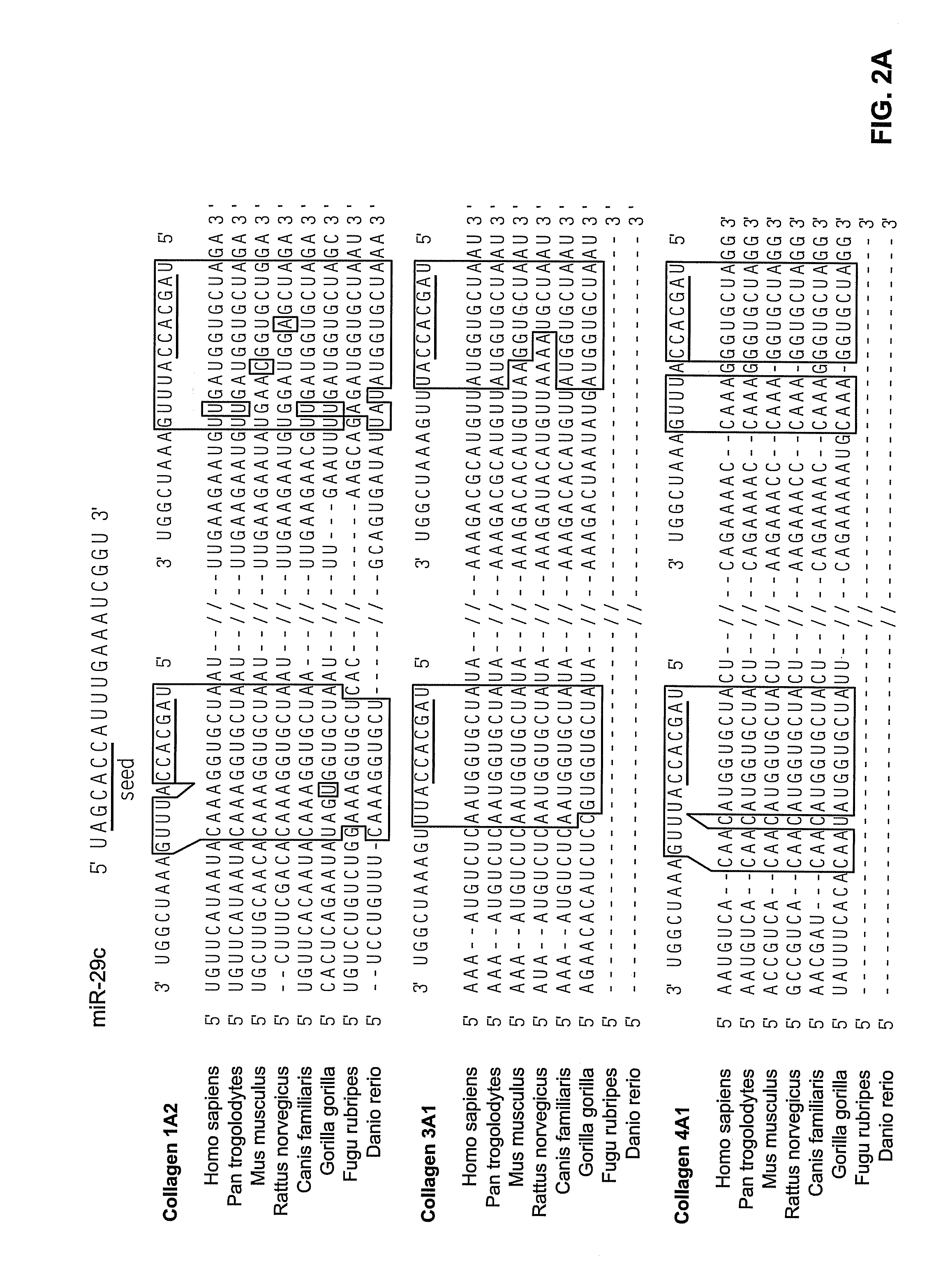
Identification of Differentially Expressed miRNAs
[0072]Cellular and viral miRNAs in EBV-associated cancers such as NPC are candidate oncogenes that may contribute to the initiation or maintenance, or both, of tumors. Accordingly, the microarray methods described above were used to screen a large number of cellular and viral miRNAs for differential expression in NPC tumors. These assays were performed using microarrays prepared as described in Example 2, comprising two hundred twenty-two human miRNAs and for five viral miRNAs, which included all miRNAs identified as of April 2005. These assays were performed substantially as described above.
[0073]The results of these assays are given in Table 2. In these experiments, background-corrected, raw-scale expression intensity values were obtained via GenePix Pro 5.0 (Molecular Devices) after some manual adjustment to align and identify spots. Data from multiple microarrays were normalized using a version of quantile normalization (Bolstad e...
PUM
| Property | Measurement | Unit |
|---|---|---|
| diameter | aaaaa | aaaaa |
| diameter | aaaaa | aaaaa |
| areas | aaaaa | aaaaa |
Abstract
Description
Claims
Application Information