SERS sensor for integrated detection of tumor protein and nucleic acid markers and preparation method thereof

A tumor protein and marker technology, applied in the field of functional nanomaterials and biological detection, can solve the problem of no SERS sensor

Active Publication Date: 2020-10-16
NANJING UNIV OF POSTS & TELECOMM
View PDF7 Cites 13 Cited by
  • Summary
  • Abstract
  • Description
  • Claims
  • Application Information

AI Technical Summary

Problems solved by technology

[0004] However, due to the different biological characteristics and detection methods of nucleic acids and

Method used


Image

  • SERS sensor for integrated detection of tumor protein and nucleic acid markers and preparation method thereof
  • SERS sensor for integrated detection of tumor protein and nucleic acid markers and preparation method thereof
  • SERS sensor for integrated detection of tumor protein and nucleic acid markers and preparation method thereof

Examples


Embodiment 1

[0080] Example 1 Preparation of SERS sensor for integrated detection of tumor protein and nucleic acid markers

[0081] 1. Preparation of silver nanorod arrays and washing with ultrapure water several times;

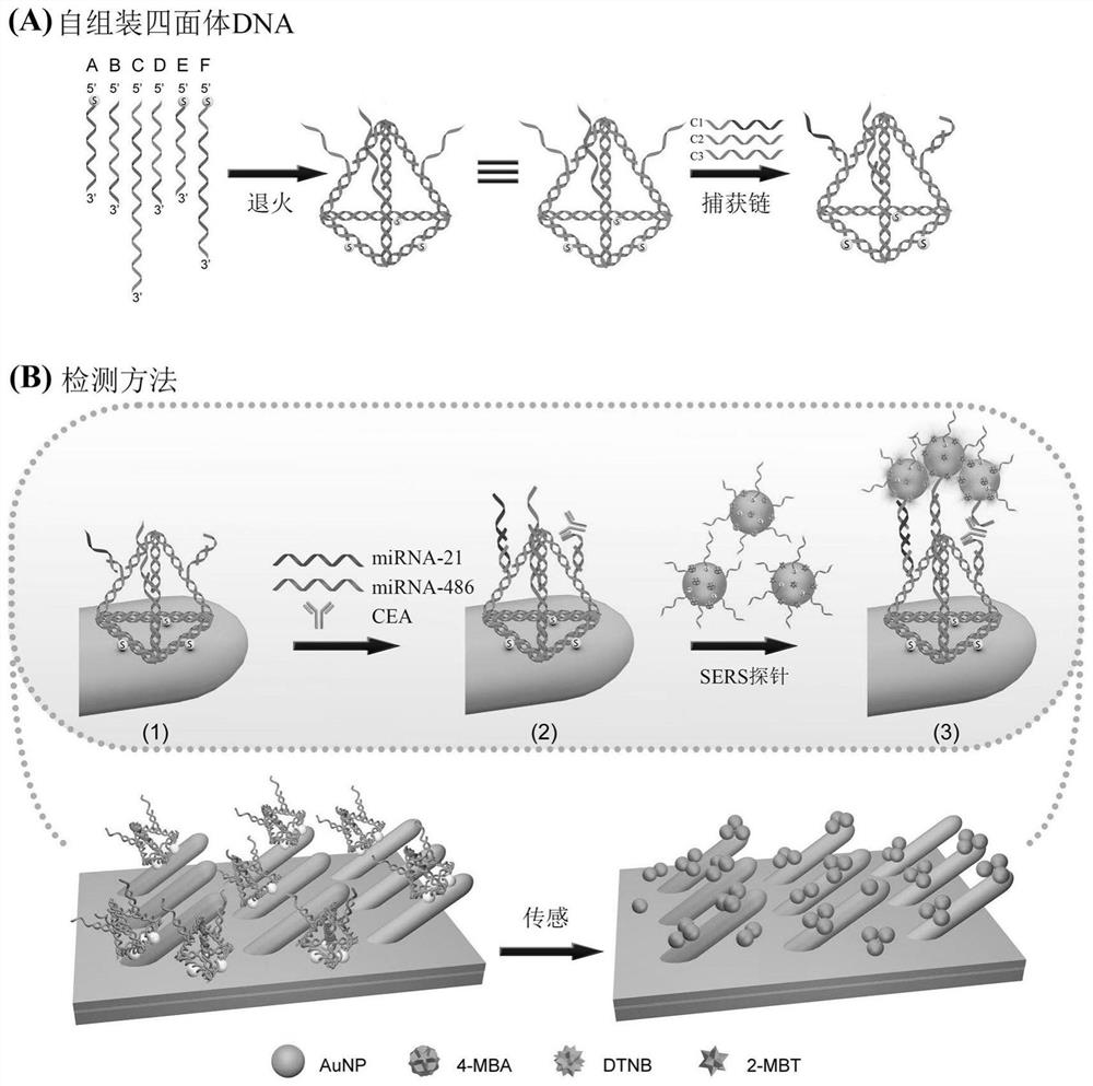
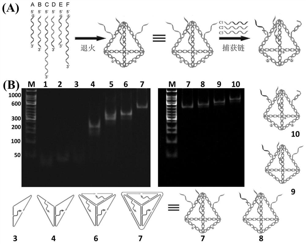
[0082] 2. Mix the amount of six specially designed DNA single strands A, B, C, D, E and F in TM buffer (20mM Tris-HCl, 50mM magnesium chloride, pH 8.0), and heat it after annealing After cooling down to 95°C and cooling down to 4°C, assemble to form tetrahedral DNA (final concentration is 1 μM), then mix and co-culture the three capture strands C1, C2 and C3 with tetrahedral DNA in the same amount of substances for 1 h to form three capture arms tetrahedral DNA probes. figure 2 (A) is a schematic diagram of tetrahedral DNA formation and hybridization of tetrahedral DNA to three capture strands. The formation of tetrahedral DNA and the hybridization of tetrahedral DNA to the three captured strands were verified by 5% polyacrylamide gel electrophoresis. figure 2 (B) i...

Embodiment 2

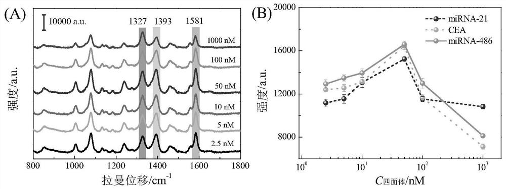
[0086] Example 2 Selection of tetrahedral DNA assembly concentration of SERS sensor on silver nanorod array substrate

[0087] Add 20 μL of tetrahedral DNA solutions (1000 nM, 100 nM, 50 nM, 10 nM, 5 nM, 2.5 nM) with different assembly concentrations dropwise into each small hole patterned on the silver nanorod array, at 25-37 ° C, 60-80 % humidity environment for 3-5 hours. Then, 20 μL of the target molecule mixture containing 100pM miRNA-21, 100pM miRNA-486 and 100pg / mL CEA was added dropwise on the surface-modified silver nanorod array substrate with tetrahedral DNA prepared in Example 1 and co-cultured for 2 hours, using PBS After the buffer solution (10mM phosphate, 100mM sodium chloride, 2mM magnesium chloride, pH 7.4) was cleaned, the mixed solution of the three SERS probes prepared according to the method of Example 1 was added dropwise on the detection chip for co-cultivation for 3 hours, and then Wash the small holes with PBS buffer and ultrapure water in sequence, ...

Embodiment 3

[0089] Example 3 Working curve and detection limit of SERS sensor for detection of three tumor markers in serum samples

[0090] In 10% normal human serum concentration of two nucleic acids miRNA-21 and miRNA-486 from 100aM to 100pM and one protein carcinoembryonic antigen (CEA) from 0.1fg / mL to 100pg / mL in 20μL of target Add the molecular mixture dropwise on the silver nanorod array substrate with tetrahedral DNA modified on the surface prepared in Example 1, let it stand for 3-5 hours at 25-37°C and 60-80% humidity environment, and clean it with PBS buffer Afterwards, the mixture of the three SERS probes prepared according to the method in Example 1 was added dropwise to the small wells for co-cultivation for 3 hours. Subsequently, the wells were washed sequentially with PBS buffer and ultrapure water. After natural air-drying, the SERS test was carried out on the silver nanorod array substrate, and the SERS spectrum and its characteristic signal intensity value were obtain...


PUM


Abstract

The invention discloses an SERS sensor for integrated detection of tumor protein and nucleic acid markers and a preparation method thereof. The SERS sensor comprises a silver nanorod array substrate and an SERS probe, wherein the surface of the silver nanorod array substrate is modified with a tetrahedral DNA structure; the tetrahedral DNA structure is formed by self-assembling six DNA single chains, and three DNA single chains are arranged on three edges of the tetrahedral DNA structure, are respectively connected with three capture chains and are used for respectively and specifically binding nucleic acid and a protein marker. The detection limits of the SERS sensor for detecting nucleic acid and protein respectively reach the Amol/L magnitude and the fg/mL magnitude; the integrated detection of various nucleic acid and protein biomarkers in complex environments such as serum can be realized. The sensor disclosed by the invention is simple to prepare, high in detection sensitivity and good in reliability, can realize high-sensitivity detection of biomarkers with extremely low abundance in the early stage of lung cancer, and has good universality for various tumor biomarkers.

Description

technical field [0001] The invention belongs to the field of functional nanometer materials and biological detection, and in particular relates to a SERS sensor for integrated detection of tumor protein and nucleic acid markers and a preparation method thereof. Background technique [0002] Lung cancer is a common malignant tumor in the world today, and has become the main cause of cancer death in most countries. Early detection of cancer can help prevent and treat cancer in time. However, the typical clinical manifestations of cancer are insidious and asymptomatic, which severely limits the effective screening of cancer at an early stage. In the early stages of cancer, the concentrations of biomarkers in the blood are extremely low, and studies have not identified a single distinct biomarker that causes a particular cancer. Usually, multiple highly correlated biomarkers (such as nucleic acids and proteins) are regulated simultaneously, and different types of markers (such...

Claims


Application Information

Patent Timeline
IPC IPC(8): G01N21/65
CPCG01N21/658
Inventor 宋春元张晶晶杨琰君汪联辉
Owner NANJING UNIV OF POSTS & TELECOMM
Why Eureka
Try Eureka