Multimodal imaging system for tissue imaging

a tissue imaging and multi-modal imaging technology, applied in the field of tissue imaging methods and apparatuses, can solve the problems of repeated biopsies, risk of underdiagnosis, and inability to accurately detect the disease, and achieve the effect of contrast enhanced area images

Inactive Publication Date: 2009-05-21
CARESTREAM HEALTH INC
View PDF9 Cites 60 Cited by
  • Summary
  • Abstract
  • Description
  • Claims
  • Application Information

AI Technical Summary

Benefits of technology

[0015]The method of the present invention is advantaged over earlier area imaging methods in that it removes the specular reflection in reflectance imaging.
[0016]The method of the present invention is also advantaged over earlier area imaging methods in that it combines polarized reflectance image and fluorescence image to obtain contrast enhanced area image.
[0017]The method of the present invention is further advantaged over earlier methods for OCT imaging in that it uses polarized reflectance image and fluorescence image to detect a region of interest to guide OCT imaging for detailed assessment over that region.

Problems solved by technology

However, biopsy is not only invasive and time-consuming; but also, a practitioner or operator may find it difficult to determine the optimal, i.e., most dysplastic, location for biopsy, leading to repeated biopsies and the risk of underdiagnosis.
This change in reflectance may not be sufficiently pronounced to be of diagnostic value when considered by itself, since the change in reflectance of light is very slight, although detectable.
For some biomedical applications, the specular component of reflectance is of no interest and is, instead, generally detrimental to obtaining an image or measurement from a sample.
Therefore, it is difficult to remove the specular reflection only without filtering out the scattering signal with the same polarization state as the specular reflection.
While autofluorescence provides a sensitive method to detect early tissue transformation, significant challenges include the low signal-to-noise ratio (SNR), difficulty in quantifying data, and limited tissue penetration.
While OCT solutions can provide very detailed image of structure beneath the surface of a tissue, OCT imaging itself can be time-consuming and its field of view is limited.
However, specular reflection in white light imaging presents an issue when identifying the suspicious region.
Fluorescence image only may lead to unnecessary OCT scan.
Optical apparatus and methods disclosed in the cited patents, co-pending applications and paper fall short of what is needed for tissue imaging that combines these imaging functions with suitable image quality and is yet compact and easy to use.

Method used


Image

  • Multimodal imaging system for tissue imaging
  • Multimodal imaging system for tissue imaging
  • Multimodal imaging system for tissue imaging

Examples


Embodiment Construction

[0033]The present description is directed in particular to elements forming part of, or cooperating more directly with, apparatus in accordance with the invention. It is to be understood that elements not specifically shown or described may take various forms well known to those skilled in the art.

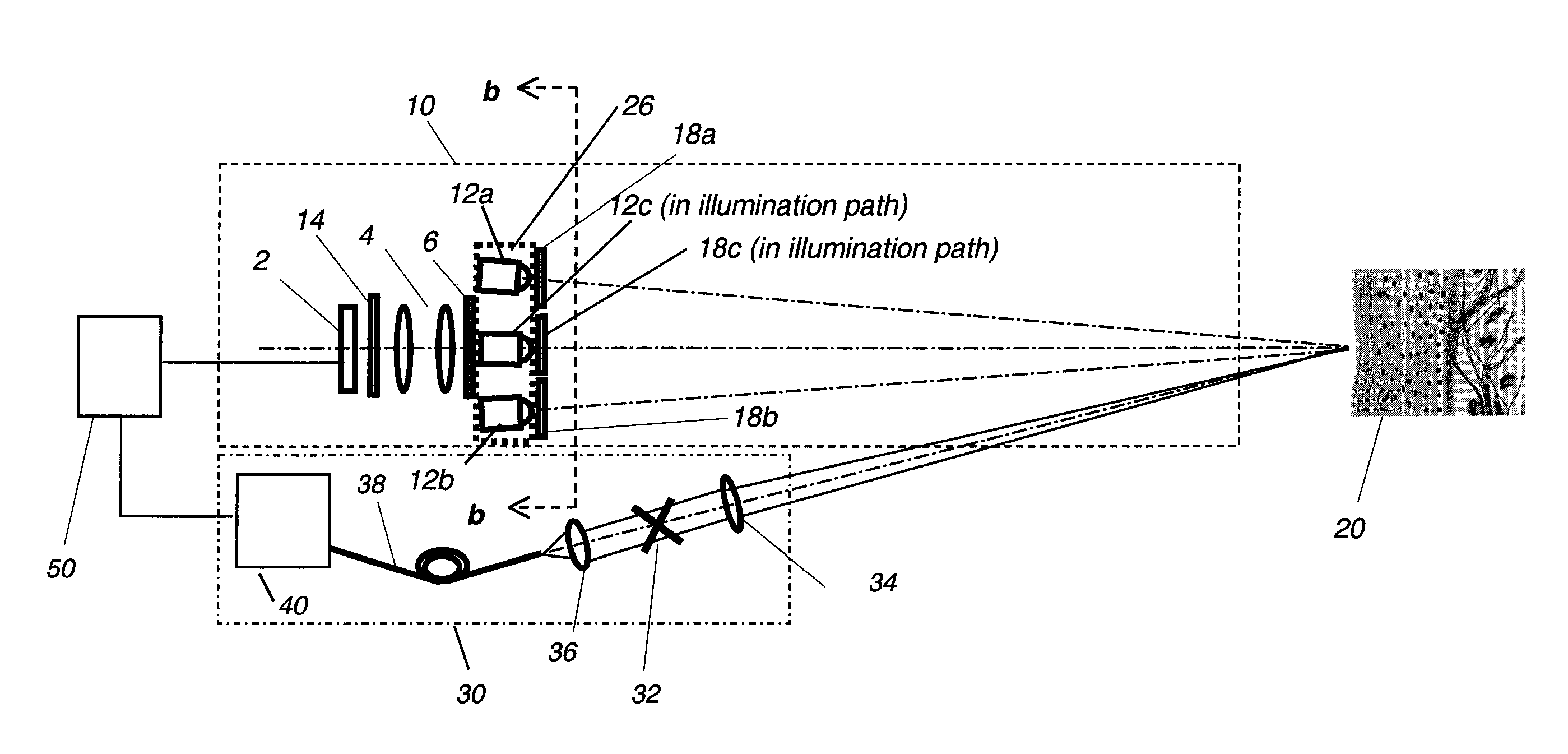
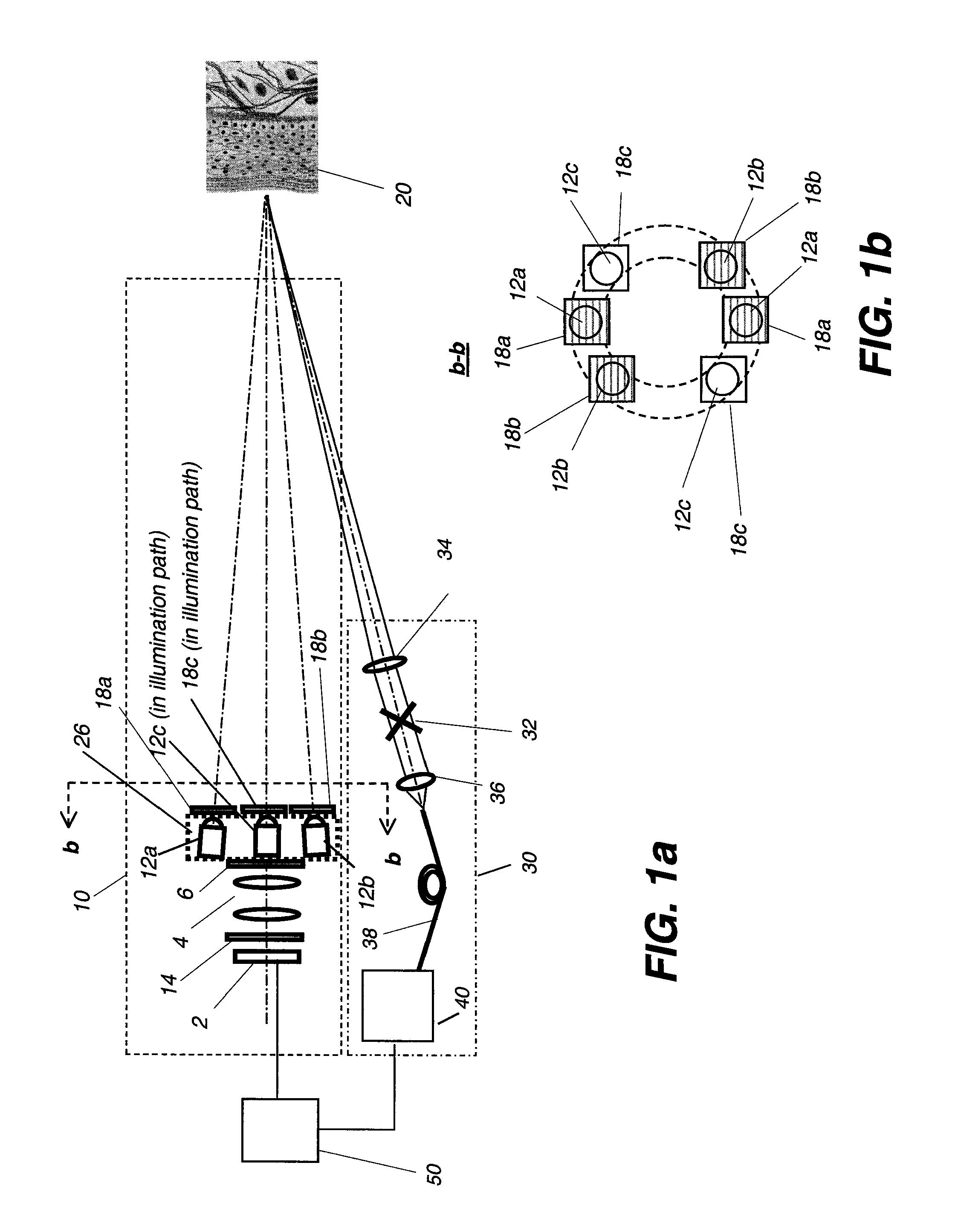
[0034]The present invention combines area imaging capabilities, namely polarized reflectance imaging and fluorescence imaging, for identifying a region or regions of interest on the tissue surface with OCT imaging capabilities for obtaining detailed OCT scan data over a specified portion of the tissue corresponding to a portion of the region of interest.

[0035]FIGS. 1a and 1b show an imaging apparatus comprising both an area imaging system 10 and an OCT imaging system 30 according to one embodiment. As part of imaging system 10, illumination means 12a, 12b and 12c provide uniform illumination on the tissue surface for area imaging. An imaging lens 4 images the surface of an area of tissue 2...


PUM


Abstract

An apparatus for obtaining area images and optical coherence tomography (OCT) image of a tissue comprises an image sensor, a first illumination means providing broadband polarized polychromatic light, a second illumination means providing narrow-band ultraviolet light, a third illumination means providing Near-Infrared light, an optical coherence tomography (OCT) imaging apparatus, one or more polarization elements, and an optical filter. An image processor identifies a region of interest according to area image, which can be a polarized reflectance image, a fluorescence light image, or both for the OCT imaging apparatus to obtain an OCT image over the region of interest.

Description

CROSS REFERENCE TO RELATED APPLICATIONS[0001]Reference is made to commonly-assigned co-pending U.S. application Ser. No. 11 / 262,869, filed Oct. 31, 2005, entitled METHOD FOR DETECTION OF CARIES, by Wong et al.; U.S. application Ser. No. 11 / 408,360, filed Apr. 21, 2006, entitled OPTICAL DETECTION OF DENTAL CARIES by Wong et al.; U.S. application Ser. No. 11 / 530,987, filed Sep. 12, 2006, entitled APPARATUS FOR CARIES DETECTION, by Liang et al.; U.S. application Ser. No. 11 / 530,913. filed Sep. 12, 2006, entitled LOW COHERENCE DENTAL OCT IMAGING, by Liang et al.; and U.S. application Ser. No. 11 / 561,971 filed Nov. 21, 2006, entitled Apparatus for Dental OCT Imaging, by Liang et al, the disclosures of which are incorporated herein.FIELD OF THE INVENTION[0002]This invention generally relates to methods and apparatus for tissue imaging using polarized visible light, ultraviolet light, polarized Near-Infrared light, and optical coherence tomography (OCT) imaging.BACKGROUND OF THE INVENTION[...

Claims


Application Information

Patent Timeline
Patent Type & Authority Applications(United States)
IPC IPC(8): A61B6/00
CPCA61B5/0066A61B5/0059
Inventor LIANG, RONGGUANG
Owner CARESTREAM HEALTH INC
Why Eureka
Try Eureka