

Trileaflet Semi-Lunar Prosthetic Tissue Valve
a semi-lunar, prosthetic tissue valve technology, applied in the field of prosthetic tissue valves, can solve the problems encroaching of the prosthetic valve, and achieve the effects of reducing the amount of blood flow allowable, saving needed space, and facilitating blood flow
- Summary
- Abstract
- Description
- Claims
- Application Information
AI Technical Summary
Benefits of technology
Problems solved by technology
Method used
Image

Examples
Embodiment Construction
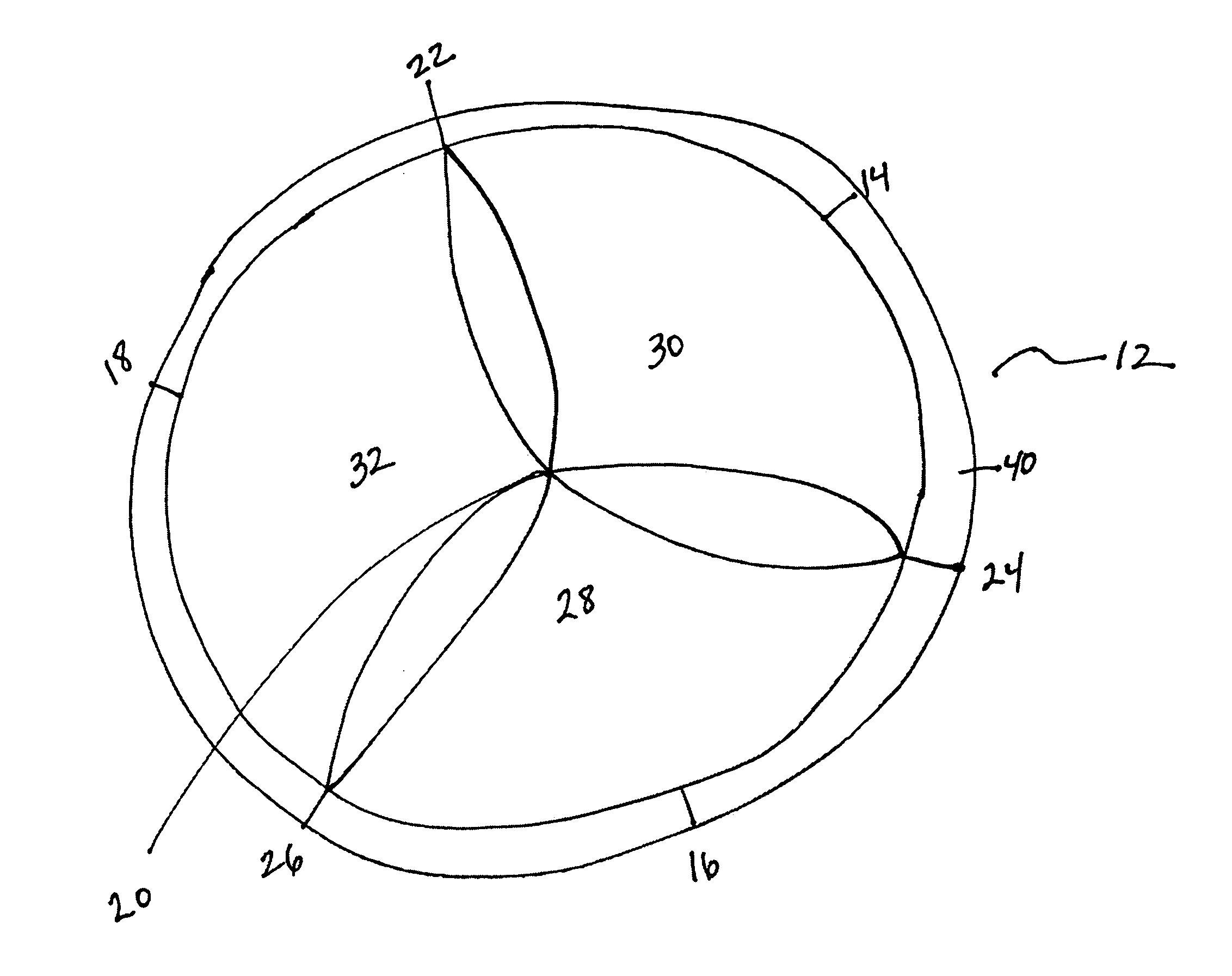
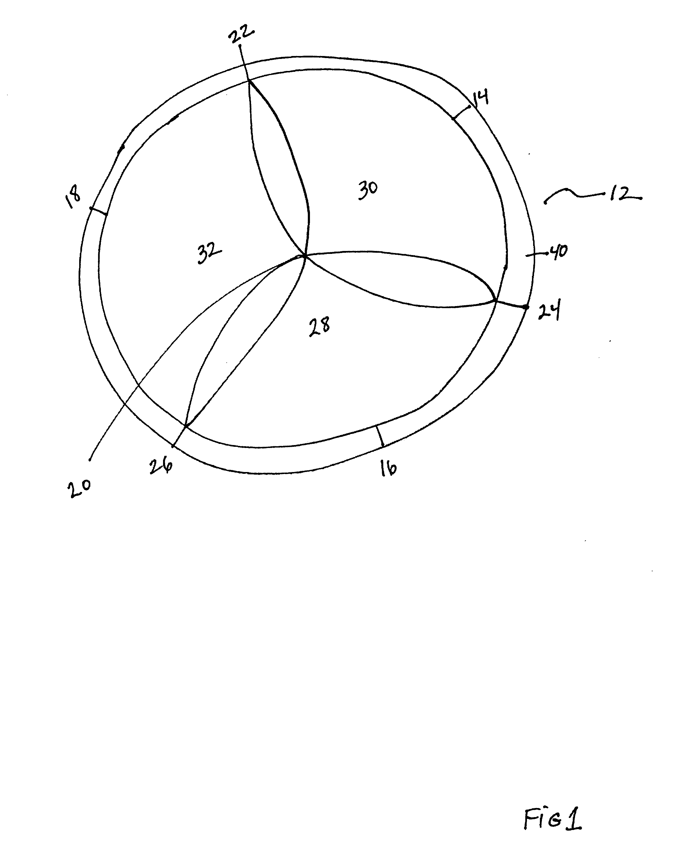
[0029]The invention is a valve for controlling fluid flow in a lumen having an annulus. The valve is suitable for replacing an aortic or pulmonary valve. The valve has a sewing ring and three equally sized leaflets. Turning now to the Figures, FIG. 1 depicts the valve as it is before attachment at an annulus. Valve 12 is semi-lunar or essentially circular having an established circumference. Leaflets 28, 30, and 32 overlap adjacent leaflets and extend to the radial center 20 of the valve. Midpoints 14, 16, and 18 of each leaflet define positions where attachment to the annulus is required for securely attaching the valve. Leaflets 28, 30, and 32 are attached to sewing ring 40.
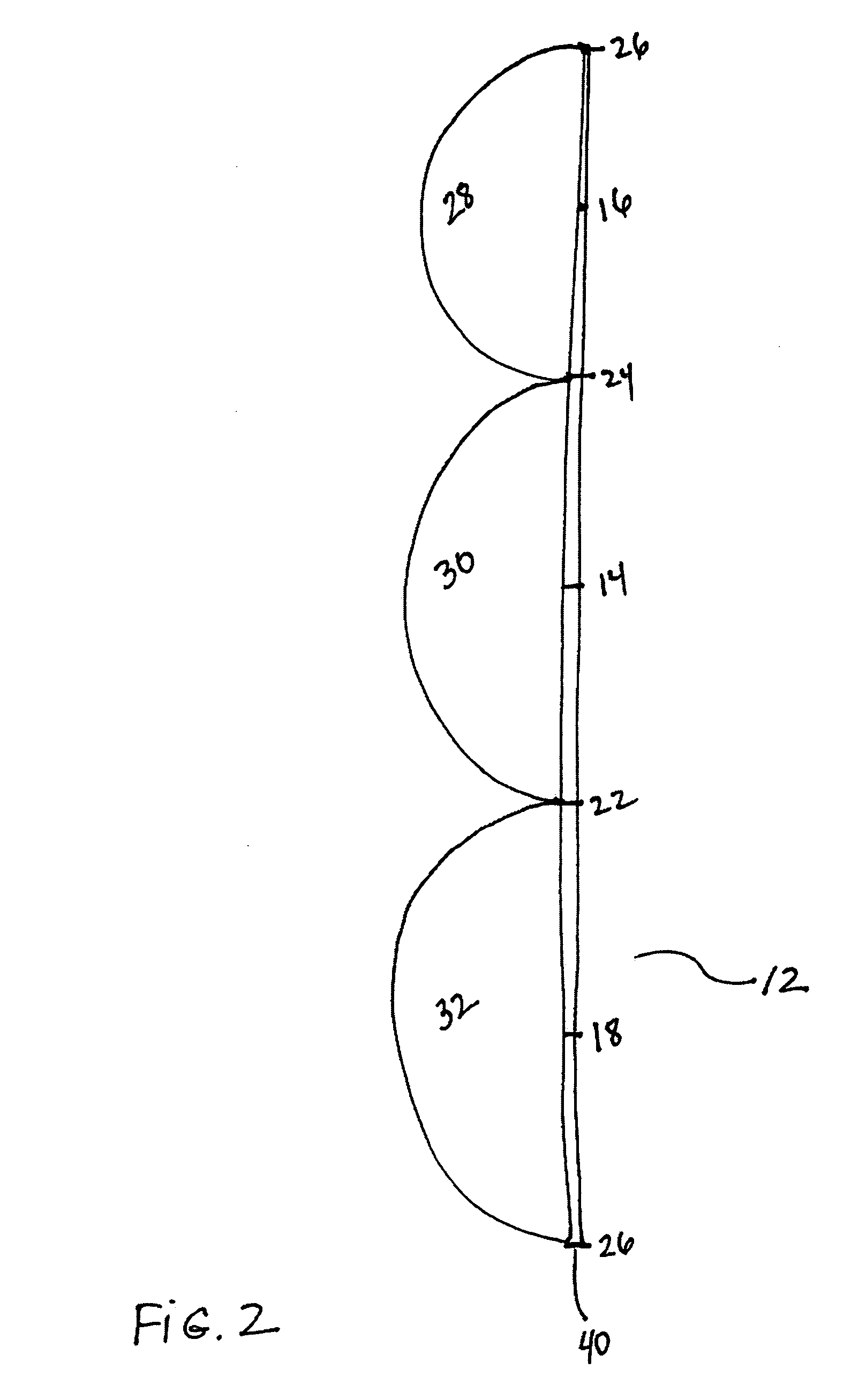
[0030]FIG. 2 depicts valve 12 if opened up at a point on the sewing ring 40. Leaflets 28, 30, and 32 are in sequence. Points 26, 22, and 24 mark the points of contact of adjacent leaflets on the sewing ring 40. Midpoints 14, 16, and 18 indicate the middle of each leaflet on the circumferencial ring to which it ...
PUM
 Abstract
Description
Claims
Application Information