
Test device for rapid diagnostics

a test device and rapid diagnostic technology, applied in the direction of fluorescence/phosphorescence, instruments, analysis using chemical indicators, etc., can solve the problems of affecting the immunochromatographic process, rare sample application (such as feces) or sampling device,

Inactive Publication Date: 2009-06-25
CORIS BIOCONCEPT
View PDF5 Cites 8 Cited by
  • Summary
  • Abstract
  • Description
  • Claims
  • Application Information

AI Technical Summary

Benefits of technology

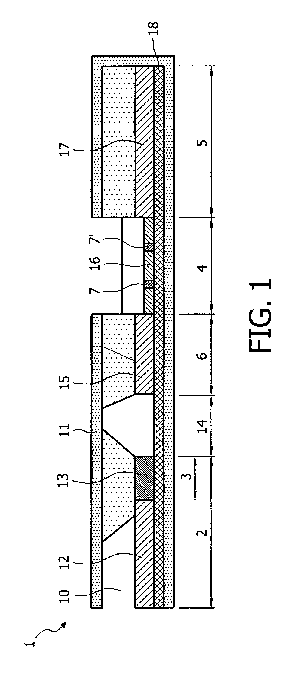
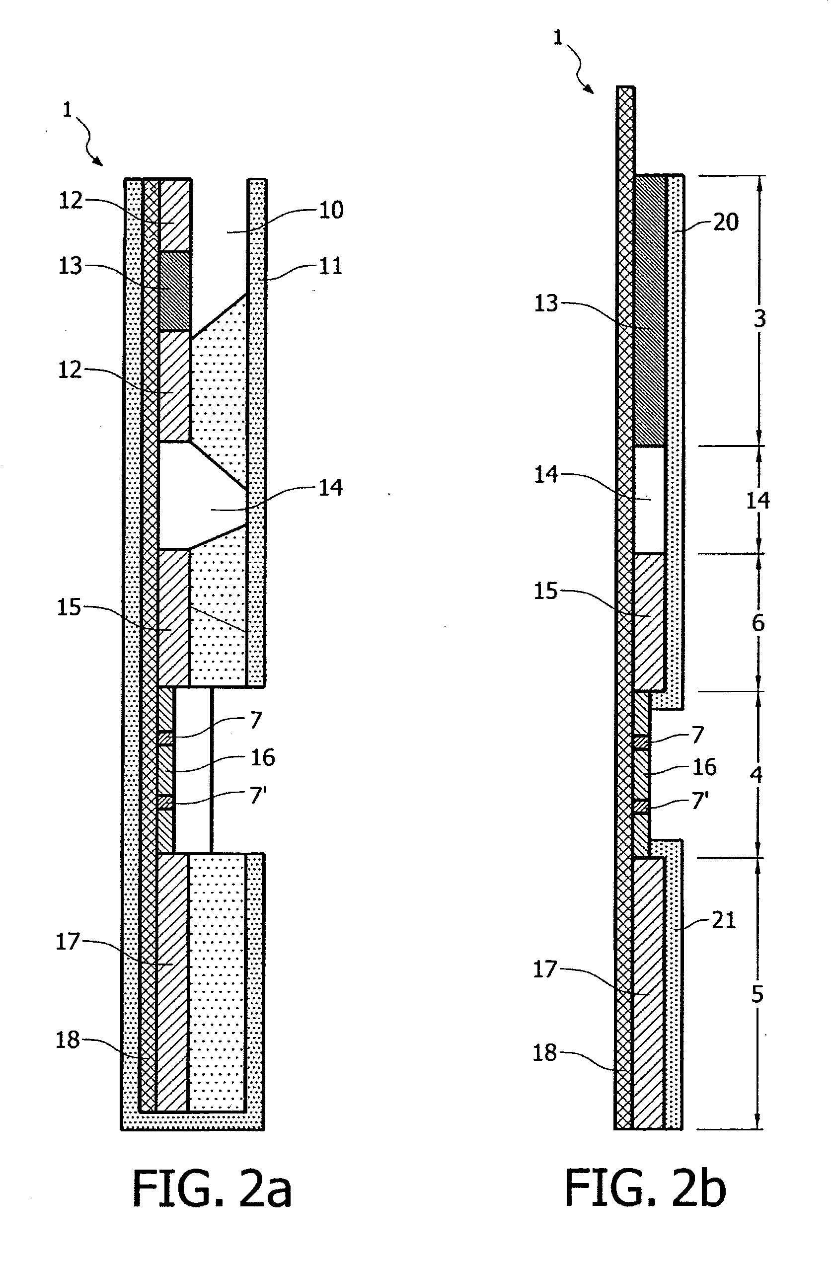
The present invention provides a gravity driven test device and method for detecting analytes in a sample. The device has multiple zones that allow the sample to move through by gravity, including a sample receiving zone, a detection zone, and an intermediate zone. The device is designed to allow for mono or multiplex detections, quantitative or semi-quantitative results, and can be used as a control to assess the accuracy and reliability of the assay. The device is easy to use, requires fewer procedures, and is highly sensitive to various levels of analytes.

Problems solved by technology

Although useful, currently available chromatographic devices using test strips have a number of drawbacks.
Many samples, such as fecal samples, contain particulate matter that can clog the pores of the chromatographic medium, greatly hindering the immunochromatographic process.
In addition it is frequently difficult with existing chromatographic test devices to apply the sample to the chromatographic medium so that the sample front moves uniformly through the chromatographic medium to ensure that the sample reaches the area where binding is to occur in a uniform, straight-line manner.
It is rarely possible to apply a sample (such as feces) or a sampling device (such as a throat swab) directly to the chromatographic medium.

Method used


Image

  • Test device for rapid diagnostics
  • Test device for rapid diagnostics
  • Test device for rapid diagnostics

Examples


example 1

[0129]Detection of Rotavirus and Enteric Adenovirus 40141 (group F, strains 40 & 41)

Preparation of Polystyrene Microspheres

[0130]Polystyrene microspheres (Estapor) are washed in a specific washing buffer (Coris BioConcept). Microspheres are then centrifuged at 13,000 RPM for 5 to 10 minutes for recovering a 1 mL volume. Pellet is then resuspended in the activation buffer and mixed for one hour. Suspension is then washed twice in the washing buffer before to be finally resuspended in the coupling buffer after a final centrifugation.

Coupling of Antibodies to Non-Activated and Amino-Activated Polystyrene Microspheres

[0131]Coupling of the reagent to the polystyrene microspheres was performed essentially following the protocol provided by the manufacturer.

[0132]First coupling was performed with a mouse monoclonal antibody directed against Rotavirus group A antigen with blue NH2-polystyrene microspheres.

[0133]Second coupling was performed with non-activated red polystyrene microspheres wi...

example 2

[0143]Detection of Legionella pneumophila Urinary Antigen

Preparation of Colloidal Gold Particles

[0144]Colloidal gold particles of about 40 nm were purchased from a commercial source (Diagam).

Coupling of Antibodies to Colloidal Gold Particles

[0145]Coupling antibodies to colloidal gold particles is well known in the art. In this example, purified rabbit antibodies directed against Legionella pneumophila urinary antigen were used. Purified polyserum was reacted with a colloidal gold particles suspension that had been buffered with a potassium carbonate solution to obtain the desired pH. This pH is predetermined and may be different for each immunological reagent. The dilution of the purified polyserum to be used in the coupling process was defined in a preliminary experiment.

[0146]In this preliminary experiment, increasing dilutions of the polyserum were reacted for three minutes with the buffered colloidal gold particles and then sodium chloride was added to reach about 1% final conce...


PUM

PropertyMeasurementUnit
diameteraaaaaaaaaa
diameteraaaaaaaaaa
diameteraaaaaaaaaa

Abstract

Devices for detecting analytes or analogues thereof in a biological sample are disclosed. The device includes a solid support. The solid support has several juxtaposed zones. The sample is able to migrate from a sample receiving zone towards a detection zone. The analyte, if present, is detected in the detection zone. Both zones have material allowing a capillary flow of the sample through the zones. In between the zones, there is an intermediate zone of transport of the sample which is free from any capillary material. This allows the ample to migrate by gravitational forces on the support laid in a vertical position. Methods for detecting analytes or analogues thereof in a biological sample using the device are also disclosed.

Description

FIELD OF THE INVENTION[0001]This invention relates to methods and devices for detecting analytes or analogues thereof in a biological sample. This invention relates to improved rapid tests such as “dipsticks” devices. The invention in particular relates to a test device made of one or more active sides, in order to allow mono or multiplex detections, quantitative or semi-quantitative detections. The devices described in this invention allow detecting or identifying various biologicals or chemicals with one manipulation.BACKGROUND OF THE INVENTION[0002]Several approaches have been developed for detection of analytes in a biological sample for routine diagnostics in diagnostic laboratories via for instance immunochromatography. EP 0 088 636, EP 0 186 799, EP 0 284 232 and WO 88 / 08534 disclose sheet-like chromatographic devices comprising at least a first and a second zone or region. Prior art devices disclosed in these documents comprise:[0003]a first region or zone containing porous ...

Claims


Application Information

Patent Timeline
Patent Type & Authority Applications(United States)
IPC IPC(8): C12Q1/70G01N21/64G01N33/558G01N33/569
CPCG01N33/54386G01N33/569G01N33/558G01N33/54391
Inventor MERTENS, PASCALDENORME, LAURENCELECLIPTEUX, THIERRY
Owner CORIS BIOCONCEPT
Learn More