Method and Apparatus to Perform Limited Two Dimensional Separation of Proteins and Other Biologicals

a technology of two-dimensional separation and proteins, applied in the direction of fluid pressure measurement, liquid/fluent solid measurement, peptide, etc., can solve the problems of time-consuming and non-quantitative, inability to automate the process nor quantify the resolved component proteins or other analytes, and the separation and ph gradient obtained in cief may be disturbed, so as to achieve superior separation resolution

Active Publication Date: 2009-08-06
PROTEINSIMPLE
View PDF16 Cites 31 Cited by
  • Summary
  • Abstract
  • Description
  • Claims
  • Application Information

AI Technical Summary

Benefits of technology

[0008]This invention describes improved method and apparatus for carrying out limited electrophoretic separation in the liquid phase. The objective of the invention is to provide a simple method and apparatus for limited “2D” separation using both capillary or channel IEF separation and capillary zone electrophoresis (CZE) separation within the same capillary or channel. The present invention also integrates real-time, whole-channel electrophoresis detection with automatic sample injection, automatic cIEF separation, separation zone manipulation and on-line electrolyte selection, to achieve a separation resolution superior to that obtained using an orthogonal capillary arrangement.

Problems solved by technology

2D gel electrophoresis is generally carried out in a polyacrylamide slab gel and although it has become a workhorse in the field of proteomics, owing to the high degree of resolution which can be obtained thereby, it is very labour-intensive, time consuming and non-quantitative.
Moreover, although 2D gel electrophoresis does afford the highest degree of molecular weight resolution of known electrophoretic separation techniques, it has not yet been possible to automate that process nor quantify the resolved component proteins or other analytes.
It is known that the separation and pH gradient obtained in cIEF may be disturbed when mobilizing focused protein zones to reach the outlet end.
A as result, it is more challenging to transfer separated zones from the first separation dimension to the second separation dimension in the orthogonal capillary or microchannel format than in apparatus for 2D gel electrophoresis.
Fluid connections and for control of nanoliter volumes are required, making for complex analytical design and operation.

Method used


Image

  • Method and Apparatus to Perform Limited Two Dimensional Separation of Proteins and Other Biologicals
  • Method and Apparatus to Perform Limited Two Dimensional Separation of Proteins and Other Biologicals
  • Method and Apparatus to Perform Limited Two Dimensional Separation of Proteins and Other Biologicals

Examples


example 1

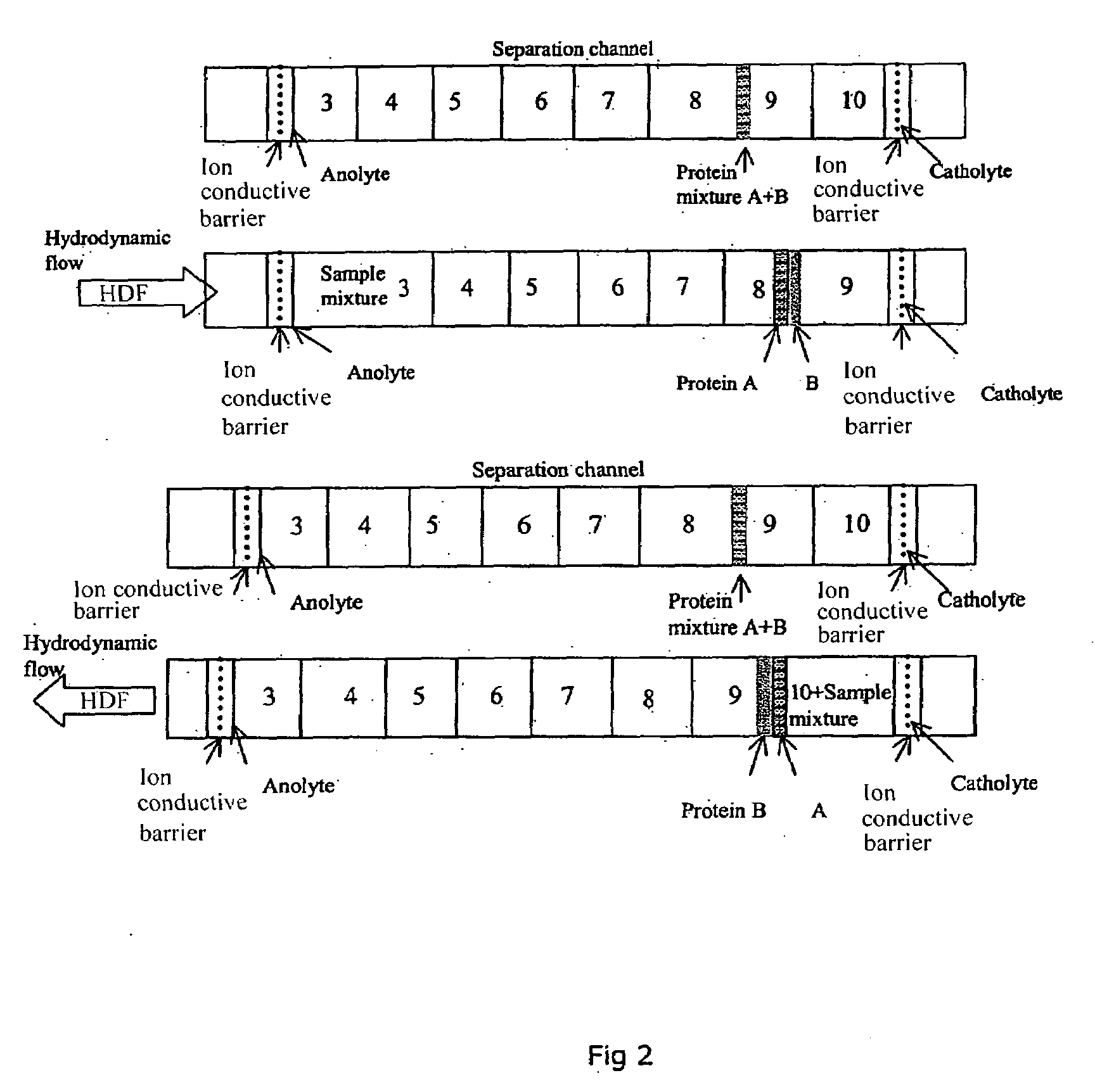
ydrodynamic Flow as Second Dimension of Separation

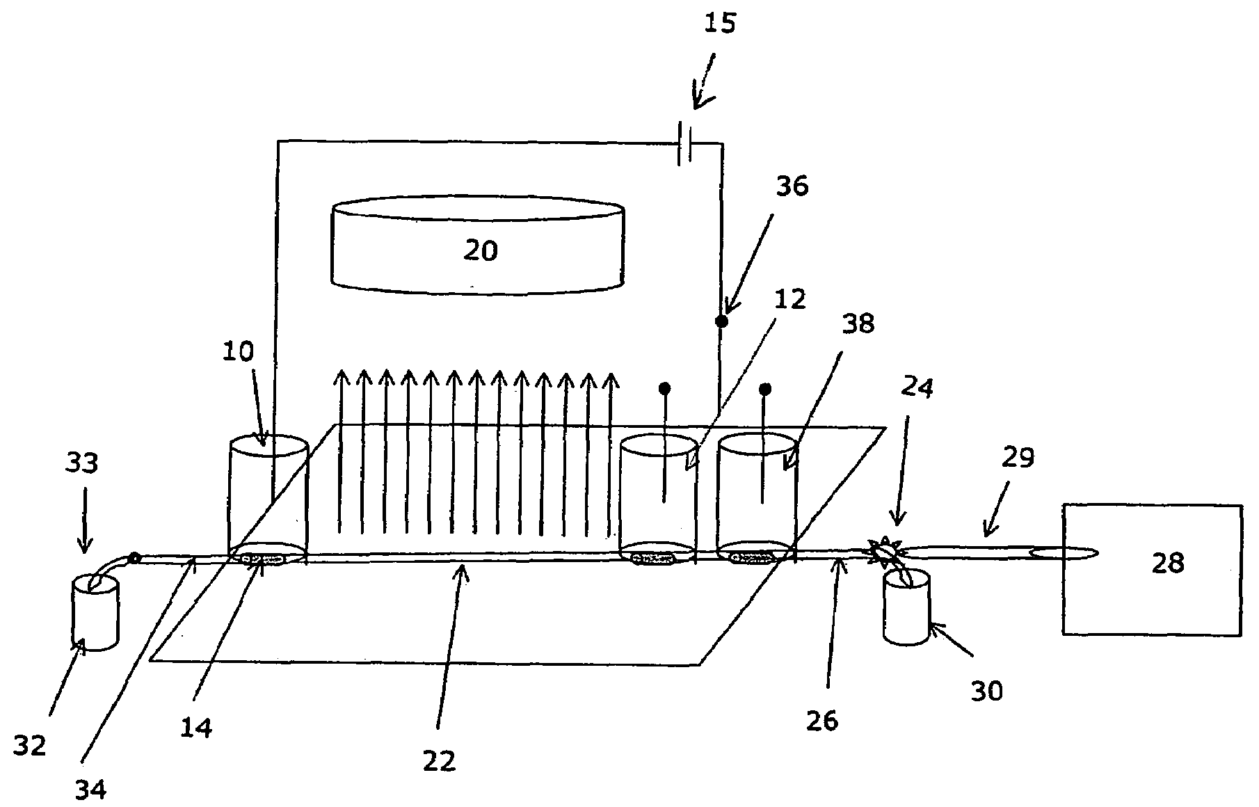
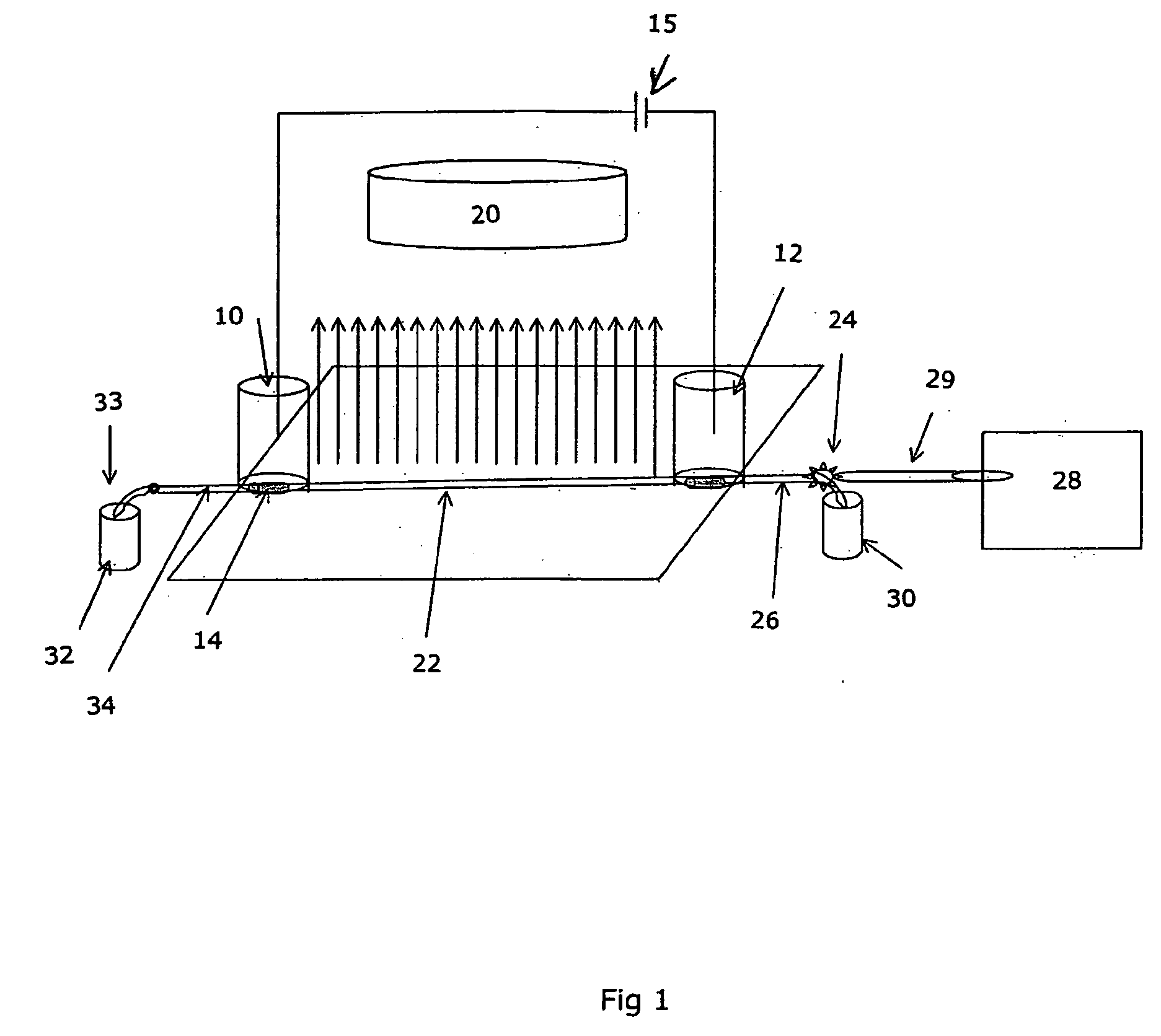
[0025]FIG. 5 illustrates hydrodynamic flow induced limited 2D separation of protein trypsinogen and a small molecular weight pI marker. In this experiment, trypsinogen and a small molecular pI marker were mixed with 8% pH 3-10 Pharmalyte and 0.35% methylcellulose. The sample mixture was injected into a 50 mm 100 μm inner diameter FC coated capillary with a micro autosampler. Focusing was conducted at a focusing voltage of 3000 V, with 80 mM H3PO4 as anolyte and 100 mM NaOH as catholyte. Detection was conducted with a real-time, whole column UV detector. The hydrodynamic flow is controlled by the water level difference in the hydrodynamic flow vial and the inlet vial.

[0026]It can be seen that when hydrodynamic flow was minimized (i.e. under first dimension cIEF separation conditions), there were two peaks in the electrophorogram (trace a). The more acidic peak to the left of the electrophorogram (egram) contains the minor component of...

example 2

Mobilization as Second Dimension of Separation

[0027]FIG. 6 illustrates chemical mobilization induced limited 2D separation of transferrin, myoglobin and a small molecular weight pI marker (pI 4.22). In this experiment, transferrin and myoglobin and the pI marker were mixed with 8% pH 3-10 Pharmalyte and 0.35% methylcellulose. The sample mixture was injected into a 50 mm 100 μm inner diameter FC coated capillary with a micro autosampler. Focusing was conducted at a focusing voltage of 3000 V, with 80 mM H3PO4 as anolyte and 100 mM NaOH as catholyte. Detection was conducted with a real-time, whole column UV detector. For anodic mobilization (trace b), the anolyte was replaced with 100 mM NaOH upon completion of cIEF focusing. For cathodic mobilization (trace c), the catholyte was replaced with 80 mM H3PO4 upon completion of focusing. In Trace a, it can be seen that when electroosmotic flow and hydrodynamic flow are well controlled (i.e. under first dimension cIEF separation conditions...


PUM

PropertyMeasurementUnit
amphotericaaaaaaaaaa
column imagingaaaaaaaaaa
hydrodynamic pressureaaaaaaaaaa

Abstract

A method and apparatus are provided for performing capillary isoelectric focusing followed by mobilization of the focused zones by induced hydrodynamic flow or chemical mobilization. These two dimensions of separation are integrated with real-time whole-channel electrophoresis detection and automatic sample injection to achieve a separation resolution superior to that obtainable using known orthogonal capillary two dimensional arrangements.

Description

RELATED APPLICATION[0001]This application claims priority to U.S. Provisional Patent Application No. 61 / 023,492 filed on Jan. 25, 2008, and entitled SIMPLIFIED METHOD AND APPARATUS FOR CARRYING OUT LIMITED TWO DIMENSIONAL SEPARATION OF PROTEINS AND OTHER BIOLOGICALS, the disclosure of which is incorporated herein by reference.BACKGROUND OF THE INVENTION[0002]The present invention is in the technical field of two-dimensional separation of proteins and other biologicals and relates particularly to apparatus and a method for the rapid and reproducible separation of species in a liquid medium.[0003]The separation and characterization of proteins is ubiquitous throughout the life sciences. Two of the most popular electrophoresis separation techniques are: 1) gel isoelectric focusing (IEF), where the separation mechanism is based on protein surface charge providing isoelectric point (pI) separation and 2) sodium dodecyl sulfate (SDS) gel electrophoresis where the separation mechanism is b...

Claims


Application Information

Patent Timeline
Patent Type & Authority Applications(United States)
IPC IPC(8): B01D57/02
CPCC07K1/285C07K1/36G01N27/44795G01N27/44773G01N27/44721
Inventor HUANG, TIEMINWU, JIAQI
Owner PROTEINSIMPLE
Why Eureka
Try Eureka