Feeding tube

a feeding tube and tube technology, applied in balloon catheters, surgery, other medical devices, etc., to achieve the effect of reducing the displacement of bacteria or cancer cells, and preventing a larger entry poin

Active Publication Date: 2009-08-27
SAINATH INTPROP
View PDF32 Cites 24 Cited by
  • Summary
  • Abstract
  • Description
  • Claims
  • Application Information

AI Technical Summary

Benefits of technology

[0040]In one example, a portion of the dilator is within the feeding tube, although the dilator end extends from the distal end of the feeding tube by a small amount and the collar covers the end of the G tube and a balloon. In one example, the small amount is about ½ inch or 1.27 cm. The dilator tip may have a flexible collar that extends back up the outer side of the feeding tube by ½ inch, for example. For example, the dilator and its flexible collar form a single unit. The single unit may be formed from a flexible plastic material, such as a polyurethane, a polyethylene, a polyvinyl, a nylon, a silicone rubber, a co-polymer, a PTFE or the like. By having only a minimal portion of the flexible collar extending over the feeding tube's distal end, the feeding tube is prevented from being held up at the dilation entry point. In addition, this unit prevents a larger dilation entry point into the stomach than necessary to fit the external tube. One advantage of the dilator of the integrated tube unit is that this prevents larger entry points than required for a feeding tube or other purpose, as distinguished from methods requiring larger entry points because the dilator must be positioned outside of the feeding tube.
[0041]Yet another advantage is that displacement of bacteria or cancer cells may be reduced, unlike previous methods which involved pulling of the feeding tube through the mouth without endoscopic visualization. Another advantage is that removal and replacement of the jejunal tube is also simplified, as the jejunal balloon collar may be deflated and the tube pulled out through the gastric sleeve. Also, using a gastrocutaneous fistula, a new external tube may be placed within the stomach. Another advantage is that the time for completion of the procedure is reduced, as compared to a surgical insertion procedure, such as the method used in the total parenteral nutrition method, for example.
[0042]The integrated gastro jejunal unit is much more easily inserted and replaced, compared to conventional jejunostomy feeding tubes. According to a method of replacement, either the jejunal tube only or both the jejunal tube and the gastric tube may be replaced by deflating a balloon collar for easy removal.

Problems solved by technology

No known jejunal tube has a balloon capable of being retained in the jejunum during jejunal feeding.

Method used


Image

  • Feeding tube
  • Feeding tube
  • Feeding tube

Examples


Embodiment Construction

[0060]The examples described and the drawings rendered are illustrative and are not to be read as limiting the scope of the invention as it is defined by the claims that eventually issue.

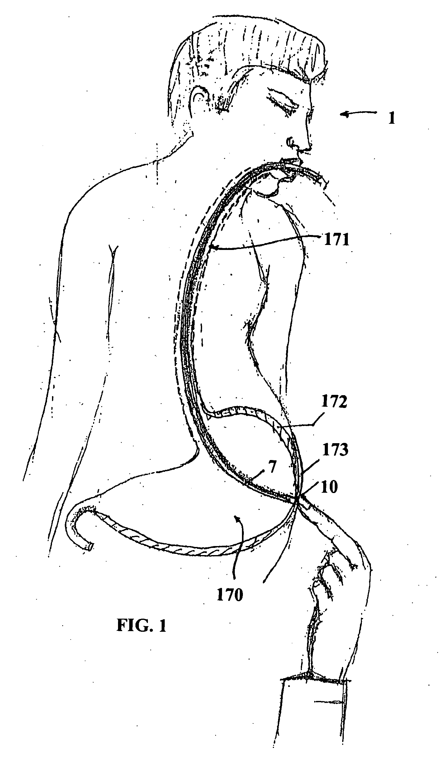
[0061]In FIG. 1, the endoscope 7 is disposed adjacent to the abdominal wall within the stomach and is used to fill the stomach with air, displacing viscera between the stomach wall 172 and the abdominal wall 173. The light on the endoscope may be used to identify the location of the endoscope, which may also be felt using a finger of a surgeon's hand, as illustrated in FIG. 1. For example, a physician may use finger pressure at a position 10 to locate a site for insertion of an external tube such as a gastrostomy tube. Once the location of the endoscope in the stomach is confirmed, by whatever means, the physician may transluminate the abdominal wall of the patient with an endoscope light, and may select a site to insert the feeding tube.

[0062]Then, a small incision may be made by the physician at t...


PUM


Abstract

An feeding tube and a kit for installation of a feeding tube includes a balloon anchored gastric tube. A protective sleeve on a dilator protects the toroidal balloon from damage during surgical insertion of the feeding tube. In one example, a jejunal tube is integrated with the kit and the gastric tube serves as a gastric sleeve forming an annular region or channels for fluid flow to and/or from the stomach and to/from the jejunum. An integrated gastro jejunal feeding tube unit may include a jejunal tube outlet port at one end, a jejunal balloon port, a gastric sleeve outlet port, one or more gastric balloon ports, one or more gastric balloons, gastric drainage holes, a jejunal balloon positioned at end of the jejunal tube and sleeve. A black silk loop may affix an end of the jejunal tube at a position in the jejunum, and insertion of the jejunal tube may be monitored continuously using an endoscope. Drainage holes may be provided in the jejunal tube and/or the gastric sleeve. Depending on the clinical state, feeding may be initiated through the gastric sleeve outlet port or via the jejunal tube outlet port and may be altered without surgical intervention, as required. Drainage holes in the gastric sleeve may be used to deflate the stomach, while nutrition is provided by a jejunal tube inserted in the jejunum, simultaneously.

Description

CROSS RELATED APPLICATIONS[0001]The Applicant claims the benefit of and priority to U.S. Pat. Appl. Ser. No. 61 / 031,442, filed Feb. 26, 2008, and U.S. Pat. Appl. Ser. No. 61 / 113,697, filed Nov. 12, 2008, and the Applicant incorporates by reference herein the entire specification of both of applications.FIELD OF THE INVENTION[0002]The field relates to total parenteral nutrition, nasogastric tubes, gastrostomy, and related methods in which feeding a patient bypasses mastication and ingestion.BACKGROUND[0003]Conventional nasogastric tubes have been in use for many years. Complications include trauma related to tube insertion and effects from use of the tube for prolonged periods of time. Occasionally, severe complications such as tracheo-esophageal fistula and perforation of various organs have been reported. Gastrointestinal bleeding of various magnitudes also can occur but less frequently. In addition, feeding tubes inserted through the nose or mouth are acceptable for short period, ...

Claims


Application Information

Patent Timeline
Patent Type & Authority Applications(United States)
IPC IPC(8): A61M25/10A61F2/958
CPCA61J15/0015A61J15/0042A61J15/0069A61B17/3415A61J15/0049A61J15/0073A61J15/0007
Inventor NATH, IYUNNI VENKATA SESHA SAYI
Owner SAINATH INTPROP
Why Eureka
Try Eureka