Generation of biological pacemaker activity
a technology of biological pacemaker and activity, which is applied in the field of generating biological pacemaker function in cells, can solve the problems of not being able to achieve this on its own, and the approach has not been shown to maintain the function of the biological pacemaker
- Summary
- Abstract
- Description
- Claims
- Application Information
AI Technical Summary
Problems solved by technology
Method used
Image
Examples
example 1
Generation of Biological Pacemaker by Kir2.1 Gene Transfer
[0095]Efforts to engineer biological pacemakers have focused either on over-expression of HCN channels or suppression of IK1 to liberate endogenous pacemaker activity. Here we report a novel strategy designed to convert IK1 into a cationic nonselective “leak” current. We utilized Kir2.1 channel mutations (E138R and R148E, Kir2.1ER) which have been shown to render the channel non-selective, conducting Na+ as well as K+.
[0096]Co-expression of wild-type and the mutant channels by Ad-Kir2.1ER-IRES-Kir2.1WT in HEK293 cells yielded Ba2+-sensitive hyperpolarization-activated inward currents (−5.5 pA / pF at −80 mV) with a Vrev of −35.1±2.1 mV (n=5). The data demonstrates that over-expression of Kir2.1ER destabilizes the resting membrane potential established by Kir2.1 WT channels.
[0097]Equipped with this data, adenovirus expressing Kir2.1ER channels (Ad-Kir2.1ER-IRES-GFP) were expressed by a direct injection into the apex of guinea pi...
example 2
Generation of Viral Vectors for Expression of Kir2.1AAA and HCN
[0107]Plasmids, Viruses and Cell Lines
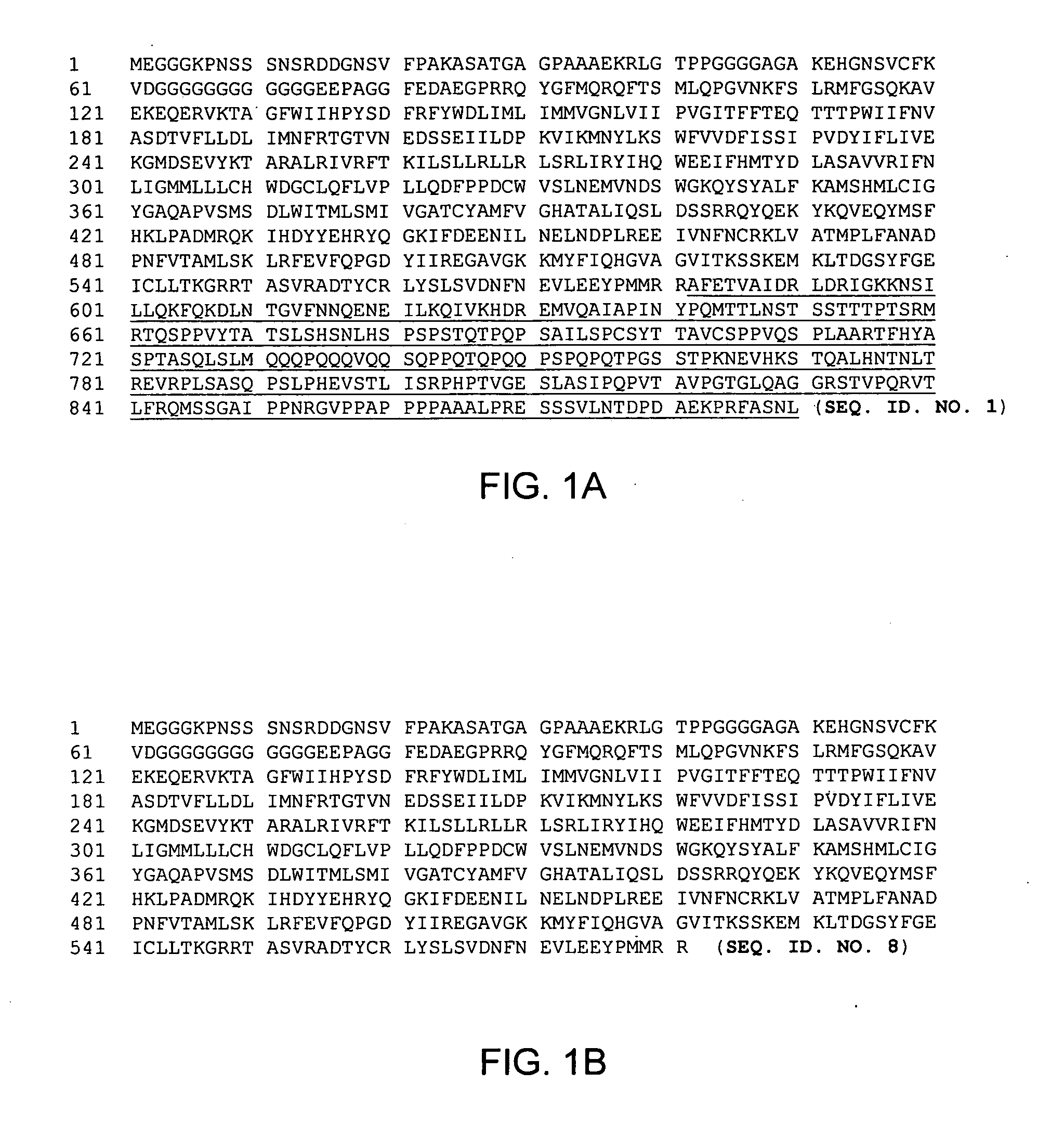
[0108]HD adenovirus vector system was provided by Microbix. HDAd-HCN1tr-IRES-Kir2.1AAAeGFP was constructed by placing a linker containing a NotI site into the AseI site of pHCN1tr-IRES-Kir2.1AAAeGFP. The resulting plasmid (see FIG. 11) was cloned into the NotI site of pC4HSU, a plasmid containing the HD adenovirus backbone and stuffer DNA. Expression of Kir2.1AAA should result in an amino acid of SEQ. ID. NO. 6, see FIG. 5B. Expression of HCN1tr should result in an amino acid comprising amino acids 1-581 of SEQ. ID. NO. 1 with amino acids 582-890 truncated (SEQ. ID. NO. 8), see FIG. 1B.
[0109]To construct HDAd-HCN4tr-IRES-Kir2.1AAAeGFP, a linker containing AscI sites was cloned into pHCN4tr-IRES-Kir2.1AAAeGFP. The AscI fragment from pHCN4tr-IRES-Kir2.1AAAeGFP containing the entire transgene was then cloned into the homologous site in pC4HSU. Expression of Kir2.1AAA should result in an...
example 3
Prophetic Example
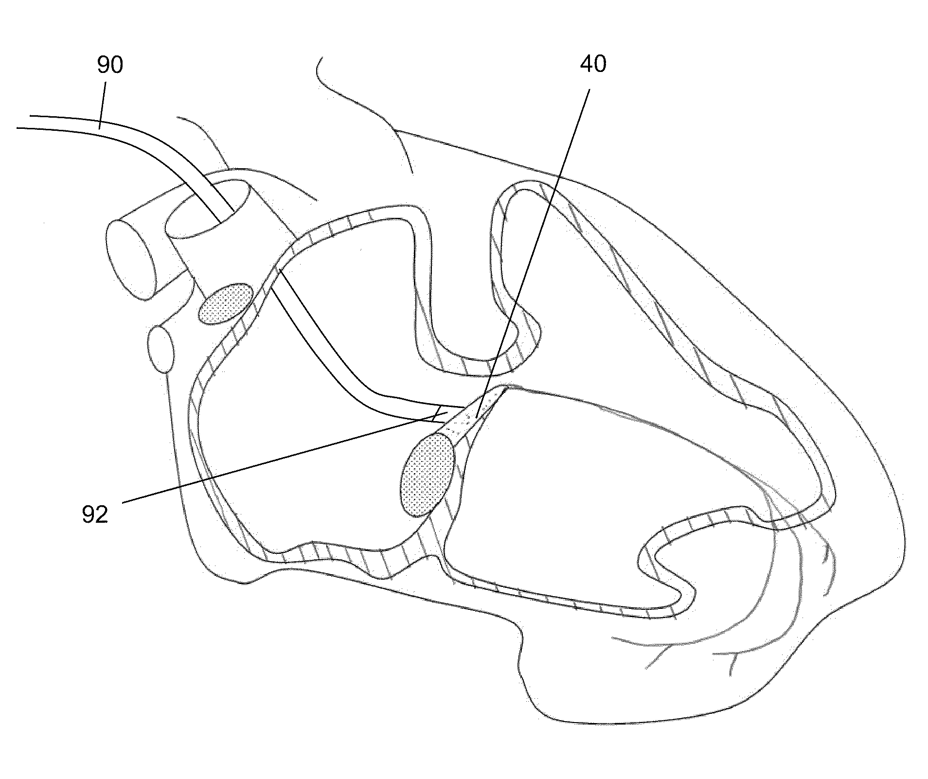
[0115]Adult guinea pigs may be infected by intramuscular injection (via catheter) of a solution of saline with a viral concentration range of approximately 3×10¹⁰ to 3×10¹⁴ plaque forming units (PFU) HDAd or AAV 2 / 9. The HDAd or AAV 2 / 9 may contain an expression vector having DNA encoding an HCN1 channel and a dominant negative Kir2.1 channel. The expression vector may contain a reporter gene, such as green fluorescence protein. DNA encoding a short polypeptide protein, such as myc-tag, which can serve as an antigen for verification of expression, may be inserted such that it will be expresses at the N- or C-terminal of the HCN1 channel or the Kir2.1 channel.
[0116]For targeted injection to the right side of the heart, the catheter may be guided to the right atrium, either via the superior vena cava or inferior vena cava, which by itself may be accessed via one of the femoral veins. The right ventricle may then be accessed by guiding the catheter through the tricuspi...
PUM
| Property | Measurement | Unit |
|---|---|---|
| volumes | aaaaa | aaaaa |
| membrane potential | aaaaa | aaaaa |
| outer diameter | aaaaa | aaaaa |
Abstract
Description
Claims
Application Information
 - R&D
- Intellectual Property
- Life Sciences
- Materials
- Tech Scout
- Unparalleled Data Quality
- Higher Quality Content
- 60% Fewer Hallucinations
Browse by: Latest US Patents, China's latest patents, Technical Efficacy Thesaurus, Application Domain, Technology Topic, Popular Technical Reports.
© 2025 PatSnap. All rights reserved.Legal|Privacy policy|Modern Slavery Act Transparency Statement|Sitemap|About US| Contact US: help@patsnap.com


