
Methods and devices for passive residual lung volume reduction and functional lung volume expansion

a technology of passive residual lung volume and functional lung volume, applied in the field of medical methods and equipment, can solve the problems of limited devices, and limited use of pulmonary passageway viewing devices, and achieve the effect of reducing residual volum

Active Publication Date: 2009-10-01
PULMONX
View PDF30 Cites 43 Cited by
  • Summary
  • Abstract
  • Description
  • Claims
  • Application Information

AI Technical Summary

Benefits of technology

[0012]The present invention provides methods and apparatus for passively reducing the residual volume (the volume of air remaining after maximal exhalation) of hyperinflated or otherwise diseased lung compartments or segments. By “passively reducing,” it is meant that air can be removed from the diseased lung region without the use of a vacuum aspiration to draw the air from the region. Typically, such passive reduction will rely on a non-implanted one-way flow element, structure, or assembly which permits air to be exhaled or exhausted from the lung region while preventing or inhibiting the inspiration of air back into the lung region. By non-implanted, it is meant that some portion of the element, structure, or assembly will be temporarily placed in an airway or bronchus leading to the lung region in a manner that allows that portion to be removed later, typically within days or hours, without the need for surgical intervention Thus, the methods of the present invention will not require the permanent implantation of valves or other structures prior to actually achieving the desired residual lung volume reduction, as with the one-way implantable valve structures of the prior art.
[0015]In a first aspect of the present invention, methods for reducing the residual volume of a hyperinflated lung compartment comprise sealingly engaging a distal end of a catheter in an airway feeding the lung compartment. Air is allowed to be expelled from the lung compartment through a passage in the catheter while the patient is exhaling, and air is blocked from re-entering the lung compartment through the catheter passage while the patient is inhaling. As the residual volume diminishes, the hyperinflated lung compartment reduces in size freeing up the previously occupied space in the thoracic cavity. Consequently, a greater fraction of the Total Lung Capacity (TLC), which is the volumetric space contained in the thoracic cavity that is occupied by lung tissue after a full inhalation becomes available for the healthier lung compartments to expand and the volume of the lung available for gas exchange commonly referred to in clinical practice as the lung's Functional Vital Capacity (FVC) or Vital Capacity (VC) increases, the result of which is effectively a functional lung volume expansion.
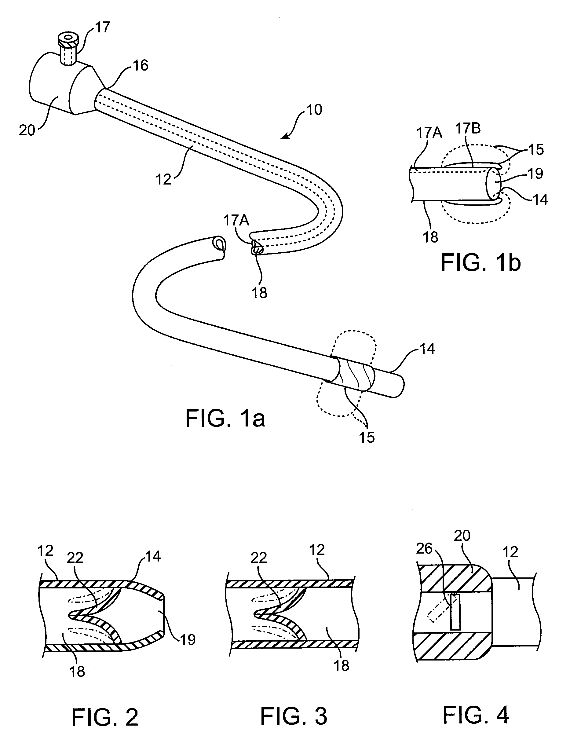
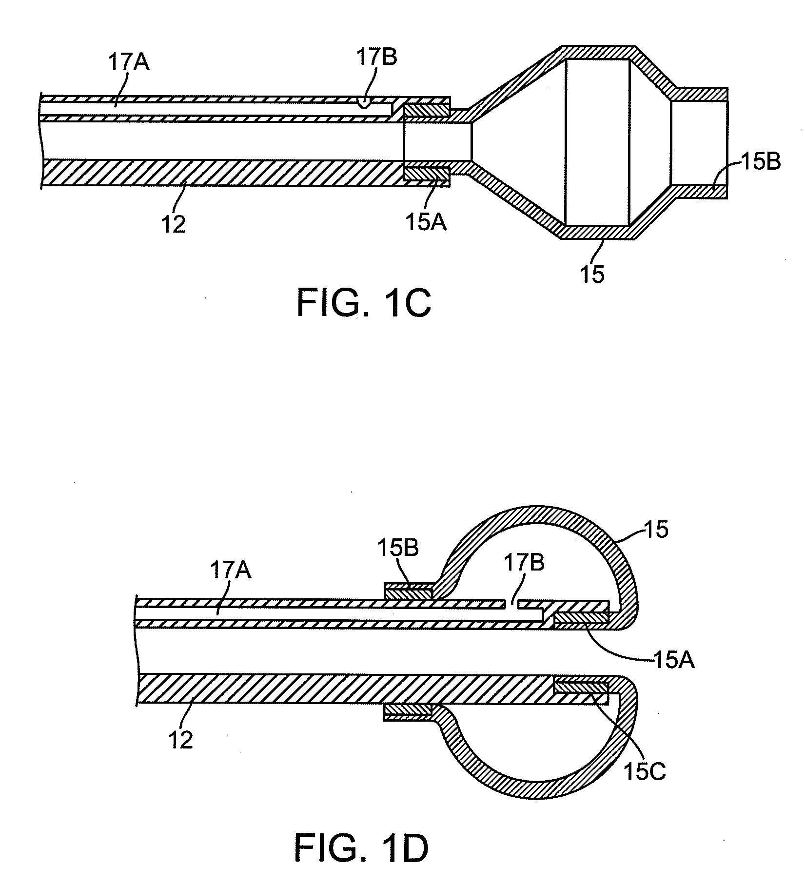
[0022]In another embodiment of the catheter, the expandable occluding element is disposed at the distal end of the catheter body, and is configured to form a cover over the rim of the lumen. This embodiment prevents or inhibits entry of mucus into the lumen, and prevents the catheter tip from contacting the airway wall. A method of manufacturing this embodiment of the catheter is also disclosed. One end of the occluding element is attached to the internal surface of the central passageway at the tip of the catheter. The occluding element is then inverted over the catheter body and a second end of the occluding element is attached to the outer surface of the catheter body. The expandable occluding element is optionally transparent to enable viewing the body passageway (for example during diagnostic or treatment procedures).
[0023]The one-way flow element is adapted to be disposed within or in-line with the lumen of the catheter body in order to allow flow in a distal-to-proximal direction so that air will be expelled from the isolated lung compartment or segment as the patient exhales. The one-way flow element, however, inhibits or prevents flow through the lumen in a proximal-to-distal direction so that air cannot enter the isolated lung compartment or segment while the patient is inhaling.

Problems solved by technology

Though these catheters utilize the balloons to view the passageways, their use for viewing pulmonary passageways is limited in several aspects.
Practically, the use of these devices in pulmonary passageways would be limited to those passageways large enough to accommodate a similarly constructed bronchoscope.
These devices are also limited by the fact that treatment is limited to the exact site of visualization, rather than at a point distal to the visualization point.
Further, the flexibility of these devices would be limited by the inherent properties of a visualization catheter.

Method used


Image

  • Methods and devices for passive residual lung volume reduction and functional lung volume expansion
  • Methods and devices for passive residual lung volume reduction and functional lung volume expansion
  • Methods and devices for passive residual lung volume reduction and functional lung volume expansion

Examples


Embodiment Construction

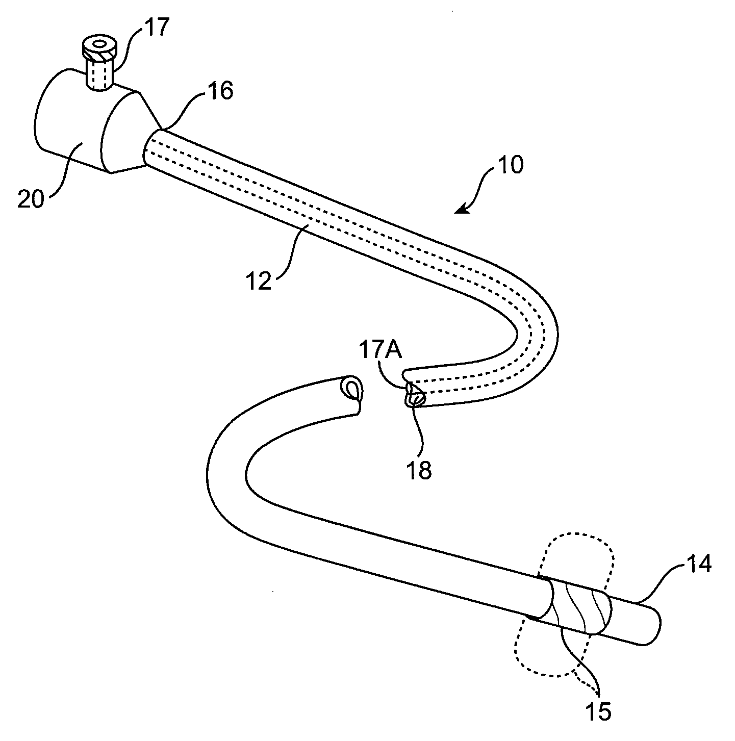
[0035]Referring to FIGS. 1a and 1b, an endobronchial lung volume reduction catheter 10 constructed in accordance with the principles of the present invention includes an elongate catheter body 12 having a distal end 14 and a proximal end 16. Catheter body 12 includes at least one lumen or central passage 18 extending generally from the distal end 14 to the proximal end 16. Lumen 18 will have a distal opening 19 at or near the distal end 14 in order to permit air or other lung gases to enter the lumen and flow in a distal-to-proximal direction out through the proximal end of the lumen. Additionally, catheter body 12 will have an expandable occluding member or element 15 at or near the distal end 14, to occlude an air passageway during treatment.

[0036]As mentioned above, in one embodiment the expandable occluding member is disposed near the distal end of the catheter body to seal the passageway, while in an alternate embodiment the expandable occluding element forms a cover of the rim...


PUM

PropertyMeasurementUnit
Luminous fluxaaaaaaaaaa
Diameteraaaaaaaaaa
Diameteraaaaaaaaaa

Abstract

The volume of a hyperinflated lung compartment is reduced by sealing a distal end of the catheter in an airway feeding the lung compartment. Air passes out of the lung compartment through a passage in the catheter while the patient exhales. A one-way flow element associated with the catheter prevents air from re-entering the lung compartment as the patient inhales. Over time, the pressure of regions surrounding the lung compartment cause it to collapse as the volume of air diminishes. Residual volume reduction effectively results in functional lung volume expansion. Optionally, the lung compartment may be sealed in order to permanently prevent air from re-entering the lung compartment. The invention further discloses a catheter with a transparent occlusion element at its tip that enables examination of the lung passageway through a viewing scope.

Description

CROSS-REFERENCES TO RELATED APPLICATIONS[0001]The present application is a continuation in part of application Serial No. PCT / US08 / 56706 (Attorney Docket No. 017534-003710PC), which claims the benefit of U.S. patent application Ser. No. 11 / 685,008 (Attorney Docket No. 017534-003700US) filed, Mar. 12, 2007, the full disclosures of which are incorporated herein by reference.BACKGROUND OF THE INVENTION[0002]1. Field of the Invention[0003]The present invention relates generally to medical methods and apparatus. More particularly, the present invention relates to methods and apparatus for endobronchial residual lung volume reduction by passive deflation of hyperinflated segments with functional lung volume expansion as a result.[0004]Chronic obstructive pulmonary disease is a significant medical problem affecting 16 million people or about 6% of the U.S. population. Specific diseases in this group include chronic bronchitis, asthmatic bronchitis, and emphysema. While a number of therapeu...

Claims


Application Information

Patent Timeline
IPC IPC(8): A61M16/00A61F2/958
CPCA61B17/12104A61B17/12136A61M16/0436A61M2025/1052A61M16/0434A61M25/10A61F2002/043Y10T29/49826A61B5/4836A61M16/0003A61B1/2676A61B5/0084A61B5/08A61M16/208A61M2016/0027A61M2016/003A61M2202/0208A61M2205/3334A61M2207/10
Inventor ALJURI, NIKOLAIPERKINS, RODNEY C.OLIVERA, RYANNGUYEN, HOANGRADHAKRISHNAN, SRIBEYHAN, NIYAZI
Owner PULMONX
Learn More