Multi-modality system for imaging in dense compressive media and method of use thereof

a dense compressive media and multi-modality technology, applied in the field of imaging in dense compressive media, can solve the problems of lack of sensitivity or resolution of other methods, clinical physical examination cannot identify the nature of lumps, and each prior art method and/or device possesses significant disadvantages, etc., to achieve superior penetration and resolution characteristics, superior penetration and detection capacity of microwave detection and imaging, and superior excitation capability of resultan

Inactive Publication Date: 2009-11-12
ULTRAWAVE LABS
View PDF8 Cites 37 Cited by
  • Summary
  • Abstract
  • Description
  • Claims
  • Application Information

AI Technical Summary

Benefits of technology

[0021]In view of the foregoing disadvantages inherent in the known devices and methods in the prior art, the present invention provides a novel multi-modality system and method for performing detection, characterization and imaging of materials and objects in dense compressive media, particularly but not exclusively in medical soft tissue applications. Specifically, the present invention involves coupling an ultrasound subsystem for exciting target tissues with a microwave subsystem for measuring the response and imaging the target tissues. The present invention combines the superior penetration and resolution characteristics of focused high-frequency ultrasound input waves, the superior excitation capability of resultant low-frequency ultrasound harmonics, and the superior penetration and detection capacity of microwave detection and imaging.

Problems solved by technology

However, each prior art method and / or device possesses significant disadvantages.
However, the clinical physical examination cannot identify the nature of the lump and lacks the sensitivity or resolution of other methods.
This results in substantial discomfort to the patient.
In each examination, the patient is exposed to destructive ionizing radiation, thus incurring a risk of realizing an induced breast tumor.
X-ray mammography is considered a health risk for women who are pregnant or breast-feeding, and it is not recommended for women under the age of fifty.
Further, X-ray mammography is a very poor method for early-stage cancer detection.
While MRI offers improved detection over X-ray mammography and eliminates the risk associated with ionizing radiation, it brings other attendant problems.
Many patients find the MRI procedure uncomfortable.
The MRI machine may induce anxiety in patients with a fear of confined spaces.
Further, the MRI machine produces loud percussive and buzzing noises which may be disconcerting to the patient.
The patient is required to lie motionless for this long period of time because excessive movement can blur MRI images and cause errors.
In addition, because the magnet is very strong, certain types of metal can cause significant errors in the images, and the strong magnetic fields created during an MRI can interfere with certain medical implants.
Patients have been harmed in MRI machines when they did not remove metal objects from their clothes or when metal objects were left in the room by others.
Finally, the high cost of procuring and operating an MRI machine, and the lack of people skilled at reading breast MRIs, suggests it will not replace X-ray mammography as a routine screening methodology anytime soon.
While this method demonstrates the usefulness of ultrasound excitation in imaging applications, the disclosed laser-based method is invasive, and more applicable to detection than diagnosis.
Acoustic methods of measurement and imaging suffer limitations in terms of noise, contrast and speckle.

Method used


Image

  • Multi-modality system for imaging in dense compressive media and method of use thereof
  • Multi-modality system for imaging in dense compressive media and method of use thereof
  • Multi-modality system for imaging in dense compressive media and method of use thereof

Examples


Embodiment Construction

[0043]The following description is provided to enable any person skilled in the art to make and use the invention and sets forth the best modes contemplated by the inventor of carrying out his invention.

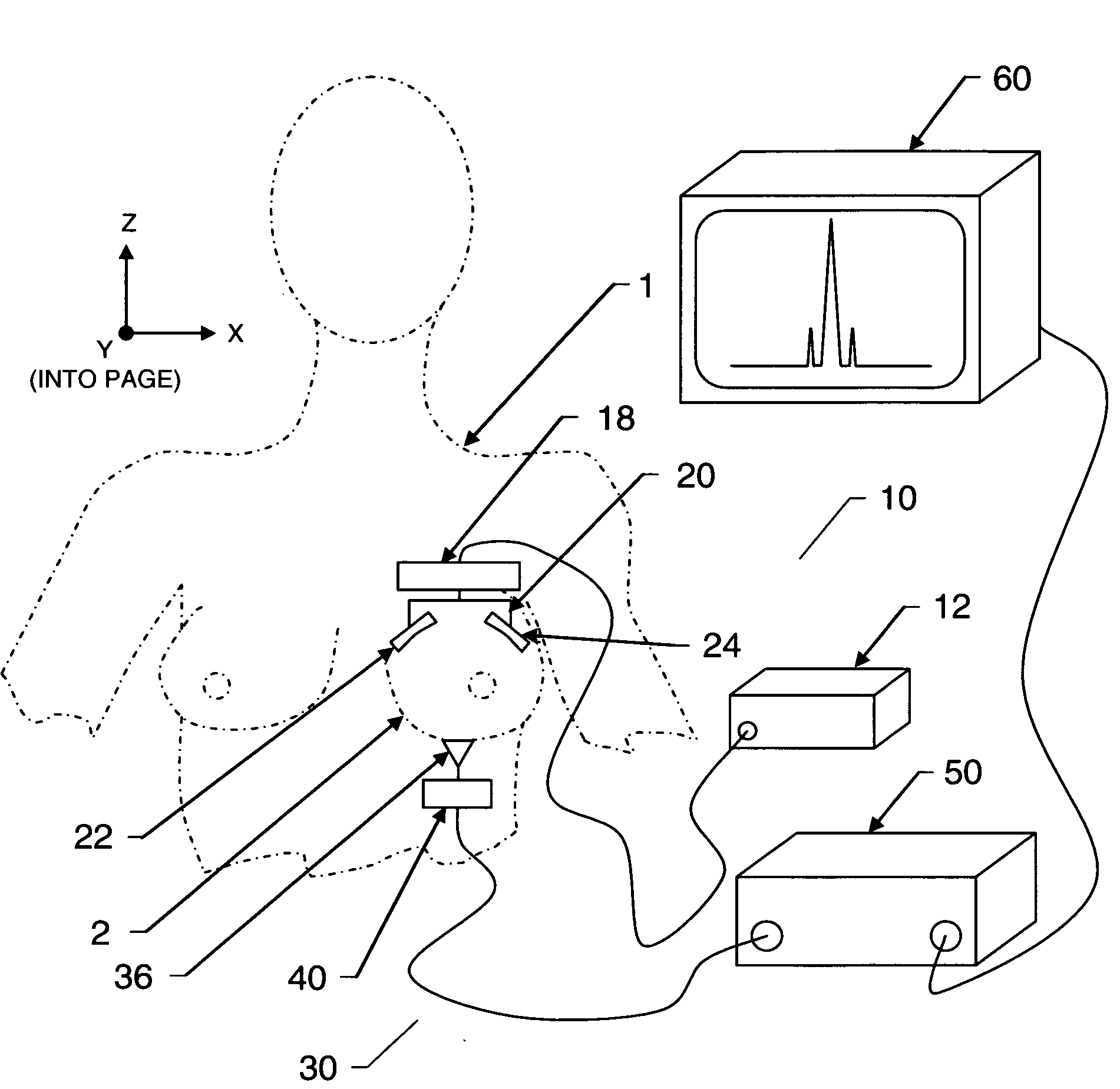
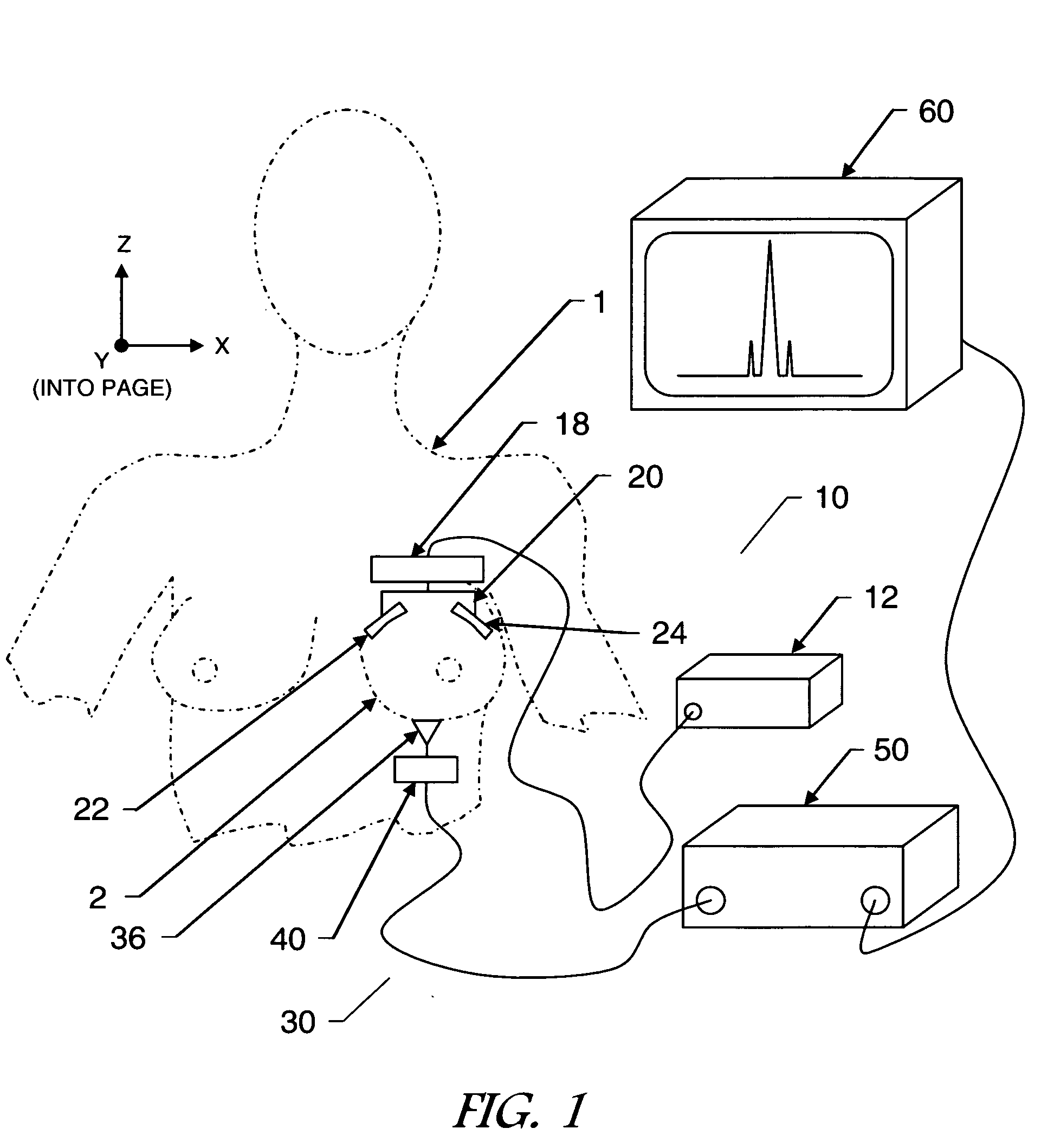
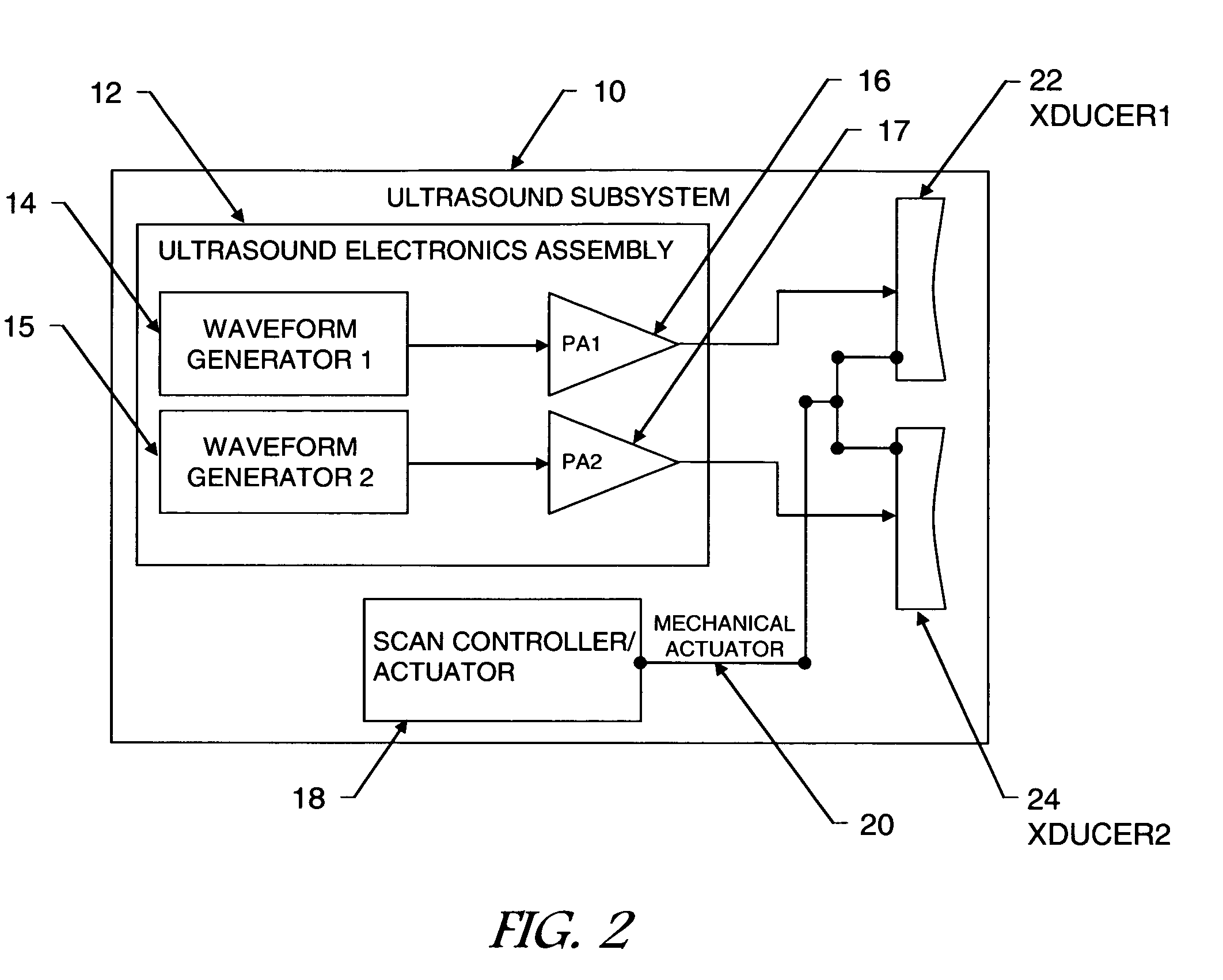
[0044]Referring now to the drawings, FIG. 1 shows the orientation of the system with respect to the patient 1 and the imaging target breast 2 in one preferred embodiment of the present invention. An ultrasound subsystem 10 and a microwave imaging subsystem 30 are employed in combination to detect and diagnose tumors in the breast 2. A first ultrasound transducer 22, a second ultrasound transducer 24 and a microwave antenna 36 are oriented with respect to the target breast 2 of the patient 1. A radio frequency transceiver 40 generates and transmits microwave signals to the microwave antenna 36. The microwave antenna 36 transmits microwaves into the target breast 2. Reflected microwaves are collected by the microwave antenna 36 and received by the radio frequency transceiver 40. A comp...


PUM


Abstract

A multi-modality system and method for performing detection, characterization and imaging of materials and objects in dense compressive media, such as in medical soft tissue applications, is disclosed. Medical tissue applications include but are not limited to the detection and diagnosis of breast tumors. Generally, an ultrasound subsystem is employed to excite a region in the dense compressive media and a microwave subsystem is employed to collect detection, characterization and imaging information from the excited region. In one preferred embodiment, multiple focused oscillating high-frequency ultrasound wave beams are transmitted into the media. The resultant low beat-frequency wave creates a force inducing motion in the materials and objects in the media. A radio-frequency microwave subsystem detects that motion and produces images based upon the Doppler effects of the excited materials and objects.

Description

CROSS REFERENCE TO RELATED APPLICATIONS[0001]Not applicableSTATEMENT REGARDING FEDERALLY SPONSORED RESEARCH OR DEVELOPMENT[0002]Not applicableINCORPORATION-BY-REFERENCE OF MATERIAL SUBMITTED ON A COMPACT DISC[0003]Not applicableBACKGROUND OF THE INVENTION[0004]1. Field of Invention[0005]The present invention relates generally to the field of imaging in dense compressive media, and more particularly to a novel apparatus and method of use thereof for imaging in medical soft tissue applications such as orthopedics, dermatology, breast tumor scanning / detection and diagnosis / characterization. For simplicity of discussion, while applications are to be found in a wide range of medical and non-medical applications, this specification primarily addresses the exemplary application of breast tumor detection and diagnosis.[0006]According to the U.S. National Library of Medicine and the National Institutes of Health, one in eight women will be diagnosed with breast cancer. One in sixteen women w...

Claims


Application Information

Patent Timeline
Patent Type & Authority Applications(United States)
IPC IPC(8): A61B6/00A61B5/00
CPCA61B5/0048A61B5/0097A61B5/4312A61B5/0507A61B5/05A61B5/0051
Inventor SALAMA, KHALED N.ISMAIL, ALY M.
Owner ULTRAWAVE LABS
Why Eureka
Try Eureka