Diagnostic Kits and Methods for Oesophageal Abnormalities

a technology for oesophageal abnormalities and diagnostic kits, which is applied in the field of methods for detecting oesophageal abnormalities, can solve problems such as thought unworkable, and achieve the effect of increasing the value of information obtained and robust combined diagnostic outpu

Inactive Publication Date: 2009-11-19
MEDICAL RESEARCH COUNCIL +1
View PDF1 Cites 73 Cited by
  • Summary
  • Abstract
  • Description
  • Claims
  • Application Information

AI Technical Summary

Benefits of technology

[0062]It is a feature of the invention that the sampling is not directed eg. visually directed to any particular part of the oesophagus. It is a further advantage of the invention that a greater proportion of the surface of the oesophagus is sampled than is achieved by prior art techniques such as endoscopic biopsy (which samples approximately 1% of the surface) or endoscopic brushing. Preferably at least 10% of the oesophageal surface is sampled, preferably at least 20%, preferably at least 30%, preferably at least 40%, preferably at least 50%, preferably at least 60%, preferably at least 70%, preferably at least 80%, preferably at least 90%. In a most preferred embodiment, preferably substantially the entire oesophagus is sampled, preferably the whole inner lumen of the oesophagus is sampled. This applies equally to the in vitro sample even when the method of the invention does not include collection of the sample.
[0063]Screening aspects of the invention relate to the detection and / or diagnosis of Barrett's oesophagus. Typically in screening embodiments of the invention, the subjects being examined, or from which the sample(s) are (or were) obtained, are of unknown status for Barrett's.
[0064]Surveillance aspects of the invention relate to the detection and / or diagnosis of dysplasia, including adenocarcinoma Although clearly dysplasia and adenocarcinoma are pathologically different conditions, adenocarcinoma can be regarded as one extreme form of dysplasia. As is discussed below, the invention may be advantageously applied to distinguish adenocarcinoma from dysplasia, depending upon the molecular markers used. However, in general the discussion of surveillance aspects of the invention relates to the detection of dysplasia, including adenocarcinoma. Typically in surveillance embodiments of the invention, the subjects being examined, or from which the sample(s) are obtained, are of unknown status for dysplasia but will typically be known to have Barrett's.
[0065]In principle the difference between screening and surveillance aspects is of little practical consequence to the working of the invention. The difference relates only to the markers chosen. The sampling and combination aspects remain the same between screening and surveillance. Indeed, it may be advantageous to combine screening and surveillance ie. to examine cell samples for markers of Barrett's as well as dysplasia including adenocarcinoma at the same time, thereby increasing the value of the information obtained and achieving a more robust combined diagnostic output.Markers
[0066]Markers that can be applied for Barrett's screening and surveillance are any markers which are not expressed in normal oesophageal tissue, preferably any markers which are not expressed in normal oesophagal surface cells. Preferably markers are markers of non-squamous cells. Preferably markers are markers of cellular proliferation.
[0067]For screening aspects (ie. for detection of Barrett's oesophagus), preferably markers that distinguish between intestinal metaplasia (Barrett's) and squamous oesophageal cells or gastric cardia are used. These markers include markers of epithelial differentiation.Screening—Columnar Markers

Problems solved by technology

From an understanding of the prior art, this would have been thought unworkable for several reasons.
Firstly, a small Barrett's oesophagus lesion occupies only about 1-2 cm of a 40 cm oesophagus.
Therefore, it is not expected that a mere sampling of the surface cells of the oesophagus can provide a diagnostically useful cell sample.
Thirdly, prior art techniques involve morphological analysis of biopsies or collected cell material.
Many attempts have been made in the art to base a diagnostic method for Barrett's abnormalities on cytology, but they have failed.

Method used


Image

  • Diagnostic Kits and Methods for Oesophageal Abnormalities
  • Diagnostic Kits and Methods for Oesophageal Abnormalities

Examples


example 1

Construction of a Sampling Device

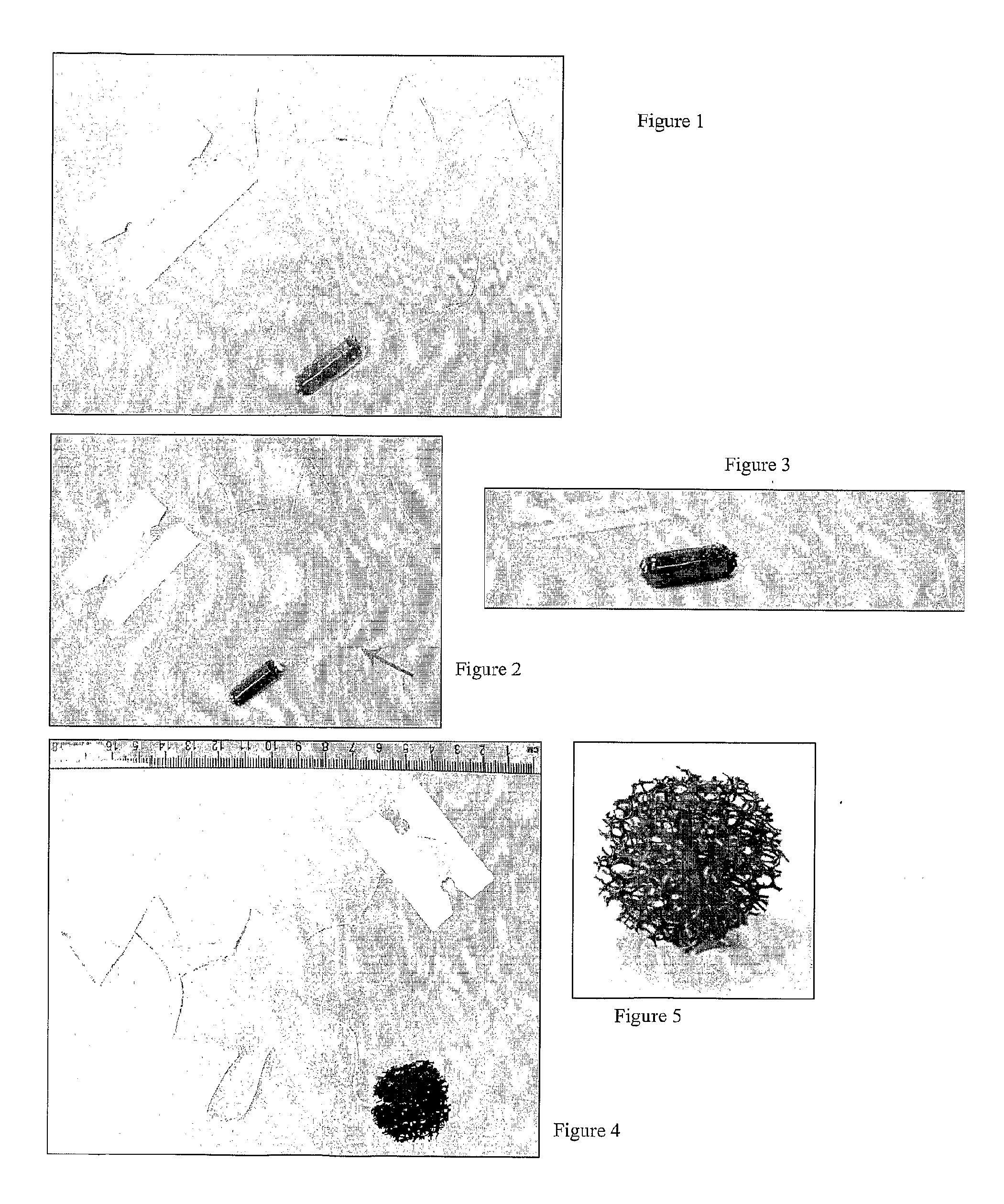
[0148]Abrasive material is cut to the appropriate size. In this example, the material is approximately the size of the internal diameter of a human oesophagus, ie. approximately 3 cm in diameter.

[0149]In this example the material is a polyurethane mesh or cloth.

[0150]A cord is stitched into the material so that it can be retrieved after swallowing. (FIG. 3 shows the device with cord attached).

[0151]The cord is sufficiently long that part of it will comfortably remain outside the buccal cavity even after the device has been swallowed and resides in the stomach. The cord and stitching is sufficiently strong and resistant to digestion so that it can be used to retrieve the device after expansion.

[0152]The material is then compressed and inserted into a gelatine capsule (FIG. 1). The cord exits the capsule (FIG. 2 shows partially disassembled capsule with cord exiting). The device is then ready for use.

example 2

Sampling of Cells from the Surface of the Oesophagus

[0153]A device according to example 1 is provided. The subject may tale a local anaesthetic in the form of a lozenge or spray by way of preparation.

[0154]The device is introduced into the subject's buccal cavity with the distal end of the cord retained outside the buccal cavity.

[0155]The device is then swallowed. A drink of warm water aids this process and wets the cord, facilitating its passage down the oesophagus.

[0156]After approximately 10-20 seconds the device arrives in the subject's stomach, the cord exiting the stomach and lying in the oesophagus and the buccal cavity and outside to the point of retention.

[0157]After 5 minutes the capsule coat has dissolved and the abrasive polyurethane material has expanded back to its uncompressed size.

[0158]The device is then withdrawn by gentle tension on the distal end of the cord, pulling the device along the oesophagus and out of the buccal cavity, collecting oesophageal cells en rou...

example 3

Assaying for Cellular Markers

[0159]The withdrawn device of example 2 is washed to collect the oesophageal cells. These are then applied to slides and fixed for visualisation.

[0160]Mcm2 is the marker in this example.

[0161]The numbers analysed for this part of the study are 18 BE patients and 22 healthy controls). The age of the BE patients was 64.5±2.1 years compared to 31.2±1.6 for the healthy volunteers and the male:female ratio was biased towards a male population in both groups (1:5 and 1:1.7 respectively).

[0162]The PAP slides were used to assess the cellularity of the samples. An expert cytopathologist assessed the cellularity of the samples and 88% of the samples had a good to very good cellularity and 22% had an average cellularity.

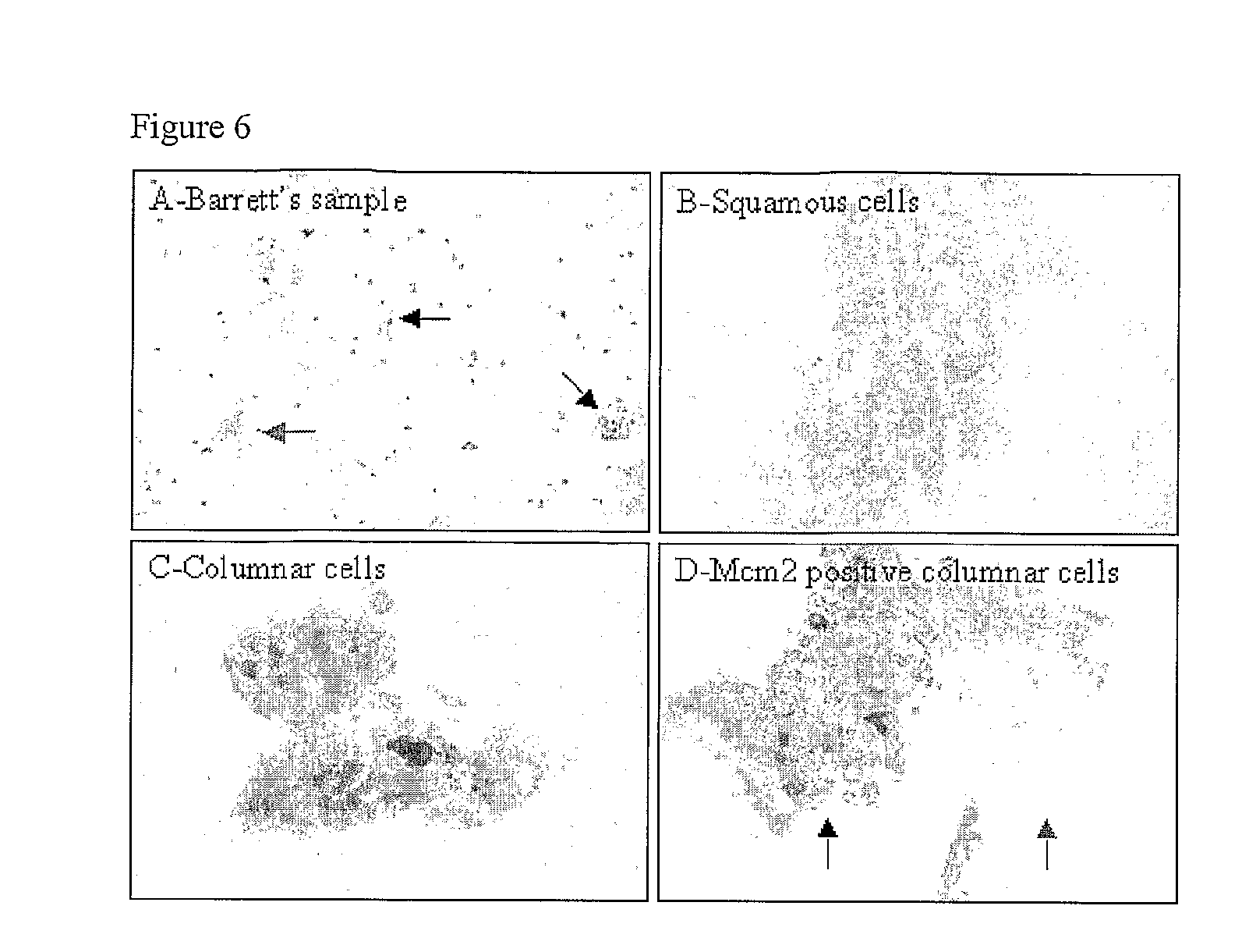
[0163]FIG. 6 shows Representative pictures of monolayers from capsule sponge samples. Pap stained samples (A-C) and Mcm2 stained sample (C). The black arrows indicate the position of columnar cells and the red arrows the position of squamous cells.

[...


PUM

PropertyMeasurementUnit
diameteraaaaaaaaaa
chromosomal compositionaaaaaaaaaa
timeaaaaaaaaaa

Abstract

The invention relates to kits and methods for aiding the diagnosis of Barrett's oesophagus or Barrett's associated dysplasia. Preferred is a method comprising assaying cells from the surface of a subject's oesophagus for a non-squamous cellular marker, wherein detection of such a marker indicates increased likelihood of the presence of Barrett's or Barrett's associated dysplasia, preferably wherein said sample of cells is not directed to a particular site within the oesophagus. The invention also encompasses a method comprising sampling the cellular surface of the oesophagus of said subject. The invention also relates to a kit comprising a swallowable device comprising abrasive material capable of collecting cells from the surface of the oesophagus, together with printed instructions for its use in detection of Barrett's oesophagus or Barrett's associated dysplasia. Preferably said device comprises a capsule sponge.

Description

FIELD OF THE INVENTION[0001]The invention relates to methods for detection of oesophageal abnormalities such as Barrett's oesophagus and Barrett's associated dysplasia including adenocarcinoma. Furthermore, the invention relates to kits for sampling oesophageal cells and detecting cellular markers associated with the above conditions.BACKGROUND TO THE INVENTION[0002]Oesophageal adenocarcinoma is rapidly increasing and is preceded by a condition called Barrett's oesophagus. Early diagnosis is crucial to improving the appalling outcome (>80% mortality at 5 years) from oesophageal adenocarcinoma. Currently the majority of patients with Barrett's oesophagus remain undiagnosed in the population. Furthermore, of those that are diagnosed it is not currently possible to accurately predict the small proportion (1% per year) that will progress to cancer. It has been suggested that endoscopic screening should be performed to detect those at risk for Barrett's oesophagus—for example endoscop...

Claims


Application Information

Patent Timeline
Patent Type & Authority Applications(United States)
IPC IPC(8): C12Q1/68C12Q1/02A61B10/02G01N33/574
CPCA61B10/02A61B2010/0216G01N33/57492G01N33/57407A61B2017/00898
Inventor FITZGERALD, REBECCA C.HARDWICK, RICHARD
Owner MEDICAL RESEARCH COUNCIL
Why Eureka
Try Eureka