Methods for treating cancer by inhibiting wnt signaling
- Summary
- Abstract
- Description
- Claims
- Application Information
AI Technical Summary
Benefits of technology
Problems solved by technology
Method used
Image
Examples
example 1
Anti-Wnt Antibody Specifically Induces Apoptosis in a Number of Different Human Cancer Cells
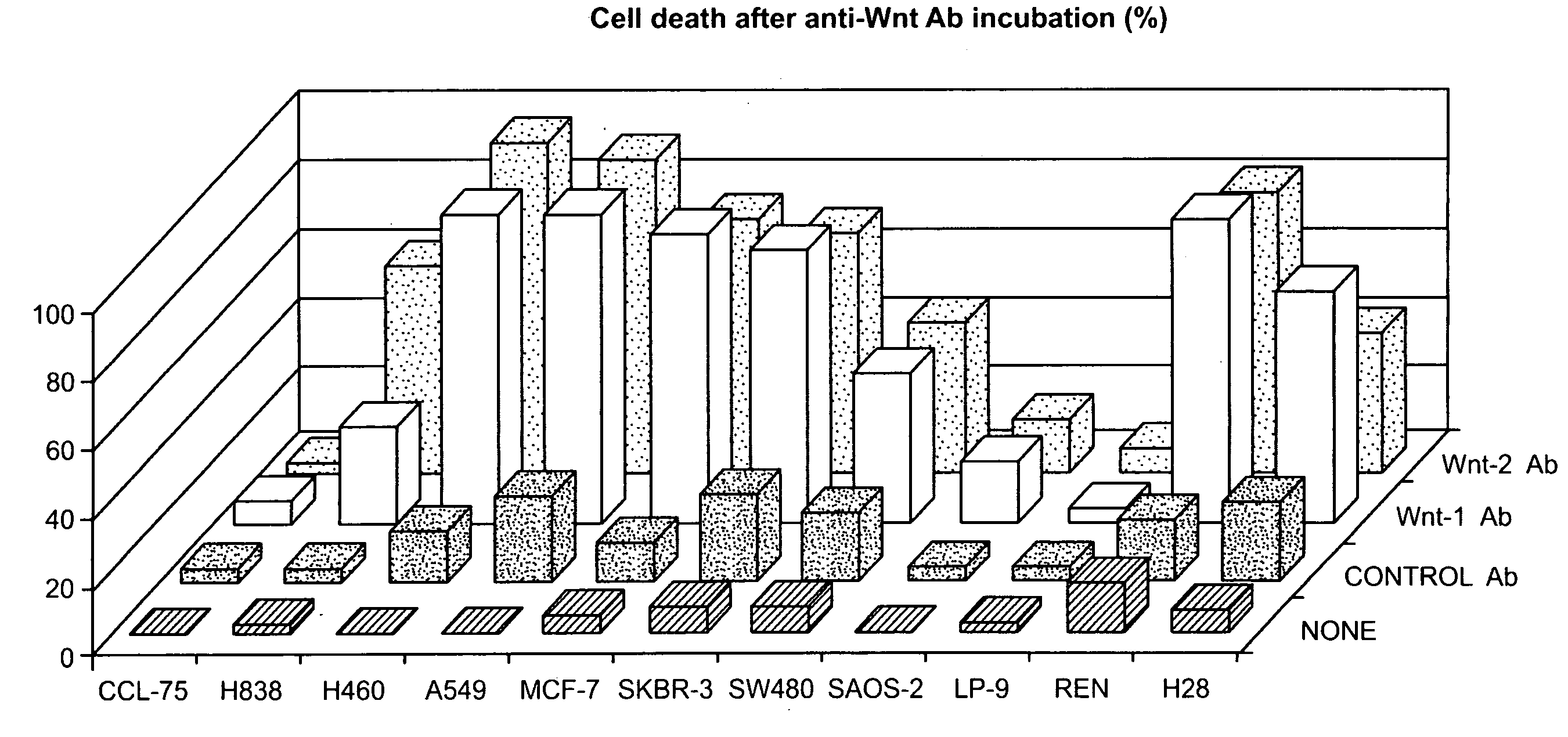
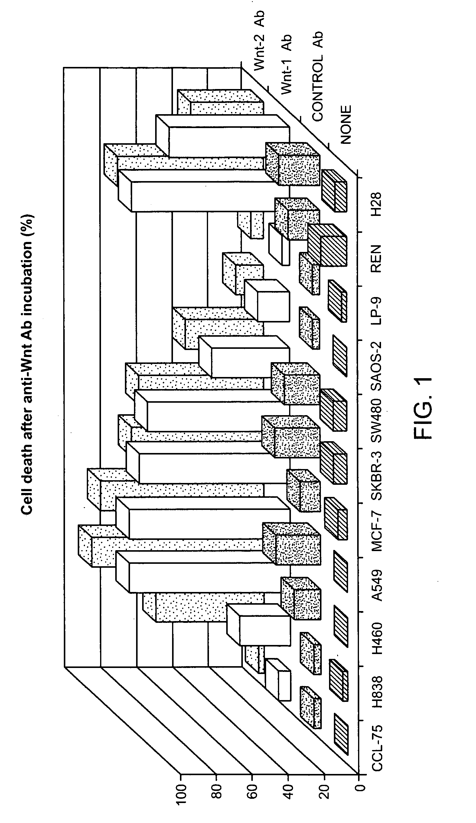
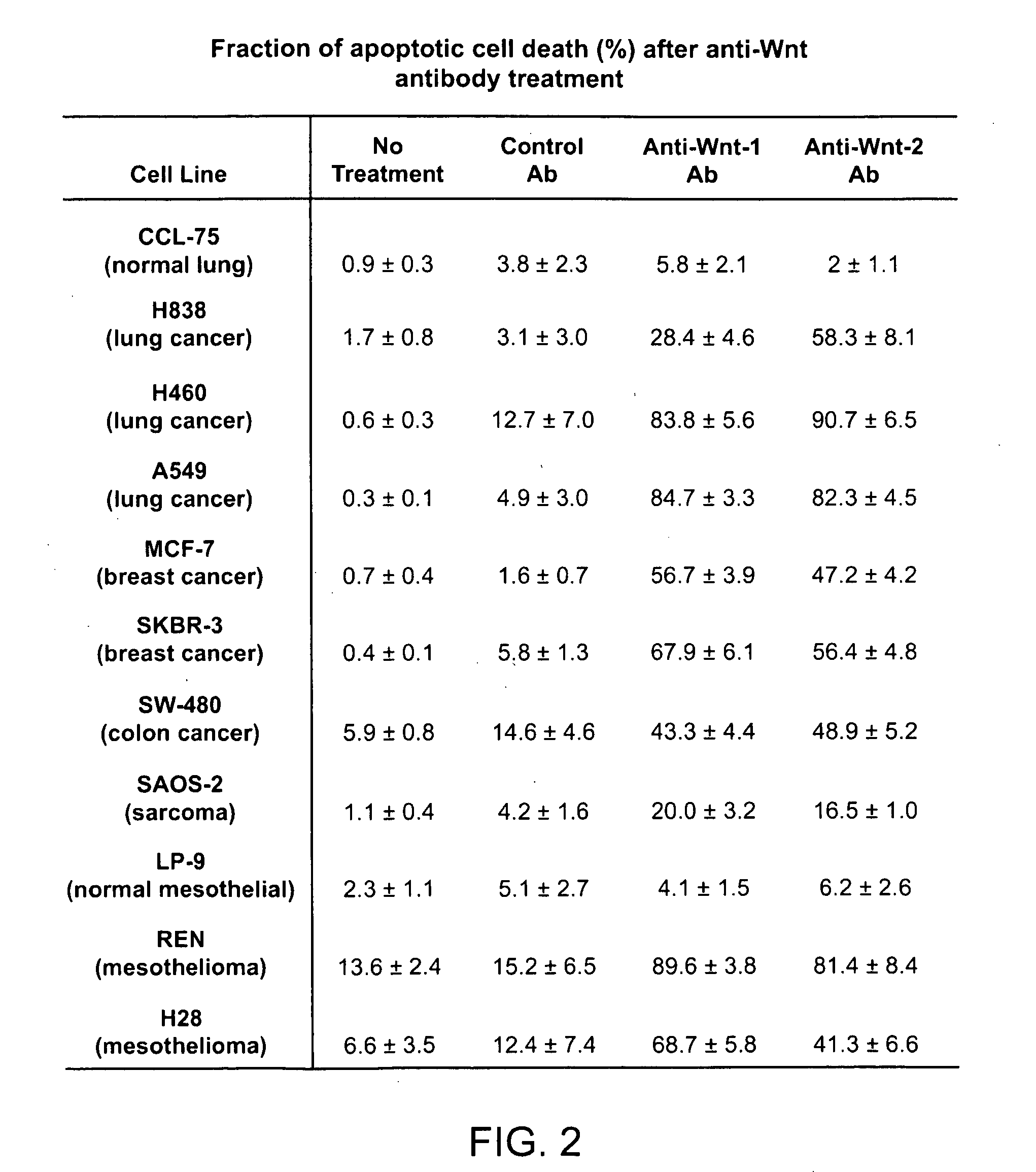
[0132]We examined whether neutralizing Wnt signaling by using anti-Wnt antibodies could inhibit cell survival in these cancers. When we incubated a number of cancer cell lines with either anti-Wnt-1 or Wnt-2 antibody (at 10 μg / ml) for about 32 hrs (we examined three non-small-cell lung cancer (NSCLC), two breast cancer, two colorectal cancer, one sarcoma, and two mesothelioma cell lines), we found that both antibodies could cause significant cell death (from 30% to 97%), except for one colorectal cancer cell line SW480 (only 4-8%)(FIG. 1). In contrast, an antibody against a cytoplasmic protein (SOCS3) (at 10 μg / ml) did not show dramatic cytotoxicity in most of those cell lines (from 4% to 45%)(FIG. 1). Interestingly, none of the antibodies had dramatic effect on the two normal cell lines that we examined (one was normal lung fibroblast (CCL-75) and the other was normal mesothelial cell line (...
example 2
Anti-Wnt Antibody-Induced Apoptosis is Correlated with the Wnt Expression
[0134]To investigate whether anti-Wnt antibody-induced apoptotic effect was associated with status of the Wnt proteins, we examined e Wnt expression in the cell lines we tested. As shown in FIG. 3A, we found that Wnt-1 had high-level expression in the cancer cell lines that were sensitive to anti-Wnt-1 antibody treatments. However, in the normal lung cell line CCL-75 that was not sensitive to the antibody treatment (see FIG. 1) only minimal Wnt-1 and expression was detected. No Wnt-1 expression was detected in two primary normal lung cells (small airway epithelial cells (SAEC) and bronchial epithelial cells (BEC)) (FIG. 3) and in normal mesothelial cell line (LP-9) (data not shown). Similar observations were made regarding Wnt-2 expression.
[0135]As a control, we examined apoptosis induction of co-incubation of anti-Wnt antibody and blocking peptide for anti-Wnt antibody in an NSCLC cell line. After about 24 hr ...
example 3
Anti-Wnt-1 Antibody-Induced Apoptosis is a Fast Process and Dose Dependent
[0136]We performed dosage and time course experiments on two NSCLC cell lines: H838 and A549 (FIG. 4A and FIG. 4B). Flow cytometry analysis after about 32 hr incubation of anti-Wnt antibody showed that 1 μg / ml antibody could induce apoptosis. A concentration of 20 μg / ml of either antibody caused dramatic apoptotic cell death. Anti-Wnt-1 antibody (at concentration of 8 μg / ml) induced apoptosis could be detected as early as after 6 hr incubation and after 50 hr incubation almost all cells were found undergoing apoptosis or necrosis. In contrast, control anti-SOCS3 antibody did not have effect on those cancer cells in the parallel experiments. Anti-Wnt-1 antibody incubation with normal lung cell line (CCL-75) was also insensitive to either time or dosage.
PUM
| Property | Measurement | Unit |
|---|---|---|
| Therapeutic | aaaaa | aaaaa |
Abstract
Description
Claims
Application Information
 - R&D
- Intellectual Property
- Life Sciences
- Materials
- Tech Scout
- Unparalleled Data Quality
- Higher Quality Content
- 60% Fewer Hallucinations
Browse by: Latest US Patents, China's latest patents, Technical Efficacy Thesaurus, Application Domain, Technology Topic, Popular Technical Reports.
© 2025 PatSnap. All rights reserved.Legal|Privacy policy|Modern Slavery Act Transparency Statement|Sitemap|About US| Contact US: help@patsnap.com


