

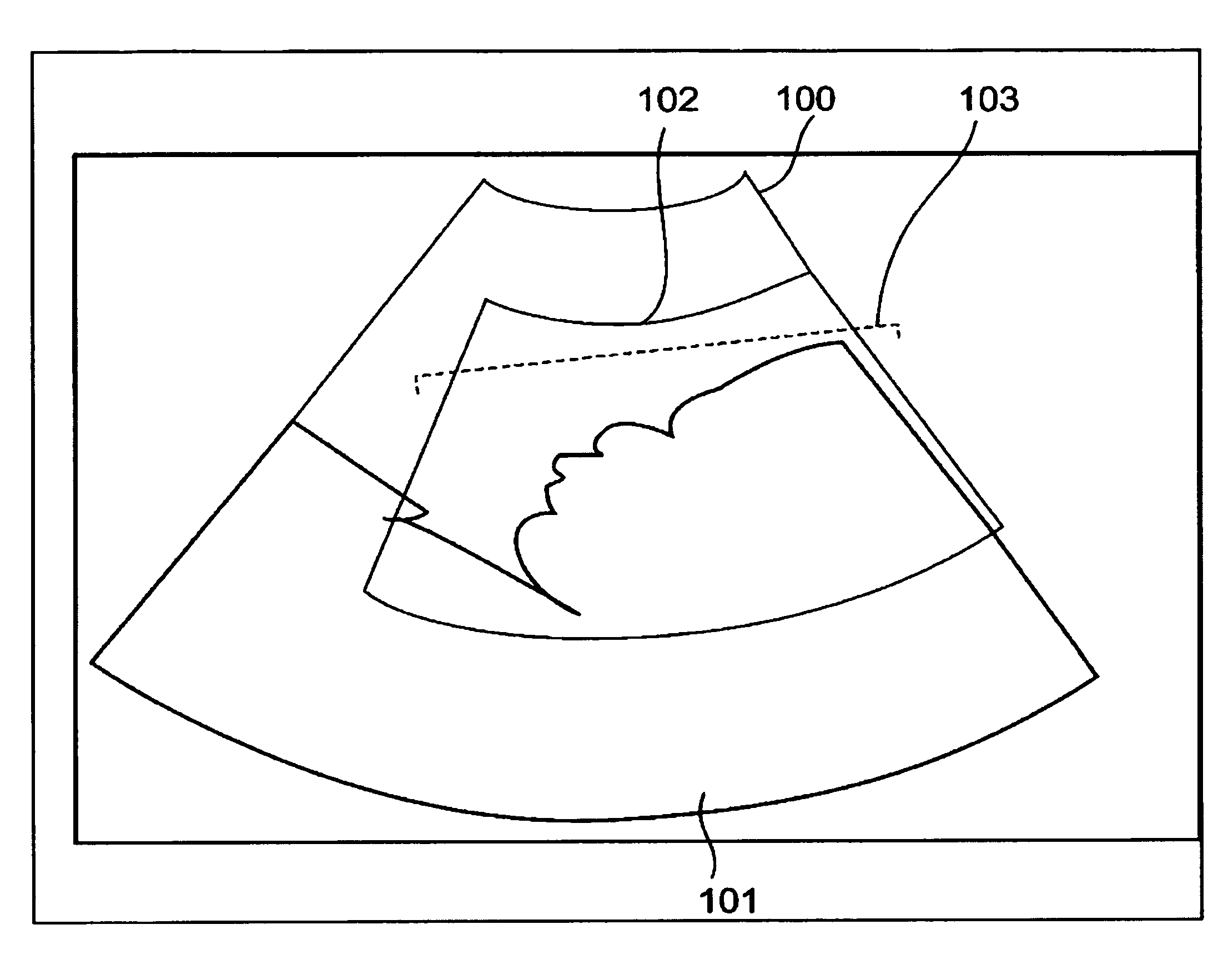
Ultrasound imaging apparatus and method for acquiring ultrasound image
a technology of ultrasound and imaging apparatus, applied in the field of ultrasound imaging apparatus, can solve the problems of difficult to appropriately display a three-dimensional image of the fetus, difficult to observe a three-dimensional image included in the region of interest, etc., and achieve the effect of removing unnecessary images, and reducing the number of unnecessary images
- Summary
- Abstract
- Description
- Claims
- Application Information
AI Technical Summary
Benefits of technology
Problems solved by technology
Method used
Image

Examples
first embodiment
(Configuration)
[0020]The configuration of an ultrasound imaging apparatus according to a first embodiment of the present invention will be described with reference to FIG. 3. FIG. 3 is a block diagram showing an ultrasound imaging apparatus according to the first embodiment of the present invention.
[0021]An ultrasound imaging apparatus 1 according to the first embodiment includes an ultrasound probe 2, a transceiver 3, a signal processor 4, a DSC 5, a first image memory 6, an image processor 7, a second image memory 8, a display controller 9, a display 10, an operation part 11, and a marker generator 12.
[0022]The ultrasound probe 2 is a two-dimensional array probe in which a plurality of ultrasound transducers are two-dimensionally arranged. The ultrasound probe 2 scans a three-dimensional range with ultrasound waves.
[0023]Moreover, the ultrasound probe 2 may be a one-dimensional array probe, which includes a plurality of ultrasound transducers aligned in a predetermined direction (...
second embodiment
[0070]Next, an ultrasound imaging apparatus according to a second embodiment of the present invention will be described with reference to FIGS. 6A through 6C. FIGS. 6A through 6C are schematic views for describing a process of obtaining a new three-dimensional scan range in the ultrasound imaging apparatus according to the second embodiment of the present invention.
[0071]Similarly to the ultrasound imaging apparatus 1 according to the first embodiment described above, the ultrasound imaging apparatus according to the second embodiment includes the ultrasound probe 2, the transceiver 3, the signal processor 4, the DSC 5, the first image memory 6, the image processor 7, the second image memory 8, the display controller 9, the display 10, the operation part 11, and the marker generator 12. The second embodiment is featured by the content of the processing by the marker generator 12.
[0072]Upon reception of an instruction to change the position, size and rotation angle of the second mark...
third embodiment
[0107]Next, the configuration of an ultrasound imaging apparatus according to a third embodiment of the present invention will be described with reference to FIGS. 9A through 9E. FIGS. 9A through 9E are schematic views for describing a process of obtaining a new region of interest (ROI) in the ultrasound imaging apparatus according to the third embodiment of the present invention.
[0108]Similarly to the ultrasound imaging apparatus according to the first embodiment described above, the ultrasound imaging apparatus according to the third embodiment includes the ultrasound probe 2, the transceiver 3, the signal processor 4, the DSC 5, the first image memory 6, the image processor 7, the second image memory 8, the display controller 9, the display 10, the operation part 11 and the marker generator 12. The third embodiment is featured by the content of processing by the marker generator 12.
[0109]In a case that the position and size of a second marker for designating a range to generate u...
PUM
 Abstract
Description
Claims
Application Information