
Multi-modality system for imaging in dense compressive media and method of use thereof

a multi-modality, dense compressive technology, applied in mammography, medical science, diagnostics, etc., can solve the problems of lack of sensitivity or resolution of other methods, clinical physical examination cannot identify the nature of lumps, and each related art method and/or device possesses significant disadvantages, etc., to achieve superior resolution and focus characteristics, superior penetration, and high diagnostic contrast capabilities

Inactive Publication Date: 2010-02-11
ULTRAWAVE LABS
View PDF8 Cites 16 Cited by
  • Summary
  • Abstract
  • Description
  • Claims
  • Application Information

AI Technical Summary

Benefits of technology

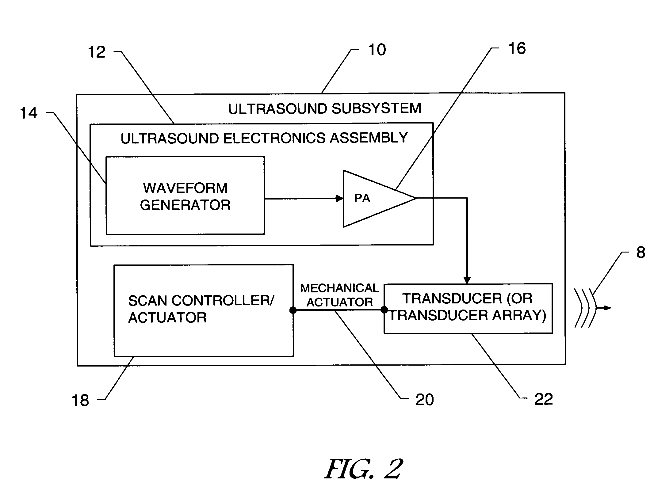
[0020]In view of the foregoing disadvantages inherent in the known devices and methods in the related art, the present invention provides a novel multi-modality system and method for performing screening / detection, imaging and diagnosis / characterization of materials and objects in dense compressive media, particularly but not exclusively in medical soft tissue applications. Specifically, the present invention involves coupling an ultrasound subsystem for stimulating target tissues with a microwave subsystem for measuring the response of the stimulated target tissues. The present invention involves a true hybrid integration of the ultrasound and microwave modalities, taking advantage of the best attributes of each subsystem modality. The superior resolution and focus characteristics of high-frequency ultrasound input waves are employed to excite Doppler displacements of materials in the target breast. At the same time, the superior penetration and high diagnostic contrast capabilities of the microwave modality are employed to perform the diagnosis an imaging function. The present invention enhances early detection and diagnosis capability without the disadvantages of the related art systems and methods.
[0022]In one embodiment of the present invention, the complexity, cost and time associated with mechanical scanning is avoided by employing an ultrasound transducer array in place of scanning ultrasound transducers.
[0024]The present invention enables three-dimensional detection and diagnosis imaging. In one alternative embodiment of the present invention, ultrasound and microwave subsystem combinations are implemented in multiple axes. These multi-axis subsystems cooperate to provide superior three-dimensional imaging capability. In yet another embodiment, phased array operation of the ultrasound subsystem allows mapping of two-dimensional planes of varying depths within the target breast. These two-dimensional maps may be integrated to create three-dimensional images.
[0026]The present invention eliminates health risks associated with related art systems and methods. The present invention eliminates the risk of short-term or long-term deleterious affects associated with ionizing radiation exposure in X-ray mammography, and the risks associated with exposure to powerful magnetic fields in MRI.

Problems solved by technology

However, each related art method and / or device possesses significant disadvantages.
However, the clinical physical examination cannot identify the nature of the lump and lacks the sensitivity or resolution of other methods.
This results in substantial discomfort to the patient.
In each examination, the patient is exposed to destructive ionizing radiation, thus incurring a risk of realizing an induced breast tumor.
X-ray mammography is considered a health risk for women who are pregnant or breast-feeding, and it is not recommended for women under the age of fifty.
Further, X-ray mammography is a poor method for early-stage cancer detection.
While MRI offers improved screening accuracy over X-ray mammography and eliminates the risk associated with ionizing radiation, it also has significant disadvantages.
Many patients find the MRI procedure uncomfortable.
The MRI machine may induce anxiety in patients with a fear of confined spaces.
Further, the MRI machine produces loud percussive and buzzing noises which may be disconcerting to the patient.
The patient is required to lie motionless for this long period of time because excessive movement can blur MRI images and cause errors.
In addition, because the magnet is very strong, certain types of metal can cause significant errors in the images, and the strong magnetic fields created during an MRI can interfere with certain medical implants.
Finally, the high cost of procuring and operating an MRI machine, and the lack of technicians skilled in reading breast MRIs present additional disadvantages to its use.
Methods that rely upon acoustic measurement alone are disadvantaged by noise, contrast and speckle limitations, and by the necessity to trade off low-frequency penetration against high-frequency resolution.
However, imaging methods that rely on microwave alone are disadvantaged by the necessity to trade off low-frequency penetration against high-frequency resolution.
Because of the limitations associated with each individual screening, imaging and diagnosis method, research is considering combining multiple imaging modalities.
This simple integration of two modalities does not take advantage of the physical interaction of the ultrasound and microwave modalities.
Therefore, this combined system possesses the disadvantages of each subsystem.
It is difficult to concurrently achieve high penetration, high resolution, fast scanning and high contrast using either subsystem alone.

Method used


Image

  • Multi-modality system for imaging in dense compressive media and method of use thereof
  • Multi-modality system for imaging in dense compressive media and method of use thereof
  • Multi-modality system for imaging in dense compressive media and method of use thereof

Examples


Embodiment Construction

[0039]The following description is provided to enable any person skilled in the art to make and use the invention and sets forth the best modes contemplated by the inventor of carrying out his invention. Further, while a breast is used in the description of these embodiments, it is to be noted that any turbid medium may be processed with this invention. Thus the present invention shall not be limited to the examples disclosed. The scope of the invention shall be as broad as the claims will allow.

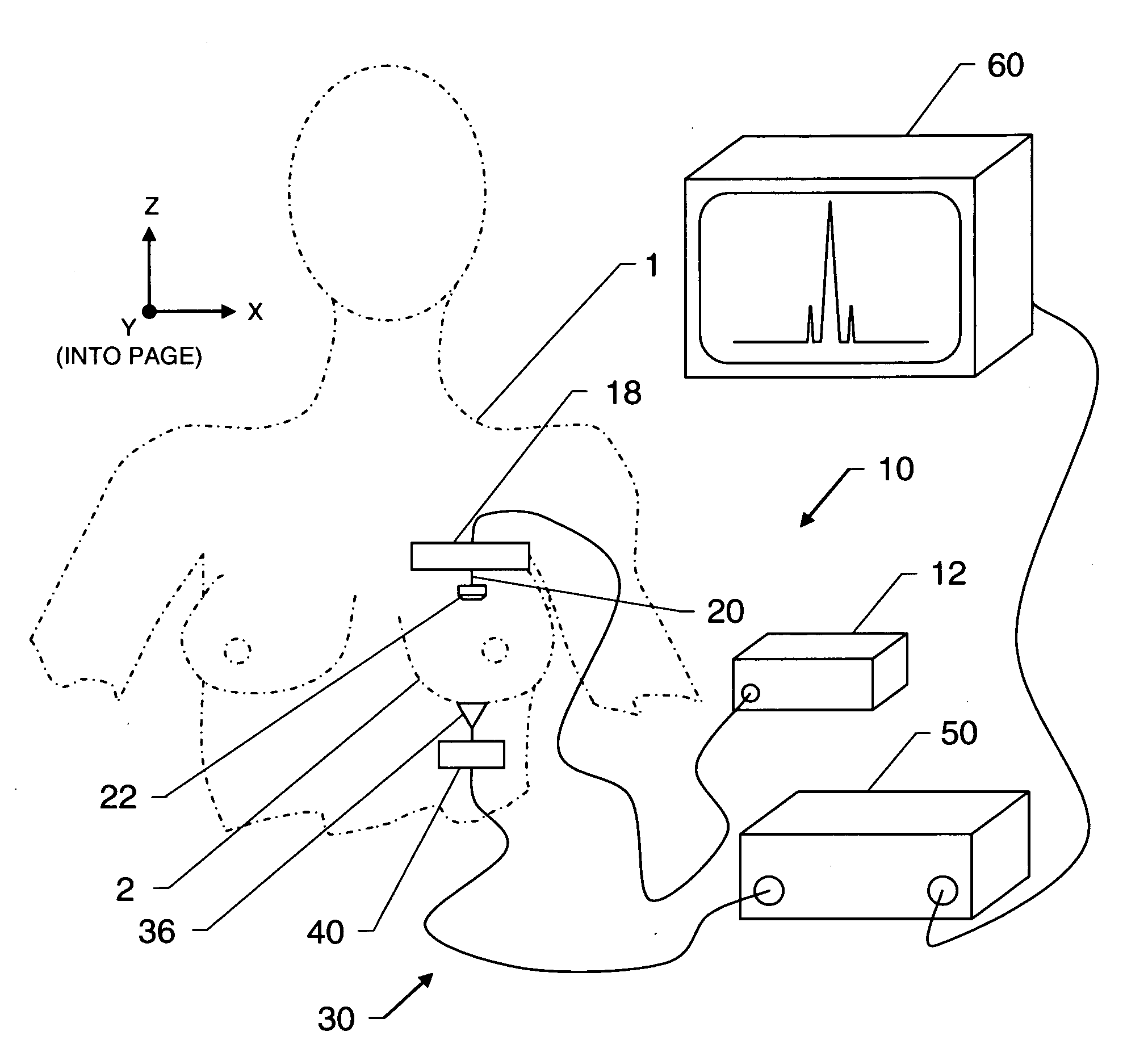
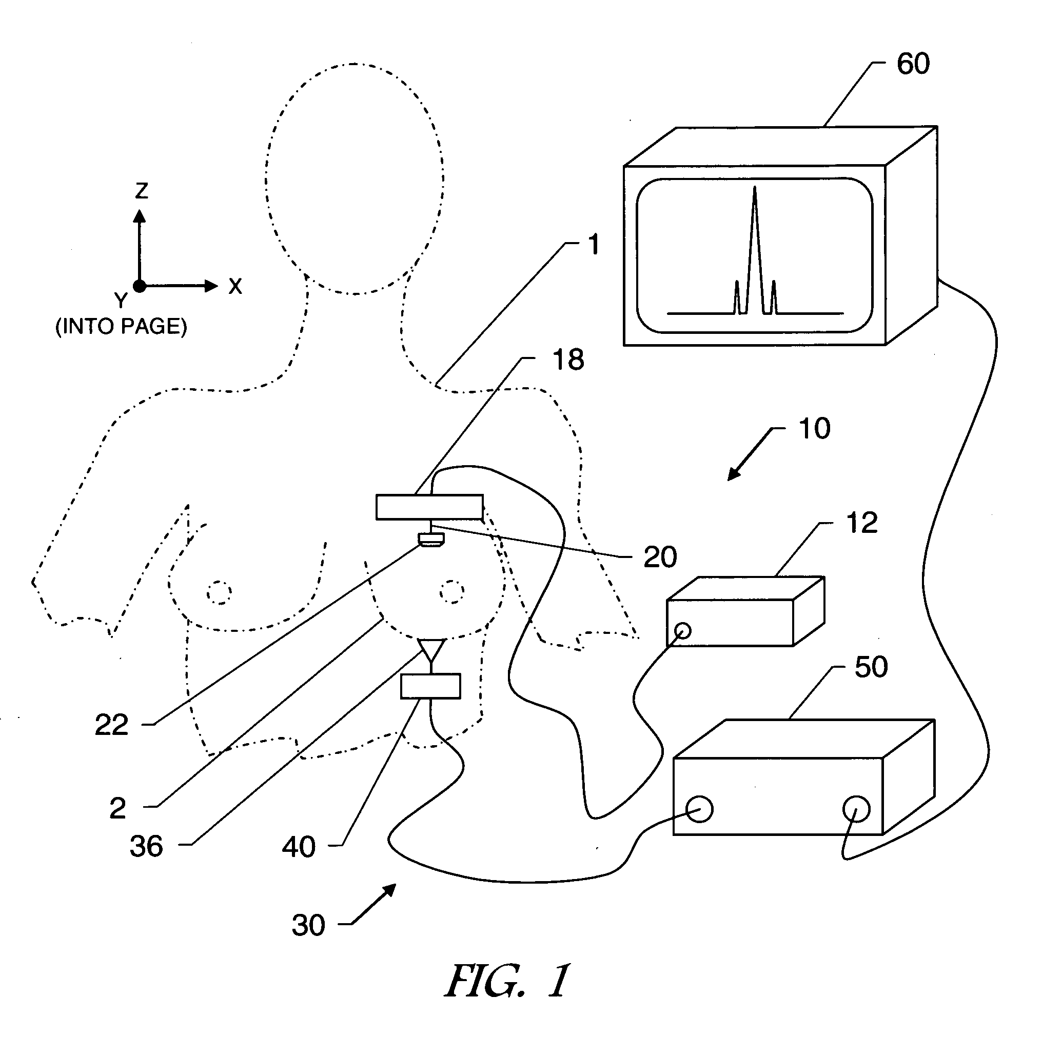
[0040]Referring now to the drawings, FIG. 1 shows the orientation of the system with respect to the patient 1 and the imaging target breast 2 in one preferred embodiment of the present invention. An ultrasound subsystem 10 and a microwave imaging subsystem 30 are employed in combination to detect and diagnose tumors in the breast 2. An ultrasound transducer 22 and a microwave antenna 36 are oriented with respect to the target breast 2 of the patient1. In one preferred embodiment, the ultraso...


PUM


Abstract

A multi-modality system and method for performing screening / detection, imaging and diagnosis / characterization of materials and objects in dense compressive media, such as in medical soft tissue applications, is disclosed. Medical tissue applications include but are not limited to the detection and diagnosis of breast tumors. Generally, the present invention involves coupling an ultrasound subsystem for exciting target tissues with a microwave subsystem for measuring the response, imaging and diagnosing the target tissues.

Description

CROSS REFERENCE TO RELATED APPLICATIONS[0001]This application claims the benefit of provisional patent Ser. No. 60 / 070,003 filed Mar. 19, 2008.[0002]Not applicableSTATEMENT REGARDING FEDERALLY SPONSORED RESEARCH OR DEVELOPMENT[0003]Not applicableINCORPORATION-BY-REFERENCE OF MATERIAL SUBMITTED ON A COMPACT DISC[0004]Not applicableBACKGROUND OF THE INVENTION[0005]1. Field of Invention[0006]The present invention relates generally to the field of imaging in dense compressive media, and more particularly to a novel system and method of use thereof for imaging in medical soft tissue applications such as dermatology, orthopedics and bone fractures, and breast tumor scanning / detection and diagnosis / characterization.[0007]According to the U.S. National Library of Medicine and the National Institutes of Health, one in eight women will be diagnosed with breast cancer. One in sixteen women will die prematurely due to breast cancer. Breast cancer is more easily treated and often curable if it i...

Claims


Application Information

Patent Timeline
IPC IPC(8): A61B6/00A61B8/00
CPCA61B5/05A61B8/4416A61B8/0825A61B5/0507A61B5/0035
Inventor ISMAIL, ALY M.COOPER, KENNETH BRIANSATAMA, KHALED N.
Owner ULTRAWAVE LABS
Learn More