Antagonist antibody for the treatment of cancer
an anti-cancer and anti-eph monoclonal antibody technology, applied in the field of new anti-cancer antibodies, can solve the problems of less tumor-effective antibodies and more complex reality, and achieve the effect of high cytotoxicity
- Summary
- Abstract
- Description
- Claims
- Application Information
AI Technical Summary
Benefits of technology
Problems solved by technology
Method used
Image
Examples
example 1
Generation of Anti-EphA2 Monoclonal Antibody Hybridomas
[0360]Four BALB / c VAF mice were immunized with human EphA2-transfected 300-19 cells, a pre-B cell line derived from a BALB / c mouse. The stably transfected cells over-expressing the antigen were generated by transfection of 300-19 cells with the full-length human EphA2 cDNA and selected for the high expression clones by flow cytometry. Clone 4-6, a clone that highly expresses the human EphA2 receptor on the cell surface, was selected as immunogen for immunization of mice and for antibody screening of hybridomas. The EphA2-transfected cells were maintained in the selection medium containing G418 at a final concentration of 1 mg / mL and were tested for the EphA2 expression regularly using a commercially available antibody.
[0361]The Balb / c mice were subcutaneously injected with approximately 5×10⁶ EphA2-transfected 300-19 cells in 200 μL of phosphate buffered saline (PBS) per mouse. The injections are performed every 2-3 weeks by sta...
example 2
Binding Characterization of Anti-EphA2 Antibodies, 37.3D7, 37.1F5, 53.2H11, EphA2-N1 and EphA2-N2
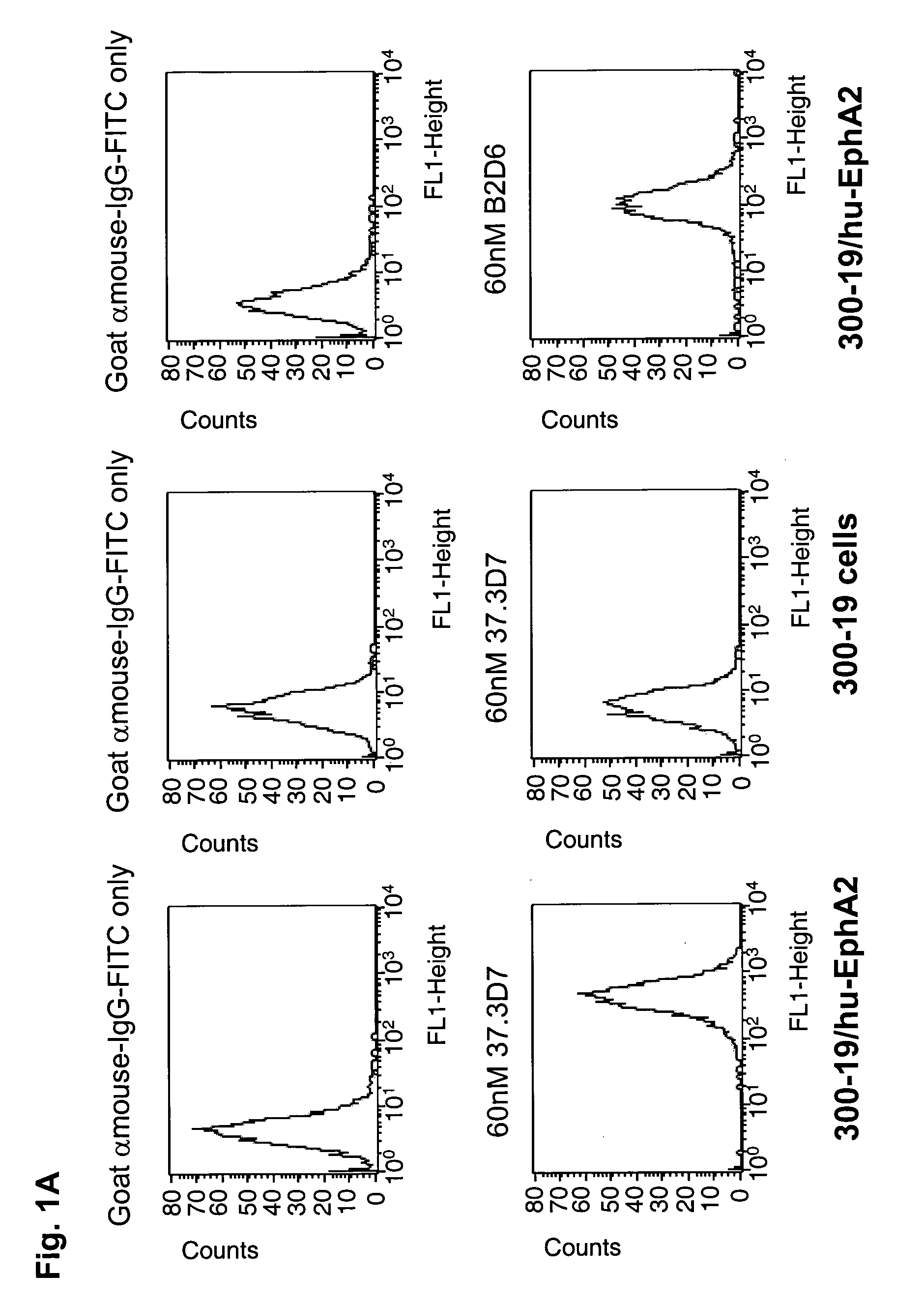
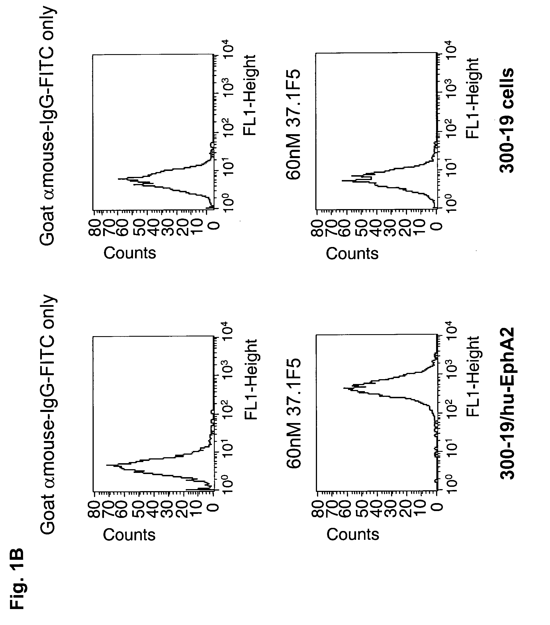
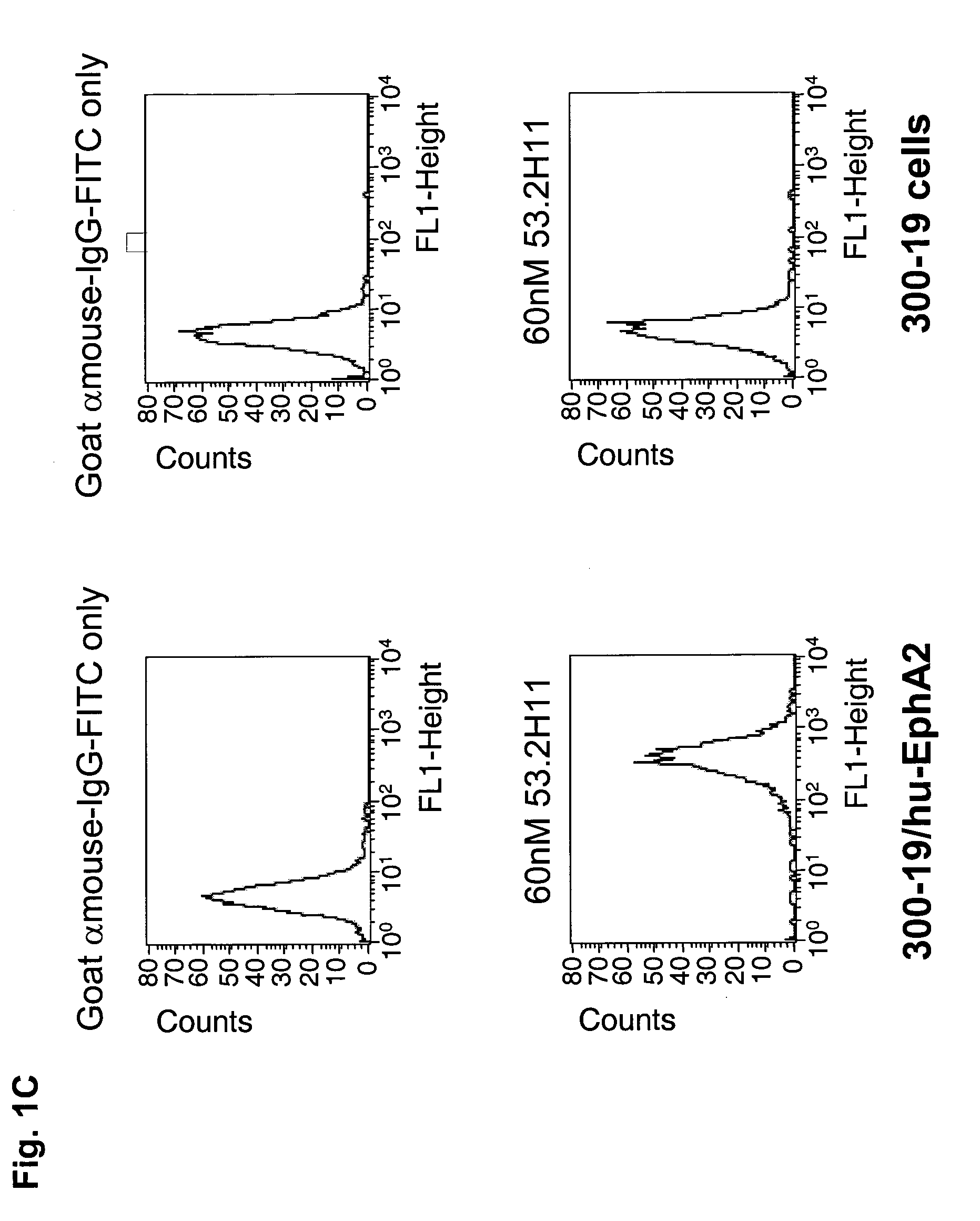
[0365]The specific binding of each of the purified anti-EphA2 antibodies, 37.3D7, 37.1F5 and 53.2H11 was demonstrated by fluorescence activated cell sorting (FACS) using cells overexpressing human EphA2 and by using cells that do not express EphA2 (FIGS. 1A, B, and C). Incubation of 37.3D7 antibody, or 37.1F5 antibody or 53.2H11 antibody (60 nM) in 100 μl cold FACS buffer (1 mg / mL BSA in Dulbecco's MEM medium) was performed using cells overexpressing EphA2 and cells that do not express EphA2 in a round-bottom 96-well plate on ice. After 1 h, the cells were pelleted by centrifugation and washed with cold FACS buffer and then incubated with goat-anti-mouse IgG-antibody-FITC conjugate (100 μL, 6 μg / ml in FACS buffer) on ice for 1 h. The cells were then pelleted, washed, and resuspended in 200 μL of 1% formaldehyde solution in PBS. The cell samples were then analyzed using a FACSCalibur read...
example 3
Inhibition of Binding of EphrinA1 to MDA-MB-231 cells by 37.3D7, 37.1F5, 53.2H11, EphA2-N1 and EphA2-N2 Antibodies
[0370]The binding of ephrinA1 to MDA-MB-231 human breast cancer cells was inhibited by 37.3D7, 37.1F5 and 53.2H11 antibodies (FIG. 6). MDA-MB-231 cells were incubated with or without 5 μg / mL 37.3D7, 37.1F5, or 53.2H11 antibody for 2 h, followed by incubation with 100 ng / mL biotinylated ephrinA1 for 30 min at 4° C. The cells were then washed twice with serum-free medium to remove unbound biotin-ephrinA1, and were then lysed in 50 mM HEPES buffer, pH 7.4, containing 1% NP-40 and protease inhibitors. An Immulon-2HB ELISA plates were coated with a mouse monoclonal anti-EphA2 antibody (D7, Upstate) and were used to capture the EphA2 and bound biotin-ephrinA1 from the lysate. The binding of the coated antibody to the cytoplasmic C-terminal domain of the EphA2 did not interfere with the binding of biotin-ephrinA1 to the extracellular domain of EphA2. The wells were washed, incu...
PUM
| Property | Measurement | Unit |
|---|---|---|
| Fraction | aaaaa | aaaaa |
| Volume | aaaaa | aaaaa |
| Mass | aaaaa | aaaaa |
Abstract
Description
Claims
Application Information
 - R&D
- Intellectual Property
- Life Sciences
- Materials
- Tech Scout
- Unparalleled Data Quality
- Higher Quality Content
- 60% Fewer Hallucinations
Browse by: Latest US Patents, China's latest patents, Technical Efficacy Thesaurus, Application Domain, Technology Topic, Popular Technical Reports.
© 2025 PatSnap. All rights reserved.Legal|Privacy policy|Modern Slavery Act Transparency Statement|Sitemap|About US| Contact US: help@patsnap.com


