Systems and methods for supporting or occluding a physiological opening or cavity

a physiological opening and cavity technology, applied in the field of physiological openings or cavities, can solve the problems of high risks of anesthesia, bleeding and infection during and following these types of procedures, and the surgical techniques for repairing septal defects, blood vessels, and other types of physiological irregularities and defects, and achieves the effects of promoting re-endothelialization and tissue growth, facilitating accurate positioning, placement and monitoring of the device, and facilitating the re-injection of the d

Active Publication Date: 2010-04-15
PULSAR VASCULAR
View PDF101 Cites 138 Cited by
  • Summary
  • Abstract
  • Description
  • Claims
  • Application Information

AI Technical Summary

Benefits of technology

[0032]Various agents, such as agents that promote re-endothelialization and tissue growth, as well as bonding agents, therapeutic agents, anti-thrombolytic agents, hydrophilic and / or hydrophobic agents, and the like may be provided to the site during or following the placement procedure and / or in association with the implantable device of the present invention. Exemplary agents that may be administered prior to, during or subsequent to device deployment, or may be associated with the implantable device, are disclosed in U.S. Patent Application Publication Nos. 2004 / 087998 A1, 2004 / 0193206 A1 and 2007 / 0191884 A1, which are incorporated by reference herein in their entireties. It will also be appreciated that radiopaque markers or radiopaque compounds may be associated with certain structures or portions of the implantable device and delivery assembly structure to facilitate accurate positioning, placement and monitoring of the device during and following deployment.
[0033]In one aspect, methods and systems of the present invention provide exclusion of a defect, such as an aneurysm, and diversion of blood flow away from the aneurysm by placement of a framework structure incorporating a membrane that restricts access to and restricts or prevents flow communication between the vessel and the interior of the aneurysm across the neck of the aneurysm, and retention of the framework structure and membrane across the opening by means of one or more anchoring structures extending from the framework structure proximally and contacting walls of a neighboring vessel, such as a parent vessel, in generally opposed regions. Methods and systems of the present invention may further promote shrinking and reabsorption of the defect, or portions of the defect, and facilitate hemostasis inside the defect. In one aspect, methods and systems of the present invention not only restore the structure and function of the parent vessel in the vicinity of the defect, but also stabilize material inside the aneurysm, prevent debris from escaping into the bloodstream, and promote a reduction in the size and mass of the aneurysm.

Problems solved by technology

Surgical techniques for closing openings and repairing defects in anatomical lumens and tissues, such as blood vessels, septal defects and other types of physiological irregularities and defects, are highly invasive.
Surgical techniques for repairing septal defects are also highly invasive.
The risks associated with anesthesia, bleeding and infection during and following these types of procedure are high, and tissue that is affected during the procedure may or may not survive and continue functioning.
Accurate and timely placement of these mixtures is crucial and very difficult.
These materials are difficult or impossible to retrieve once they have been placed in the vasculature.
Migration or projection of a vaso-occlusive device from the cavity may interfere with blood flow or nearby physiological structures and poses a serious health risk.
One type of aneurysm, commonly known as a “wide neck aneurysm” is known to present particular difficulty in the placement and retention of vaso-occlusive coils.
The placement of coils, or other structures or materials, in the internal space of an aneurysm or other defect has not been entirely successful.
The placement procedure may be arduous and lengthy, requiring the placement of multiple devices, such as coils, serially in the internal space of the aneurysm.
Longer procedures, in general, involve higher risks of complication from anesthesia, bleeding, infection, and the like.
Moreover, because placement of structures in the internal space of an aneurysm doesn't generally completely occlude the opening, recanalization of the original aneurysm is more likely to occur, debris and occlusive material may escape from within the aneurysm and present a risk of stroke, vessel blockage or other undesirable complications.
Blood may also flow into aneurysm and other blood vessel irregularities after the placement of embolic devices, which increases the risks of complication and further enlargement of the aneurysm.
Furthermore, some aneurysms, vessels and other passageway defects are not well-suited to placement of coils or other conventional occlusive devices.
Despite the numerous devices and systems available for placing embolic materials in an aneurysm and for occluding physiological defects using minimally invasive techniques, these procedures remain risky and the results rarely restore the physiological structure to its normal, healthy condition.
Neither the anchoring structures, nor the support structure, nor the membrane interferes substantially with normal or desired fluid flow in the lumens in proximity to the opening.

Method used


Image

  • Systems and methods for supporting or occluding a physiological opening or cavity
  • Systems and methods for supporting or occluding a physiological opening or cavity
  • Systems and methods for supporting or occluding a physiological opening or cavity

Examples


Embodiment Construction

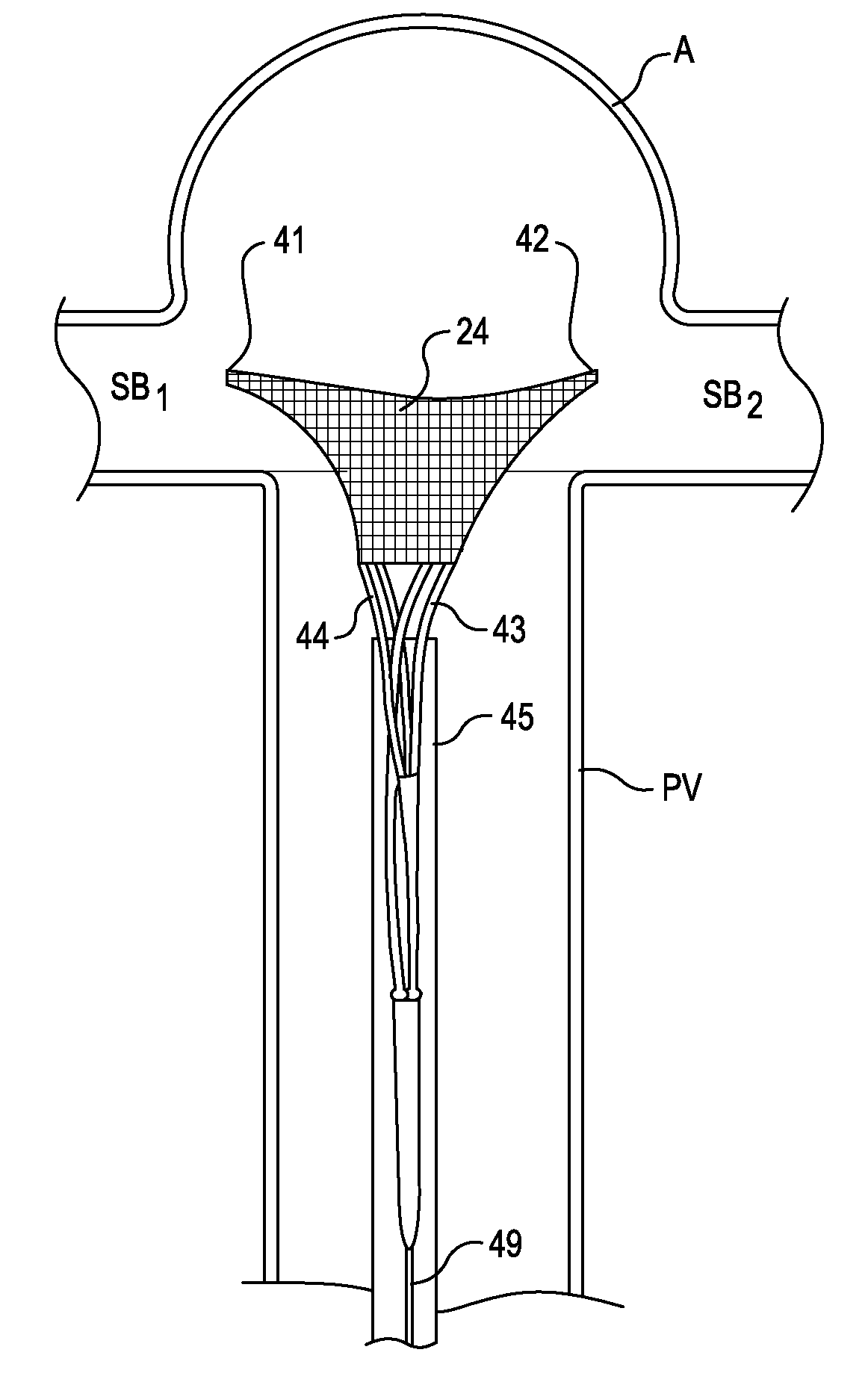
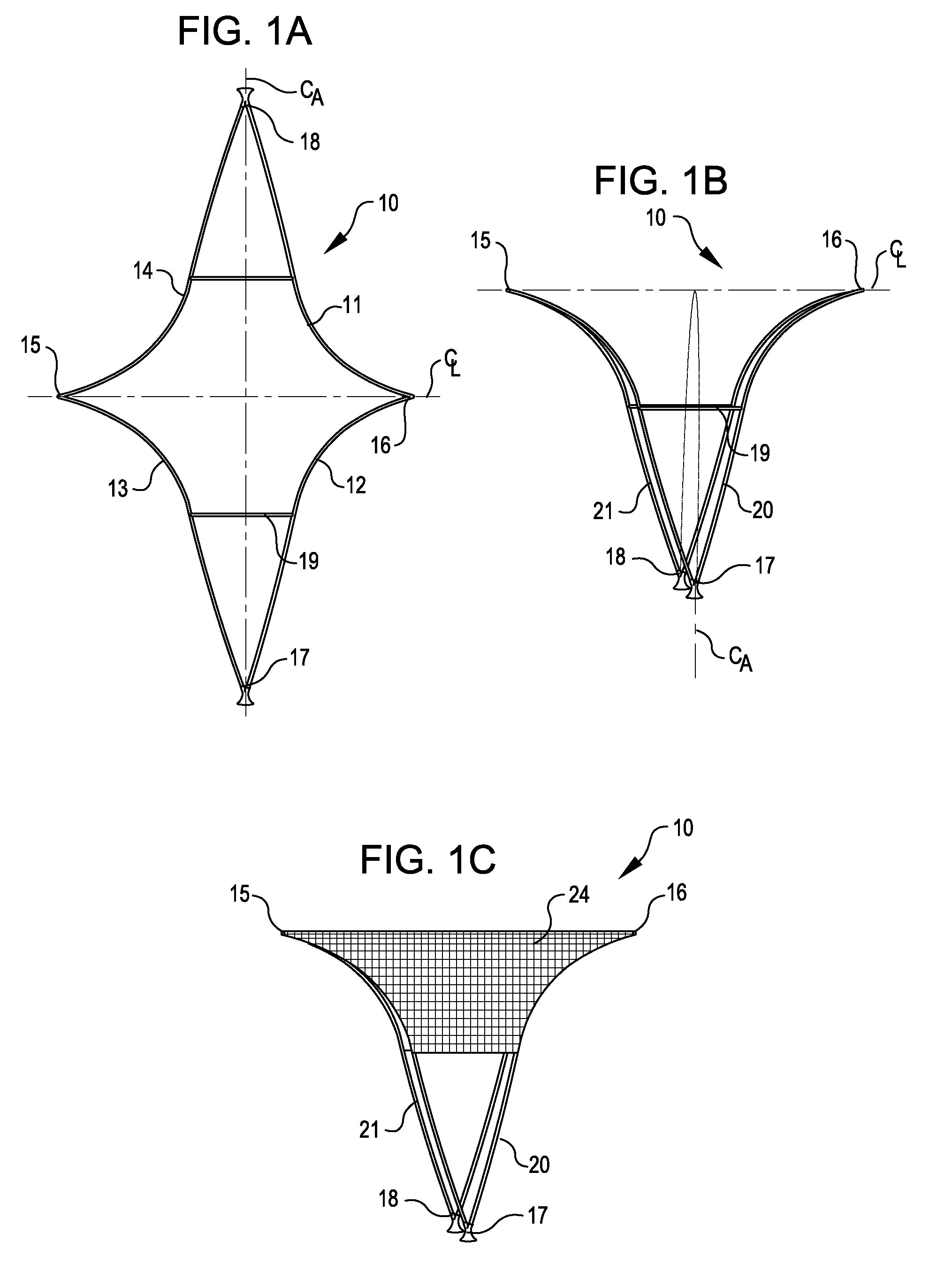
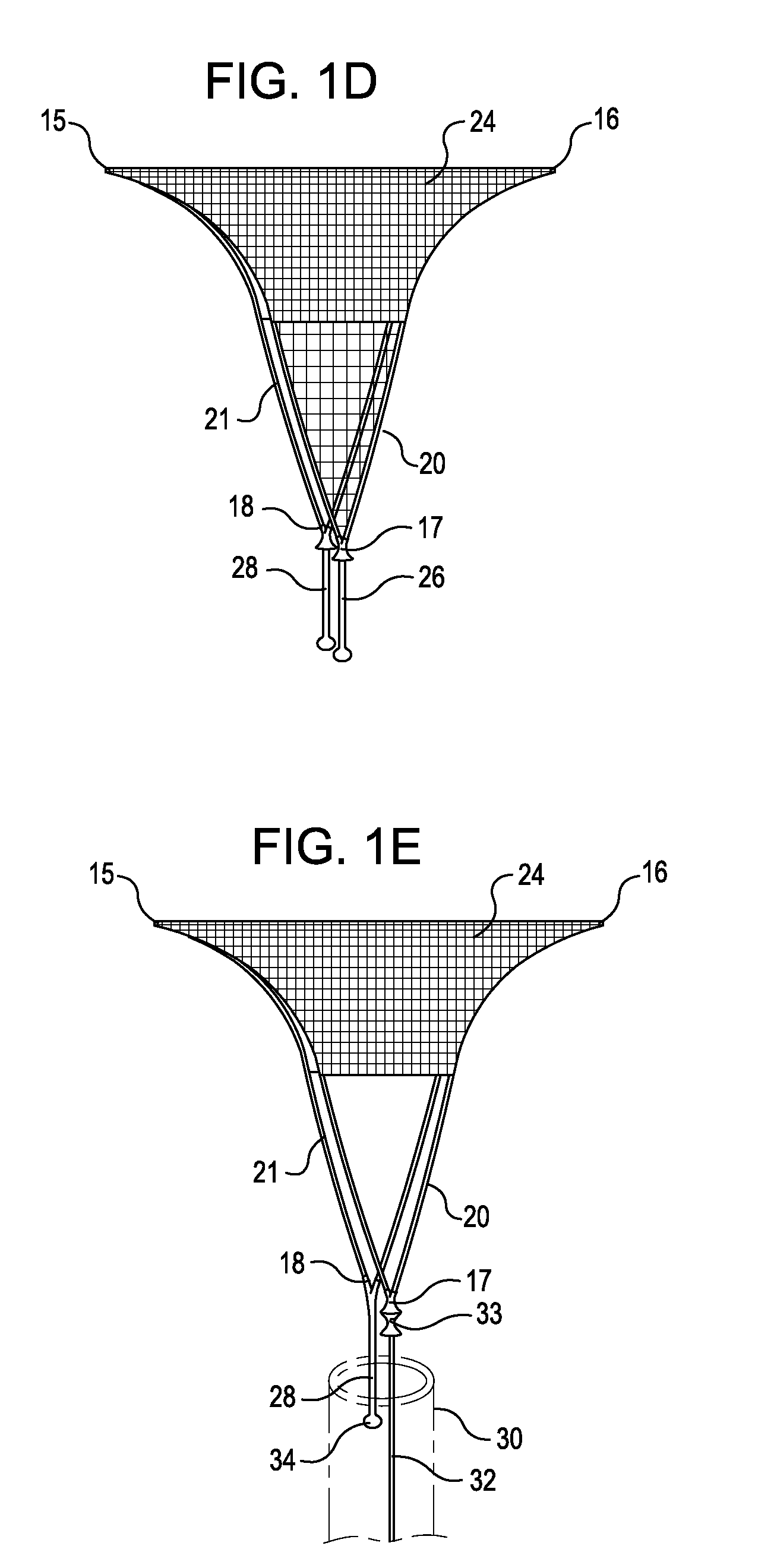
[0058]In general, implantable assemblies of the present invention comprise an implantable device attached to at least one delivery wire or tube and loaded in a catheter or a sheath for delivery to a target site in a human body, such as in the neurovasculature at a site in proximity to a wide mouth, termination or bifurcation aneurysm. The implantable device is delivered to the target site in a small diameter, constrained condition and is deployed, at the site, to its larger diameter deployed condition. The device, in the deployed condition, comprises a generally inverted U-shaped three-dimensional framework support structure having a perimeter structure configured to be positioned in close proximity to, and generally contacting tissue at the neck of the aneurysm along at least a portion of its perimeter.

[0059]The framework support perimeter structure may incorporate substantially opposed lateral corners, or wing tip structures, lying on a longitudinal centerline of the framework sup...


PUM


Abstract

Implantable devices for placement at a cavity or opening such as an aneurysm are disclosed. The implantable devices, in a deployed condition, have a generally inverted U-shaped profile with a curved or angled framework support structure sized and configured for placement in proximity to tissue surrounding the opening and anchoring legs extending proximally from the framework structure sized and configured to contact the wall of a neighboring lumen at opposed locations. Occlusive and semi-occlusive membranes may be associated with the framework support structure and deployed over the opening to provide exclusion of the opening and flow diversion. Proximal anchoring segments providing additional lumen wall surface area contact for the implantable device following deployment may be incorporated.

Description

REFERENCE TO PRIORITY APPLICATION[0001]This application claims priority to U.S. provisional patent application No. 61 / 094,693 filed Sep. 5, 2008. The disclosure of this priority application is incorporated by reference herein in its entirety.TECHNICAL FIELD OF THE INVENTION[0002]The present invention relates generally to implantable structures for placement in proximity to an opening or cavity in a physiological structure, such as the neck of an aneurysm, using minimally invasive techniques, and to methods of making and deploying such structures. In one aspect, the implantable structures described herein contact and support tissue in proximity to the opening or cavity. In another aspect, the implantable structures are at least partially occlusive and, when deployed across an opening in a physiological structure (e.g., aneurysm neck), provide flow diversion from the opening and may provide substantial occlusion of the opening. The structures described are particularly useful for plac...

Claims


Application Information

Patent Timeline
Patent Type & Authority Applications(United States)
IPC IPC(8): A61B17/08
CPCA61B17/12022A61B17/12113A61B17/12118A61B17/12172A61B17/12177A61B2017/12054A61B2017/00893A61B2017/00898A61F2/06A61F2/95A61B2017/00867A61F2240/001A61F2/04A61F2/78A61B17/08A61B17/12A61F2/00A61F2/02A61B2017/00526A61B2017/1205A61F2/86A61B17/12036A61B2017/00778
Inventor GERBERDING, BRENT C.ABRAMS, ROBERT M.CLARKE, GILBERT
Owner PULSAR VASCULAR
Why Eureka
Try Eureka