Quantitative lateral flow system and assay

a quantitative and lateral flow technology, applied in the field of quantitative and quantitative assay and system, can solve the problems of low efficiency in dissolving a volume variation, long assay time, etc., and achieve the effect of dissolving the conjugate or detectable agent and good cell filtering

Inactive Publication Date: 2010-04-22
RELIA DIAGNOSTIC SYST
View PDF13 Cites 29 Cited by
  • Summary
  • Abstract
  • Description
  • Claims
  • Application Information

AI Technical Summary

Benefits of technology

[0015]It is a further one of the objects of the present invention to provide a lateral flow assay and system as above that is efficient in dissolving the conjugate or detectable agent.
[0016]It is yet another one of the objects of the present invention to provide a lateral flow assay and system as above that provides good filtering for cells, such as red blood cells.

Problems solved by technology

Many have tried to design a lateral flow assay for determining the presence and quantity of analytes in biological samples, such as blood samples that contain whole blood, red blood cells, or white blood cells, but have failed.
The reasons for failure are many but may be attributable primarily to factors such as hemolysis of the red blood cells creating high background noise, low filtering efficiency, for example, resulting in leakage of the red blood cells onto the chromatographic strip, requirement for a relatively large sample volume (such as requiring 100 μl of sample or more), low efficiency in dissolving a conjugate or detectable agent, volume variation because of variation in cell volume when cells are present, long assay time, and inefficiency in dissolving the conjugate for detection of the analyte.
While the methods and system described in these patents are useful for detecting and quantifying most analytes, these patents do not teach how the methods and system can be used to analyze samples containing cells, including red blood cells and / or white blood cells or other cell types.
However, until now, few strategies have worked well.
Up to the present, there is lacking a rapid, effective and efficient quantitative lateral flow assay and system that can be used for determination of analytes in biological samples, such as in a blood sample, in a point-of-care setting, or a lateral flow assay and system that can be used for determination of analytes that are present in a small volume of sample, such as from a finger prick, or a lateral flow assay and system that can be used for determination of analytes that is volume independent, or that would address other problems in the prior art lateral flow assays and systems.

Method used


Image

  • Quantitative lateral flow system and assay
  • Quantitative lateral flow system and assay
  • Quantitative lateral flow system and assay

Examples


example 1

Filtering Capability of Membranes in Absence of Agglutinins

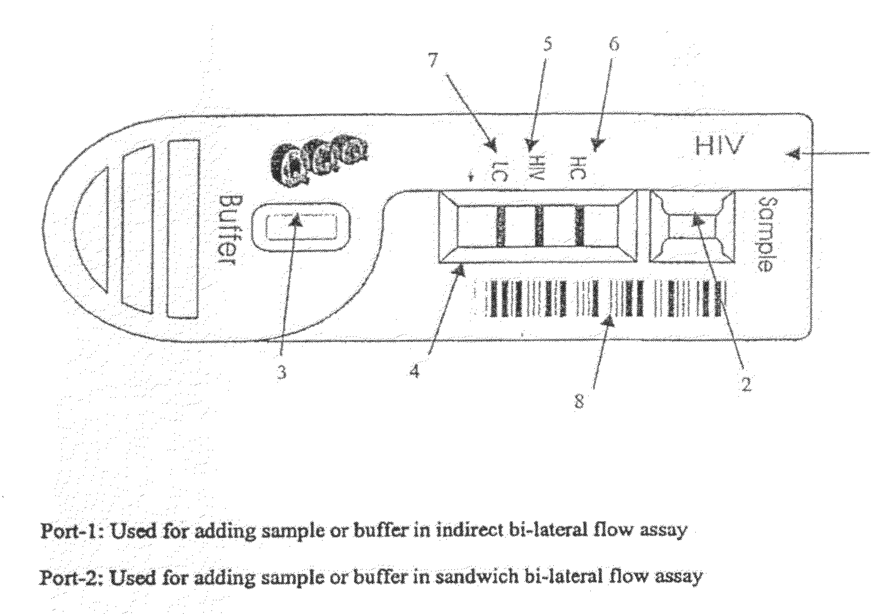
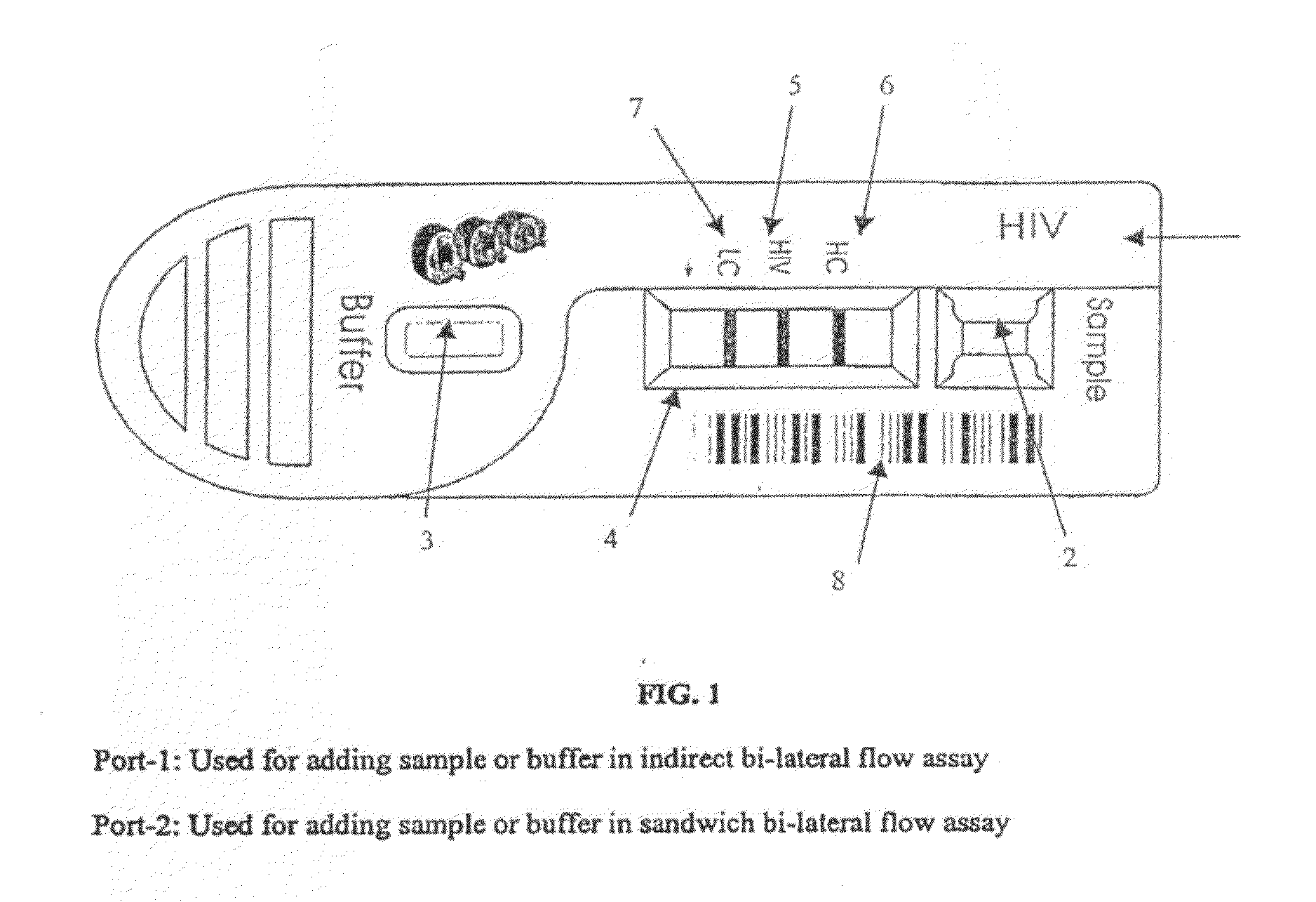
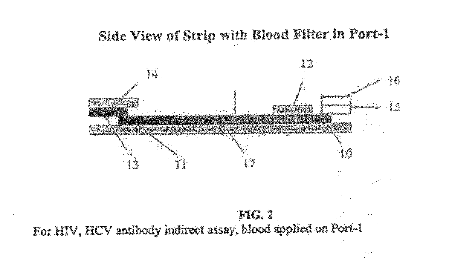
[0195]The test strip as shown in FIG. 2 was constructed with different membranes as the sample filter. The filtering membranes tested were all obtained from Ahlstrom Filtration, Inc. (USA) and include: cellulose absorbent grade 111, glass fibers grade #141 and grade #142, Cytosep grades 1660, 1661, 1662 and 1663. A sample containing whole blood was applied in Port-1 as shown in FIG. 1. The migration speed of plasma on the nitrocellulose chromatographic strip was observed. Results were obtained as set forth in Table 2.

TABLE 2Blood Filtering Membrane Without Anti-hRBCTime of RBCVolumeMigrateappearanceMembraneSampleμlRBC Leakingmm / minBackgroundon NCGrade 111Whole Blood100Yes≦16Red8 minCellulose100Yes≦16Red8 min100Yes≦16Red8 min100Yes≦16Red8 min100Yes≦16Red8 minGrade 141Whole Blood100Yes≦16Red3 minGlass Fiber100Yes≦16Red3 min100Yes≦16Red3 min100Yes≦16Red3 min100Yes≦16Red3 minGrade 142Whole Blood100Yes≦16Red2 minGlass Fiber100Yes...

example 2

Blood Filtering Capability of Membranes Pre-Treated with Anti-RBC

[0196]A ten percent (10%) TWEEN 20 solution was prepared by adding 1 g of TWEEN 20 to 9 ml of deionized water, mixing the solution, and storing the solution for about a week at room temperature.

[0197]A rabbit anti-human red blood cells antibody solution was prepared by adding 9.0825 g of Trizma Base (final concentration of 6.055 g / L), 1.7625 ml of HCl (final concentration of 1.7625 ml / L) and 1.8 g of EDTA.Na2 (final concentration of 1.2 g / L) to 1.35 liters of deionized water. The mixture was stirred slowly until the chemical reagents were dissolved completely, about an hour. The solution was kept at room temperature for 4 hours or overnight at 4° C. The pH of the solution was adjusted to pH 8.5±0.1 by adding HCl. Rabbit anti-human red blood cell antibody (anti-hRBC) was added to the solution to a final concentration of about 0.25 mg / ml. About 0.3 ml of 10% Tween-20 solution was added to the anti-hRBC to a final concent...

example 3

Comparison Between Using Whole Blood Versus Using Plasma

[0199]The test strip as in Example 2 was prepared and whole blood or plasma was added to the sample filter in Port-1 and the results were compared, as shown in Table 4.

TABLE 4Comparison of Testing Results Between Whole Blood and PlasmaRI =Anti-TESThRBCRBCTimeHCLCTESTDr / LCMembraneMg / mlSampleVolumeLeakingminDrDrDrDrS / COResult#1420.25HIV (−)50 μlNo30 min0.17770.0862000(−)0.25Blood50 μlNo30 min0.22680.0967000(−)0.2550 μlNo30 min0.0730.1083000(−)0.2550 μlNo30 min0.1830.0728000(−)0.2550 μlNo30 min0.32770.109000(−)#1420.25HIV (−)50 μlNo15 min0.40330.1926000(−)0.25Plasma50 μlNo15 min0.35870.1945000(−)0.2550 μlNo15 min0.360.1919000(−)0.2550 μlNo15 min0.38220.1955000(−)0.2550 μlNo15 min0.37590.1964000(−)#1420.25HIV (+)50 μlNo30 min0.12130.17810.20141.130811.311(+)0.25Blood50 μlNo30 min0.14190.14410.21811.513515.1325(+)0.2550 μlNo30 min0.17810.14080.21531.529115.2911(+)0.2550 μlNo30 min0.0740.0750.10321.376013.7626(+)0.2550 μlNo30 min0.16...


PUM

PropertyMeasurementUnit
volumeaaaaaaaaaa
volumeaaaaaaaaaa
volumeaaaaaaaaaa

Abstract

The present invention relates to a lateral flow assay and system, including a test strip, for detection and quantification of analytes in samples, such as samples containing cells and fluid, where the assay is volume independent, and the sample size is less than about 100 μl, where the test strip includes a first membrane such as a sample filter, that is in capillary contact with an optional second membrane, such as a fluid collector, the second membrane, if present is in capillary contact with an optional third membrane, such as a conjugate pad containing a mobilizable detectable agent, or with a fourth membrane, which is a chromatographic strip, which optionally contains a mobilizable detectable agent, all such membranes being in fluid contact with a fifth membrane, such as a buffer pad, a sixth membrane, such as an absorbent pad, optionally a seventh membrane, which is also an absorbent pad, a capture band for capturing the analyte and at least one control band, or alternatively, the chromatographic strip contains the mobilizable detectable agent in place of a conjugate pad, where the test strip is configured to support removal of red blood cells from the sample and to allow uni-directional or bi-directional fluid flow of fluid from the sample filter to the capture band to be retained therein and detected thereon.

Description

CROSS-REFERENCE TO RELATED APPLICATIONS[0001]This application hereby claims priority from U.S. Provisional Patent Application Ser. No. 60 / 576,327, filed Jun. 2, 2004 by Zhou et al. and entitled “Quantitative Lateral Flow System and Assay,” as well as from U.S. Provisional Application Ser. No. 60 / 592,202, filed Jul. 29, 2004 by Zhou et al. and also entitled “Quantitative Lateral Flow System and Assay.” The disclosures of these provisional applications are hereby incorporated herein in their entirety by this reference.TECHNICAL FIELD[0002]This application generally relates to a qualitative and quantitative assay and system for detecting the presence of at least one analyte in biological samples, particularly samples that contain whole blood, red blood cells, white blood cells, or other cell types, and determining or quantifying the amount of the at least one analyte present.BACKGROUND OF THE INVENTION[0003]Many have tried to design a lateral flow assay for determining the presence and...

Claims


Application Information

Patent Timeline
Patent Type & Authority Applications(United States)
IPC IPC(8): G01N33/53C12M1/34G01N33/558
CPCG01N33/558G01N33/54388
Inventor ZHOU, SILIANGRUTTER, WILLIAM J.LIU, NING
Owner RELIA DIAGNOSTIC SYST
Why Eureka
Try Eureka