Analyzing biological cell material based on different interactions between illumination light and cell components

a technology of illumination light and cell components, applied in the field of analyzing biological cell materials, can solve the problems of time-consuming and expensive cell sorting procedure before the real cytometry measurement may become obsolete, and achieve the effects of significant improvement of diagnostic accuracy, time-consuming and expensive, and strong change of optical properties

Inactive Publication Date: 2010-04-29
KONINKLIJKE PHILIPS ELECTRONICS NV
View PDF10 Cites 3 Cited by
  • Summary
  • Abstract
  • Description
  • Claims
  • Application Information

AI Technical Summary

Benefits of technology

[0057]This aspect of the invention is based on the idea that by combining two independent measurement parameters, which are both indicative for a particular cell defect and / or a particular disease, the diagnostic accuracy for identifying the type of these cells can be significantly increased. This holds in particular for identifying cells as to be benign or malign because as compared to normal benign cells malign cancer cells show a comparatively strong change of optical properties in different spectral regimes. This change of optical properties may be based on structural and / or chemical changes of cancer cells as compared to normal cells.
[0058]The described combined analysis of the cell material under study may make a selection of suspicious and normal cells by a pathologist no more necessary. Therefore, a time consuming and expensive cell sorting procedure before the real cytometry measurement may become obsolete. This may provide the advantage that tissue having been withdrawn from a patient can be analyzed much faster and also the result of the diagnostic procedure can be obtained much faster. Therefore, the time span, during which a patient has to lie open on an operation table, can be reduced significantly.
[0059]It has to be mentioned that the described method can only be used with biological cell material, which has been irreversibly withdrawn from a patient's body. Therefore, when carrying out the described method there are no direct interactions with the living body of the patient. Further, the described method is not able to directly result in a diagnosis a patient is suffering. The described method can only help a physician to find a diagnosis based on his medical knowledge. Thereby, the physician can take into account also other diagnostic methods, which, in combination with the described method, can help the physician to make a diagnosis more reliable.

Problems solved by technology

Therefore, a time consuming and expensive cell sorting procedure before the real cytometry measurement may become obsolete.

Method used


Image

  • Analyzing biological cell material based on different interactions between illumination light and cell components
  • Analyzing biological cell material based on different interactions between illumination light and cell components
  • Analyzing biological cell material based on different interactions between illumination light and cell components

Examples


Embodiment Construction

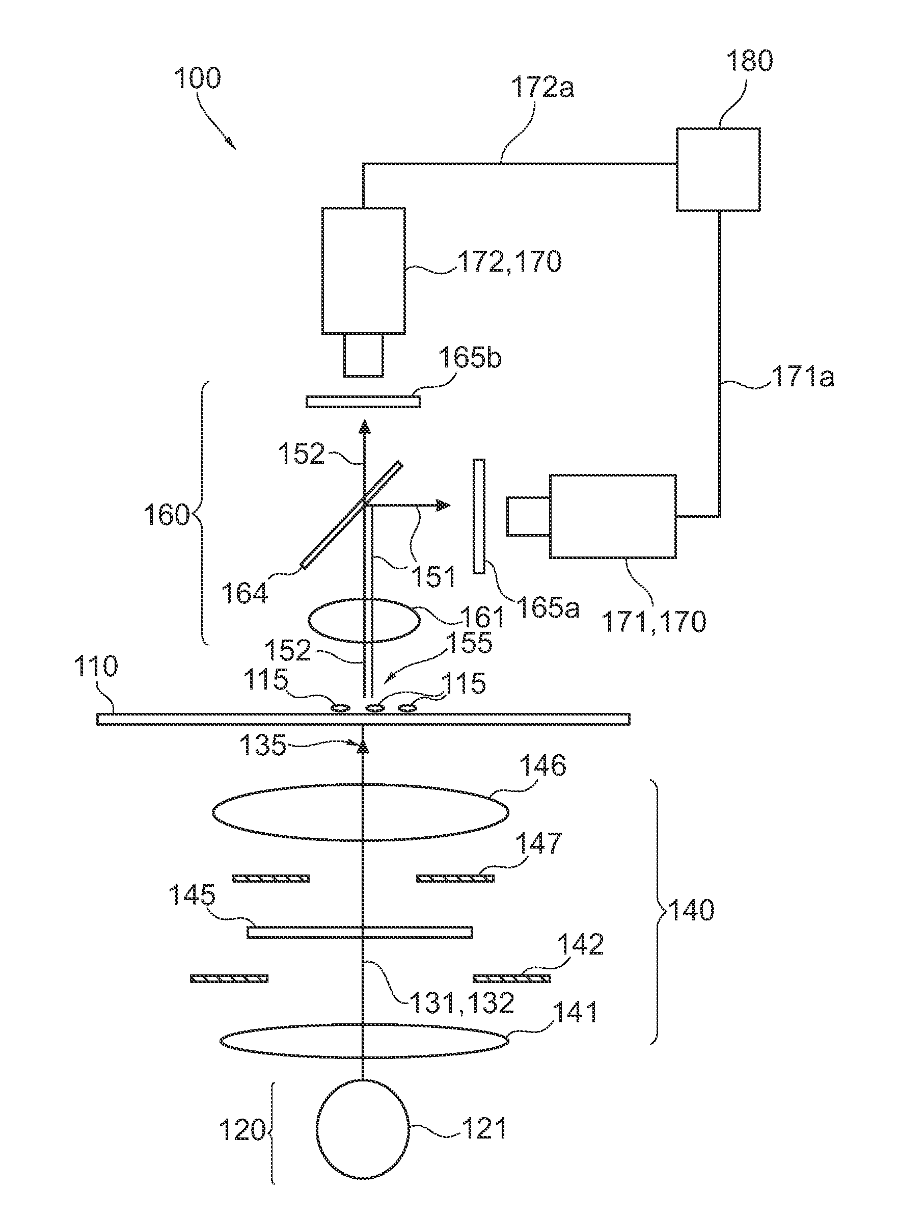
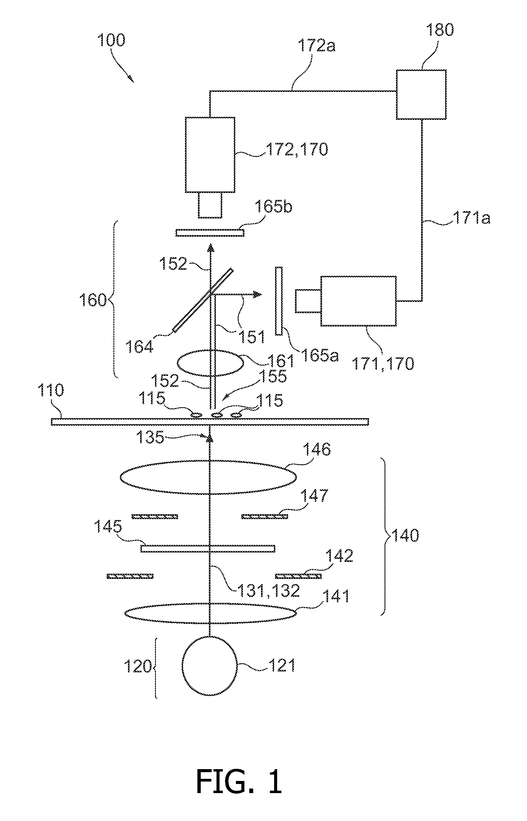
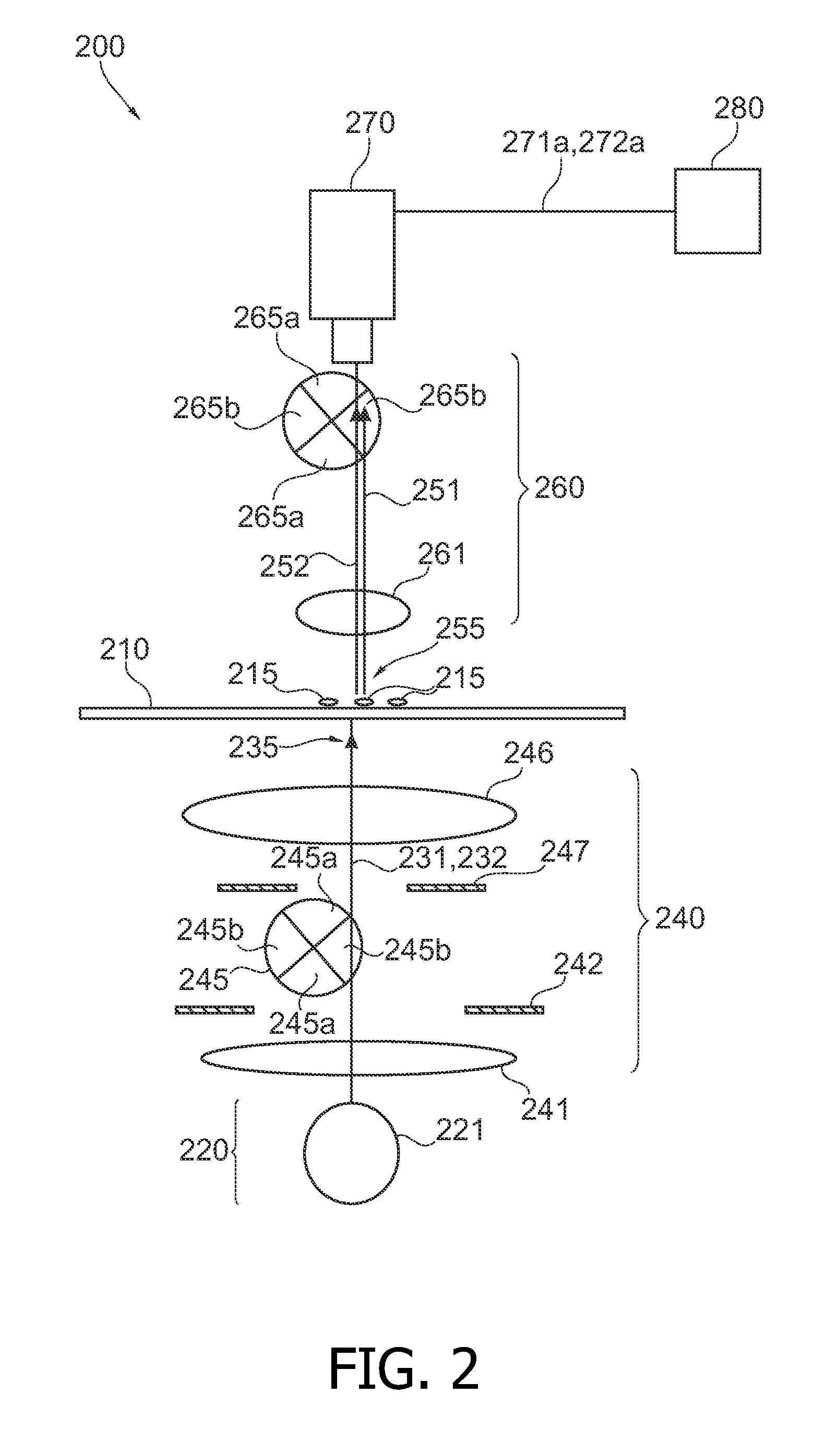
[0068]The illustration in the drawing is schematic. It is noted that in different figures, identical elements or similar elements having the same function are provided with reference signs, which are different from the corresponding reference signs only within the first digit.

[0069]FIG. 1 shows a cell-analyzing device 100, which is adapted for carrying out a UV DNA absorption cytometry in combination with autofluorescence of NAD(P)H. The device comprises a carrier element 110, which is realized by means of an object holder known for instance from optical microscopy. The carrier element 110 supports cell material 115, which may contain both benign respectively normal cells and malign respectively cancer cells. The device 100 is adapted to facilitate an identification of the type of cells 115 such that compared to known devices for cell sorting both the speed and the reliability of the cell identification can be increased.

[0070]The device 100 comprises a light source arrangement 120, ...


PUM

PropertyMeasurementUnit
wavelengthaaaaaaaaaa

Abstract

It is described a device (100) for analyzing biological cell material (115). The device (100) comprises a light source arrangement (120), which is adapted for directing a first (131) and a second illumination light (132) towards the cell material (115), wherein the first (131) and the second (132) illumination light comprises a first and a second spectral radiation component, respectively. The device (100) further comprises a detector arrangement (170), which is adapted for receiving a first measurement light (151) based on a first interaction of the first illumination light (131) with the cell material (115) and a second measurement light (152) based on a second interaction of the second illumination light (152) with the cell material (115). Further, the device (100) comprises an evaluation unit (180), which is coupled the detector arrangement (170) and which is adapted to evaluate a first signal (171a) and a second signal (171b) being indicative for the first (151) and the second measurement light (151), respectively. The device (100) may be used for accomplishing ultraviolet DNA image cytometryin combination with autofluorescence measurements of NAD(P)H.

Description

FIELD OF INVENTION[0001]The present invention relates to a device and to a method for analyzing biological cell material for instance for the purpose of distinguishing cancerous tumor cells from benign tumor cells.ART BACKGROUND[0002]Early detection of tumors is the most important prerequisite for fighting them effectively. This will also decisively improve tumor patient's prospects of being cured. In order to avoid multiple surgeries it is desirable to identify tumor tissue during one single. For further removing tissue from the patient the surgeon can than take into account reliable information about the type of tumor cells.[0003]At present, tumor border demarcation is performed by analysis of so-called ‘frozen sections’ by a pathologist. This technique takes approximately one hour; often, no intra-operative investigation is performed because of the long waiting time during which the patient lies open on the operation table. An alternative is to perform DNA cytometry for tumor bor...

Claims


Application Information

Patent Timeline
Patent Type & Authority Applications(United States)
IPC IPC(8): C12Q1/00C12M1/34
CPCG01N21/31G01N21/645G01N21/6428
Inventor KUIPER, STEINHENDRIKS, BERNARDUS HENDRIKUS WILHELMUSDE BOER, RUTH WILHELMINE IREENHOOFT, GERT
Owner KONINKLIJKE PHILIPS ELECTRONICS NV
Why Eureka
Try Eureka