
Method and device for activating stem cells

a stem cell and activation method technology, applied in the field of stem cell activation devices and methods, can solve problems such as unintended side effects, and achieve the effects of enhancing an osteoblast phenotyp

Inactive Publication Date: 2010-05-13
DEPUY SYNTHES PROD INC
View PDF16 Cites 69 Cited by
  • Summary
  • Abstract
  • Description
  • Claims
  • Application Information

AI Technical Summary

Benefits of technology

[0012]The solid support can include a column matrix material, a filter, an culture plate, tube or dish, a microtiter plate, a bead, a disk, or a combination thereof. The solid support can be a container. The solid support can include plastic, cellulose, cellulose derivatives, magnetic particles, nitrocellulose, glass, fiberglass, latex, or a combination thereof. The solid support can also include an affinity matrix to remove the at least one active agent. When a filter is present in the solid support, the filter can retain cell and bone graft substitute materials but allow passage of the at least one active agent. Alternatively, the filter can retain the at least one active agent but allow passage of the stem cells. In some embodiments, the solid support does not bind or adversely interact with stem cells. In other embodiments, the solid support can bind the stem cells without adversely interacting with the stem cells.
[0013]The device can further include a timer for controlling the time for incubating the stem cells with the at least one active agent. For example, the timer can trigger separation of the at least one active agent from the stem cell after the incubating step (i). In some embodiments, the device with the timer controls the time for incubating the stem cells with the at least one active agent to 24 hours or less. In other embodiments, the device with the timer controls the time for incubating the stem cells with the at least one active agent to 5 minutes to 1 hour.
[0020]In one embodiment, a method comprising exposing a stem cell source (e.g., bone marrow aspirate, including autologous bone marrow aspirate) to an active agent, wherein mesenchymal stem cells in the stem cell source are stimulated to differentiate into osteoblasts. In one embodiment, the bone marrow aspirate is drawn intraoperatively and / or used in a form as drawn from the patient. Alternatively, the bone marrow aspirate can further be concentrated after it is drawn from the patient. The method can also include mixing stimulated or activated stem cells (e.g., mesenchymal stem cells) with a synthetic bone graft substitute to form a mixture, wherein the exposing comprises transiently exposing the mixture to a fixed or tethered active agent in a manner effective for triggering mesenchymal stem cells to enhance an osteoblast phenotype.
[0023]In another embodiment, a method is provided for forming progenitor cells capable of stimulating bone formation. Method operations include mixing bone marrow aspirate (BMA) with an active agent, and using the active agent to trigger mesenchymal stem cells (MSCs) to enhance an osteoblast phenotype. Such a method can trigger mesenchymal stem cells to enhance or develop an osteoblast phenotype. The method can also include mixing a bone graft substitute and a bone marrow aspirate drawn from a patient to form a mixture; and transiently exposing the mixture to a fixed or tethered active agent in a manner effective for triggering mesenchymal stem cells to enhance or develop the osteoblast phenotype.

Problems solved by technology

Moreover, the use of osteogenic medium involves addition of components to the cells (e.g., growth factors) that can have unintended side effects if those components are administered to a patient along with the cells.

Method used


Image

  • Method and device for activating stem cells
  • Method and device for activating stem cells
  • Method and device for activating stem cells

Examples


example 1

Materials and Methods

[0098]The following materials and methods were used to develop certain aspects of the invention.

Differentiation of Cells

[0099]Human mesenchymal stem cells (Lonza, Walkersville, Md.) at passage 3 were seeded in basal medium (Stem Cell Technologies, Vancouver, Canada) at a density of 6×10⁴ cells / 35 mm well and incubated for 2 days at 37° C. Cells were rinsed with PBS, activated with 100 ng / ml of growth factor (FGF-2 or TGFβ3, R&D Systems, Minneapolis, Minn.) in fresh basal medium for 1 hour, rinsed with PBS and then incubated in either fresh basal medium or osteogenic differentiation medium (Stem Cell Technologies, Vancouver, Canada) for 7-14 days at 37° C.

Real time PCR

[0100]Total RNA was prepared from cells treated with various active agents using RNeasy Plus Mini Kit and QIA shredder Mini Spin columns (Qiagen). Total RNA was also prepared from untreated cells as a control. cDNA was generated using random hexamer and Oligo dT following the TaqMan Reverse Transcri...

example 2

Results

[0106]Primary human mesenchymal stem cells are capable of differentiating down an osteoblastic lineage. This is demonstrated in vitro by culturing cells in an osteogenic cocktail containing, but not limited to, dexamethasone, ascorbic acid and β-glycerophosphate. Under these culture conditions cells show an upregulation of osteoblast differentiation markers, the most common of which is the early marker alkaline phosphatase.

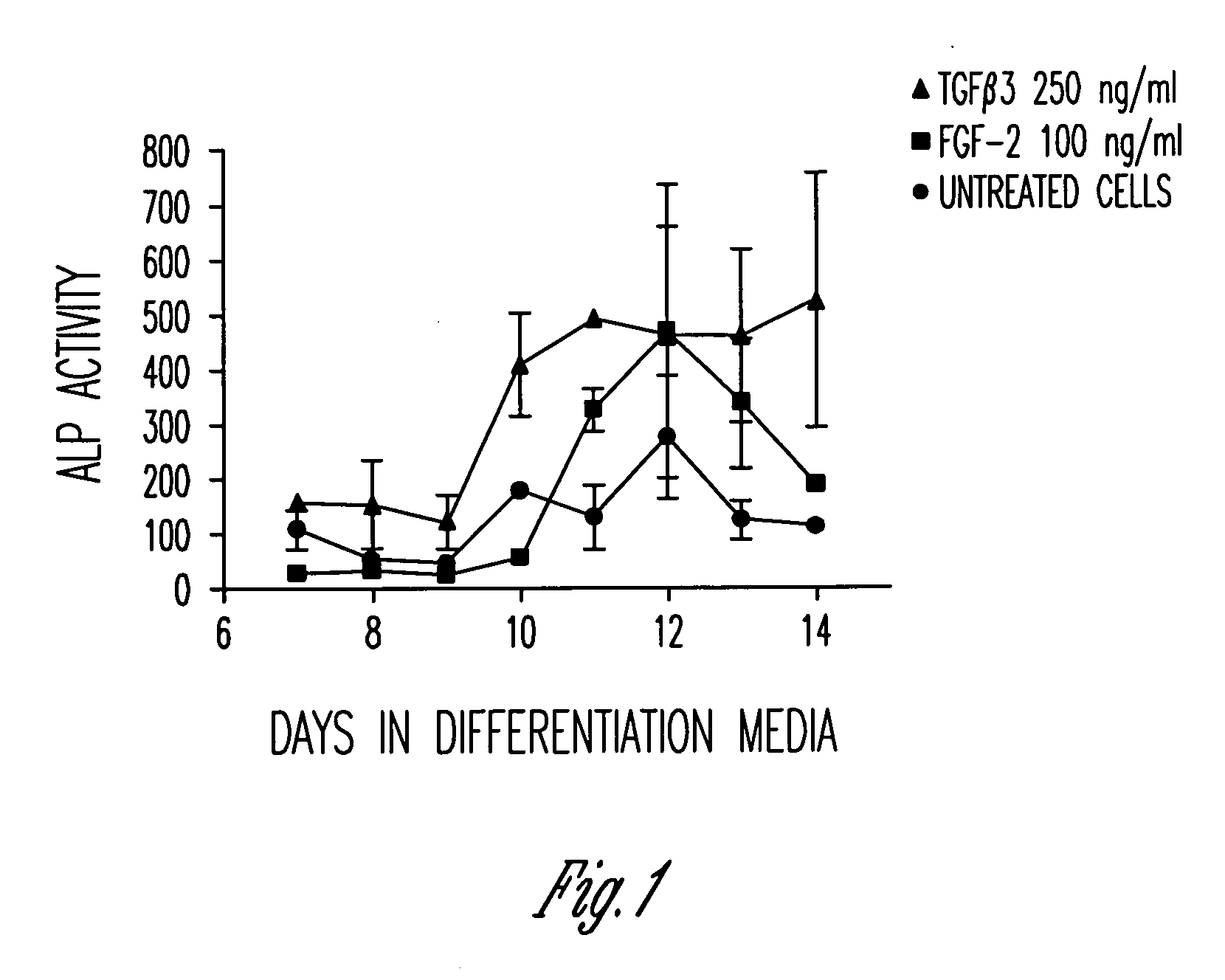
[0107]FIG. 1 shows a slight upregulation in alkaline phosphatase activity in the untreated mesenchymal stem cells (circles) from days 10 to 12, as expected. However, when the mesenchymal stem cells were pretreated with TGFβ3 (triangles) or FGF-2 (squares) for 1 hour, followed by removal of the agents and culture in differentiation media, the level of alkaline phosphatase activity increased significantly 2-3 fold.

[0108]These data indicate that just a 1 hour treatment of mesenchymal stem cells with either TGFβ3 or FGF-2 on day 0 can impact osteoblast differen...


PUM

PropertyMeasurementUnit
Timeaaaaaaaaaa
Timeaaaaaaaaaa
Timeaaaaaaaaaa

Abstract

Invention embodiments described herein include methods and devices for stimulating mesenchymal stem cells in a stem cell source to differentiate into osteoblasts capable of forming bone. Devices and methods described include exposing a stem cell source, such as bone marrow aspirate, adipose tissue and / or purified allogenic stem cells, to an active agent, in a manner effective to form activated stem cells.

Description

RELATED APPLICATIONS[0001]This patent application claims the benefit of the filing date of U.S. Provisional Patent Application Ser. No. 61 / 110,096, filed Oct. 31, 2008, and entitled, “DEVICE FOR ACTIVATING BONE MARROW ASPIRATE USING EX VIVO STIMULATION BY GROWTH FACTORS FOR IMPROVED BONE FORMATION”, and of U.S. Provisional Patent Application Ser. No. 61 / 152,335, filed Feb. 13, 2009, and entitled, “METHOD AND DEVICE FOR FORMING A BONE MARROW ASPIRATE PRODUCT”, the contents of which are incorporated herein by reference in their entirety.FIELD[0002]Inventive subject matter described herein relates to devices and methods for activating stem cells, including activating stem cells in bone marrow aspirate using ex vivo stimulation. The inventive subject matter also relates to implants containing such activated stem cells.BACKGROUND OF THE INVENTION[0003]In order to provide for maximum bone formation, it is desirable to transplant cells that already exhibit an osteoblastic phenotype, becaus...

Claims


Application Information

Patent Timeline
IPC IPC(8): A61K35/12A61P19/00C12M1/00C12M1/24
CPCA61L27/12A61L27/18A61L27/20A61L27/3834A61L27/3847C12N5/0663A61L27/46A61L27/3895C08L67/04C08L1/02A61P1/02A61P19/00A61P19/02A61F2/28A61L27/38A61L27/3821A61L27/44A61K35/32C12N5/0654C12N2501/115C12N2501/135C12N2501/15C12N2501/155C12N2506/1346
Inventor HANS, MEREDITHBUECHTER, DOUGGRUSKIN, ELLIOTTHORNSBY, STEPHENBROWN, MELISSA
Owner DEPUY SYNTHES PROD INC
Learn More