
System and Method For Ghost Magnetic Resonance Imaging

a magnetic resonance imaging and system technology, applied in the field of magnetic resonance imaging systems and methods, can solve the problems of reducing renal function, allergic reaction, and persisting risks of angiography

Active Publication Date: 2010-06-03
NORTHSHORE UNIV HEALTHSYST
View PDF3 Cites 13 Cited by
  • Summary
  • Abstract
  • Description
  • Claims
  • Application Information

AI Technical Summary

Benefits of technology

[0016]The present invention overcomes the aforementioned drawbacks by providing a system and method for MR imaging of a subject indirectly. Contrary to the widely held notions that ghosts are artifacts that int

Problems solved by technology

DSA has a high specificity and sensitivity, but despite advances in DSA and catheter technology, risks associated with angiography persist and include allergic reaction, reduced renal function, and complications related to the arteriotomy and intravascular catheter manipulation.
Because of its invasiveness and risk, it is unsuitable as a routine screening test for vascular disease.
If the central lines of k-space are acquired prior to the arrival of contrast, severe image artifacts can limit the diagnostic information in the image.
Thus, many MRA studies can be difficult to implement without including some degree of unwanted venous signal.
Short TR acquisition sequences severely limit the signal-to-noise ratio (SNR) of the acquired images relative to those exams in which longer TRs are possible.
The rapid acquisitions required by first pass CEMRA methods thus impose an upper limit on either spatial or temporal resolution.
Although the paramagnetic contrast agents in clinical use have excellent safety profiles, the recent discovery of nephrogenic system fibrosis (NSF) as a potential side effect of gadolinium administration has dampened enthusiasm for contrast-enhanced MRA and led to a “black box” warning from the United States Food and Drug Administration.
The risk of NSF relates to the presence of severely impaired renal function, as well as to the dose and stability of the contrast agent.
Unfortunately, peripheral MRA studies typically entail the administration of high doses of contrast agent.
Each of these non-contrast-agent-based imaging techniques has drawbacks or is not suitable for particular imaging studies.
However, time-of-flight and phase contrast methods are inadequate for major body MRA applications, such as imaging of the renal or peripheral arteries.
In addition, all of these imaging techniques are susceptible to motion artifacts.
This is especially true for image subtraction techniques, where even motion at a sub-pixel level can cause subtraction artifacts that degrade diagnostic quality.
For example, in contrast-enhanced breast MRI, subtraction artifacts routinely impede the detection of small enhancing tumors and can lead to false positive diagnoses.

Method used


Image

  • System and Method For Ghost Magnetic Resonance Imaging
  • System and Method For Ghost Magnetic Resonance Imaging
  • System and Method For Ghost Magnetic Resonance Imaging

Examples


Embodiment Construction

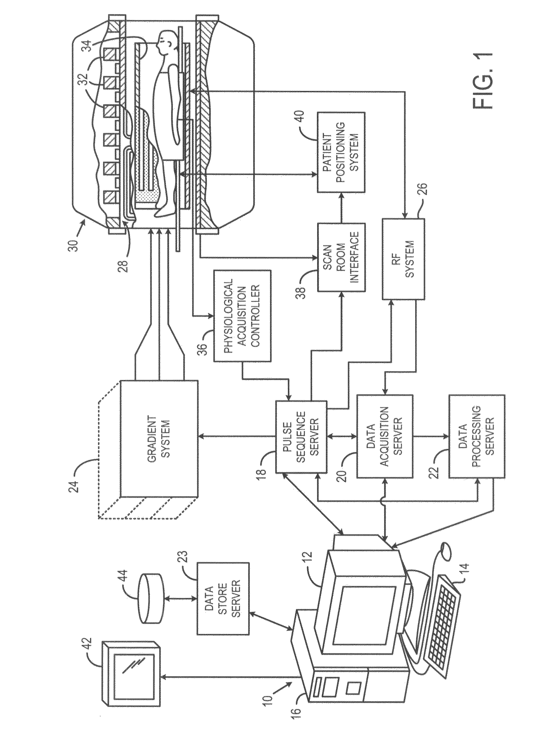
[0025]Referring to FIG. 1, the present invention is employed in an MRI system. The MRI system includes a workstation 10 having a display 12 and a keyboard 14. The workstation 10 includes a processor 16 that is a commercially available programmable machine running a commercially available operating system. The workstation 10 provides the operator interface that enables scan prescriptions to be entered into the MRI system. The workstation 10 is coupled to four servers including a pulse sequence server 18, a data acquisition server 20, a data processing server 22, and a data store server 23. The workstation 10 and each server 18, 20, 22 and 23 are connected to communicate with each other.

[0026]The pulse sequence server 18 functions in response to instructions downloaded from the workstation 10 to operate a gradient system 24 and an RF system 26. Gradient waveforms necessary to perform the prescribed scan are produced and applied to the gradient system 24 that excites gradient coils in ...


PUM


Abstract

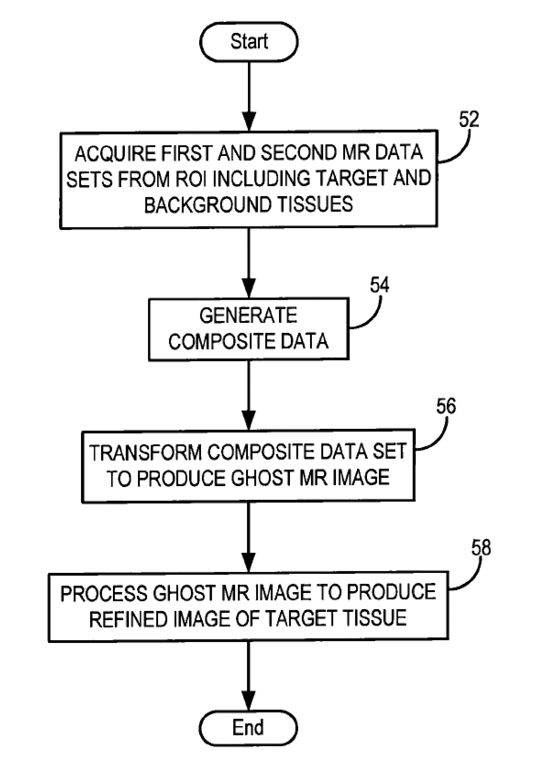
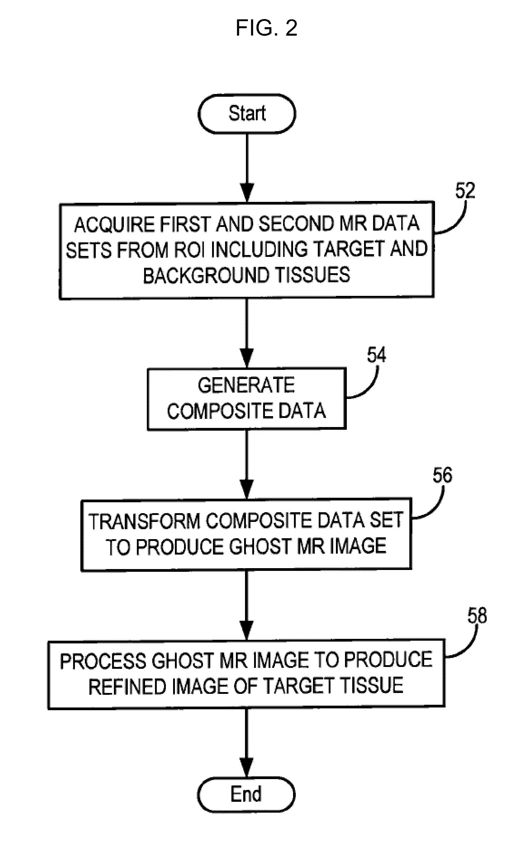
A system and method enables the creation of medical images using data related to ghost artifacts. The method thus allows components of an imaged subject to be segmented based on state changes in the components that lead to the controlled production of ghost artifacts. This is achieved in MR by performed a pulse sequence so that multiple sets of MR data are acquired in which the signals from a target tissue vary across the data sets while the signals from a background tissue do not vary across the data sets. A composite data set is generated by populating selected k-space lines of the composite data set with information from a first MR data set and populating the remaining k-space lines of the composite data set with information from a second MR data set. An MR image is then reconstructed from the composite data set. The MR image contains ghost artifacts that faithfully reproduce the 2D or 3D anatomic detail of the target tissues without signal contributions from the background tissues, allowing for background-suppressed or segmented MR images of a target tissue without the need for image subtraction.

Description

CROSS-REFERENCE TO RELATED APPLICATIONS[0001]This application claims the benefit of U.S. Provisional patent application Ser. No. 61 / 083,627, filed on Jul. 25, 2008, and U.S. Provisional patent application Ser. No. 61 / 102,197, filed on Oct. 6, 2008, both entitled “SYSTEM AND METHOD FOR GHOST MAGNETIC RESONANCE IMAGING.FIELD OF THE INVENTION[0002]This invention relates generally to systems and methods for magnetic resonance imaging (“MRI”) and, more particularly, to a system and method for utilizing ghost artifacts to reconstruct a medical image of a subject.BACKGROUND OF THE INVENTION[0003]When a substance such as human tissue is subjected to a uniform magnetic field (polarizing field B0), the individual magnetic moments of the excited nuclei in the tissue attempt to align with this polarizing field, but precess about it in random order at their characteristic Larmor frequency. If the substance, or tissue, is subjected to a magnetic field (excitation field B1) that is in the x-y plan...

Claims


Application Information

Patent Timeline
IPC IPC(8): G01R33/48
CPCG01R33/5635G01R33/4838
Inventor EDELMAN, ROBERT R.KOKTZOGLOU, IOANNIS
Owner NORTHSHORE UNIV HEALTHSYST
Learn More