Medical Image Processing

Inactive Publication Date: 2010-06-24
D V P TECH
View PDF10 Cites 166 Cited by
  • Summary
  • Abstract
  • Description
  • Claims
  • Application Information

AI Technical Summary

Benefits of technology

[0018]A broad aspect of some embodiments of the invention relates to image processing which increases visibility of a physiologically important region of a field of view in a medical image. In an exemplary embodiment of the invention, the image is acquired using white light. Optionally, the image can be an internal body image acquired via an intrabody camera, such as using an endoscope, laparoscope, colonoscope, swallowed camera or oth

Problems solved by technology

Unfortunately, normal tissue and adjacent diseased tissue may often appear similar.
This can make it difficult for medical personnel to select an informative sample based on visual analysis.
Described methods are suitable for use in analysis of grey values and are not applicable to RGB color data.
. . the white light illumination fo

Method used


Image

  • Medical Image Processing
  • Medical Image Processing
  • Medical Image Processing

Examples


example 1

Detection of Ectopic Abnormalities

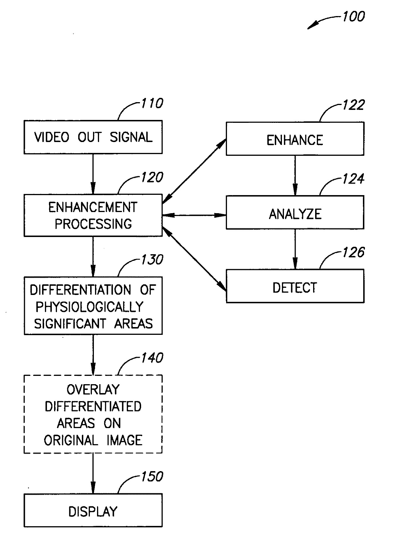
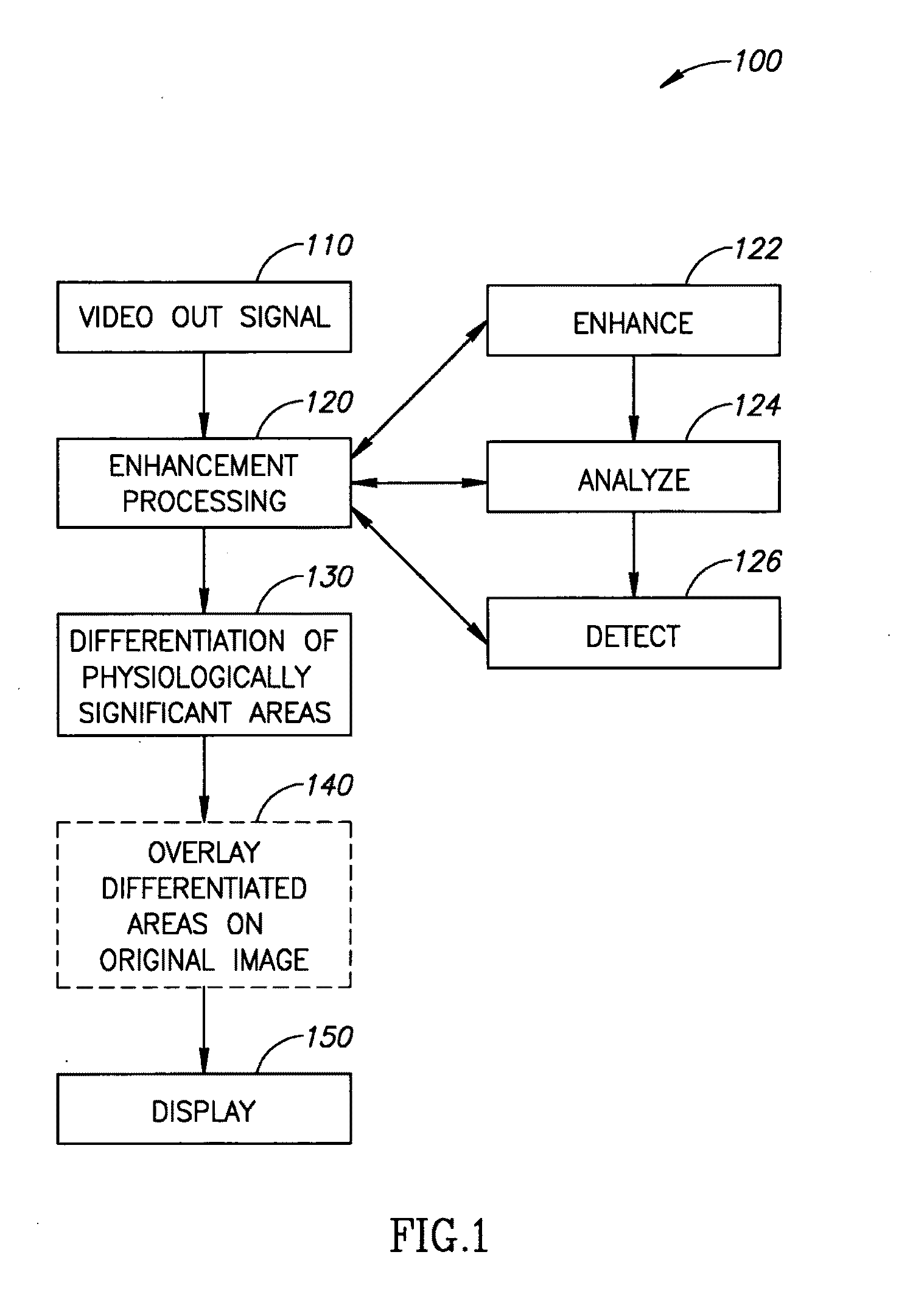
[0285]FIGS. 9, 10 and 11 illustrate application of an exemplary enhancement processing method 120 to detection and / or analysis of ectopic skin lesions.

[0286]FIGS. 9A; 9B; 9C; 9D; 9E and 9F are a series of images illustrating a sequence of intermediate results of a high resolution mole / lesion detection method which relies upon an exemplary embodiment of enhancement processing 120. All panels in this and subsequent figures are presented here as grayscale images, although the actual image capture and analysis employs RGB images. The sequence of images follows exemplary image processing approach 1 (300) as detailed hereinabove and depicted in FIG. 3.

[0287]FIG. 9A depicts the “G” channel of an RGB image resulting from PRP 312.

[0288]FIG. 9B depicts the same field of view with the PCF (R−G) applied to produce PCFI 320.

[0289]FIG. 9C depicts the same field of view segmented with PCF (R−B) applied, a bi-modal histogram H distribution (350) and thresholding T ...

example 2

Exemplary Embodiment of Invention Compared to Autofluorescence in Detection of Flat Adenoma (Tumor)

[0336]FIGS. 12A, 12B, 12C, 12D, 12E, 12F, 12G and 12H illustrate that an exemplary enhancement processing 120 algorithm according to embodiments of the invention can serve in place of auto-fluorescence detection for flat adenoma. Flat adenoma (e.g., in colon tissue) presents special problems in detection because it typically lies slightly below the surface. Use of Red light is often inappropriate as it penetrates too deeply. Use of blue light is often problematic because it does not penetrate sufficiently.

[0337]FIG. 12A is an image acquired using an endoscope and auto florescence detection of a field of view including a flat adenoma. The flat adenoma is discernible in the central part of the image as a large dark region.

[0338]FIG. 12B is an un-enhanced RGB image of the same field of view as FIG. 12A. As expected, the flat adenocarcinoma is not discernible.

[0339]FIGS. 12C and 12D depict...

example 3

Exemplary Embodiment of Invention Compared to Narrow Band Imaging (NBI) in Detection of Colorectal Lesions

[0345]FIGS. 13A, 13B, 13C, 13D, 13E and 13F illustrate that an exemplary enhancement processing 120 algorithm according to embodiments of the invention can serve in place of auto-fluorescence detection for adenoma. In an exemplary embodiment of the invention, the lesion being searched for is a view of a faint (redish) lesion with loss of capillary pattern, and of diameter 7 mm, in the normal sigmoid colon.

[0346]FIG. 13A is an unenhanced image of a flat adenoma acquired with a narrow band infrared (NBI) endoscope. The colorectal lesion is barely discernible near the center of the field of view.

[0347]FIG. 13B is an RGB image of the same field of view as FIG. 13A with no enhancement processing applied. The colorectal lesion is not discernible.

[0348]FIG. 13C depicts the Lab a-component of the same field of view as FIG. 13A after application of an ABPF according to an exemplary embod...


PUM


Abstract

A method of image processing, including:
  • (a) calculating at least one pixel color feature (PCF) value for each pixel in a color medical image to generate a set of PCF data; and
  • (b) filtering the PCF data with at least one spatial adaptive bandpass filter (ABPF) to sort the pixels into physiologically significant regions;
    • wherein the at least one PCF value for at least one pixel depends on at least 2 color components of the medical image.

Description

RELATED APPLICATIONS[0001]The present application claims the benefit under 35 USC 119(e) of U.S. Provisional Application No. 60 / 715,572 filed on Sep. 12, 2005 entitled “BODY PHOTOGRAPHIC IMAGING, REFERENCING AND ANALYSIS FOR MEDICAL AND AESTHETIC APPLICATIONS”; and of U.S. provisional application Ser. No. 60 / 825,309, filed on even date as this application with same inventors and same title and including a set of color figures, the disclosures of which is incorporated herein by reference. These color figures are also being provided herewith to the PCT receiving office (IB) and are to be considered a part of the application as filed.FIELD OF THE INVENTION[0002]The present invention relates to systems, apparatus and / or methods for image processing, especially medical image processing.BACKGROUND OF THE INVENTION[0003]In medical procedures it is common for medical personnel to select a sample (e.g. biopsy) based on tissue appearance. For external tissue (e.g. skin) selection may be based...

Claims


Application Information

Patent Timeline
IPC IPC(8): G06K9/62G06V10/56
CPCG06K9/00369G06K9/4652G06K2209/05G06T7/0012G06T2207/30088G06T7/408G06T2207/10068G06T2207/20012G06T2207/30028G06T7/0081G06T7/11G06T7/90G06V40/103G06V10/56G06V2201/03A61B5/0077A61B5/1032A61B5/444A61B5/445A61B5/725G06T2207/30092
Inventor GUISSIN, AVRAHAM RAMILAVI, EITAN
Owner D V P TECH
Why Eureka
Try Eureka