Gene expression signature for classification of cancers

a gene expression and classification technology, applied in the field of methods for classification and identification of cancers, can solve the problem that the diagnostic accuracy of tumor markers has not yet been defined

Inactive Publication Date: 2010-07-15
TEL HASHOMER MEDICAL RES INFRASTRUCTURE & SERVICES +1
View PDF1 Cites 55 Cited by
  • Summary
  • Abstract
  • Description
  • Claims
  • Application Information

AI Technical Summary

Benefits of technology

[0012]According to another aspect, the present invention provides a method of classifying a cancer or hyperplasia, the method comprising: obtaining a biological sample from a subject; measuring the relative abundance in said sample of nucleic acid sequences selected from the group consisting of SEQ ID

Problems solved by technology

The concept of CUP comes from the limitation of present methods to identify cancer origin, despite an often complicated and costly process which can significantly delay proper treatment of such patients.
These studies used machine-learning methods that a

Method used


Image

  • Gene expression signature for classification of cancers
  • Gene expression signature for classification of cancers
  • Gene expression signature for classification of cancers

Examples


example 1

Samples and Profiling

[0199]Since formalin-fixed paraffin-embedded (FFPE) archival samples are an important source for tumor material, we developed a method for extracting RNA from FFPE blocks which preserves the microRNA fraction. We compared RNA extracted from fresh-frozen, formalin-fixed, or FFPE samples, and demonstrated that the RNA quantity and quality was similar for all preservation methods. Furthermore, the microRNA profile was stable in FFPE samples for as long as 11 years of storage.

[0200]MicroRNA profiling was performed on Rosetta Genomics' miRdicator™ microarrays19, containing probes for all microRNA in miRBase (version 9)3.

[0201]333 FFPE samples and 3 fresh-frozen samples were collected and profiled, including 205 primary tumors and 131 metastatic tumors, representing 22 different tumor origins or “classes” (see Table 1 for a summary of samples). Tumor percentage was at least 50% for more than 90% of the samples. 83 of the samples (approximately 25% of each class) were ...

example 2

Comparison of Primary and Metastatic Tumors

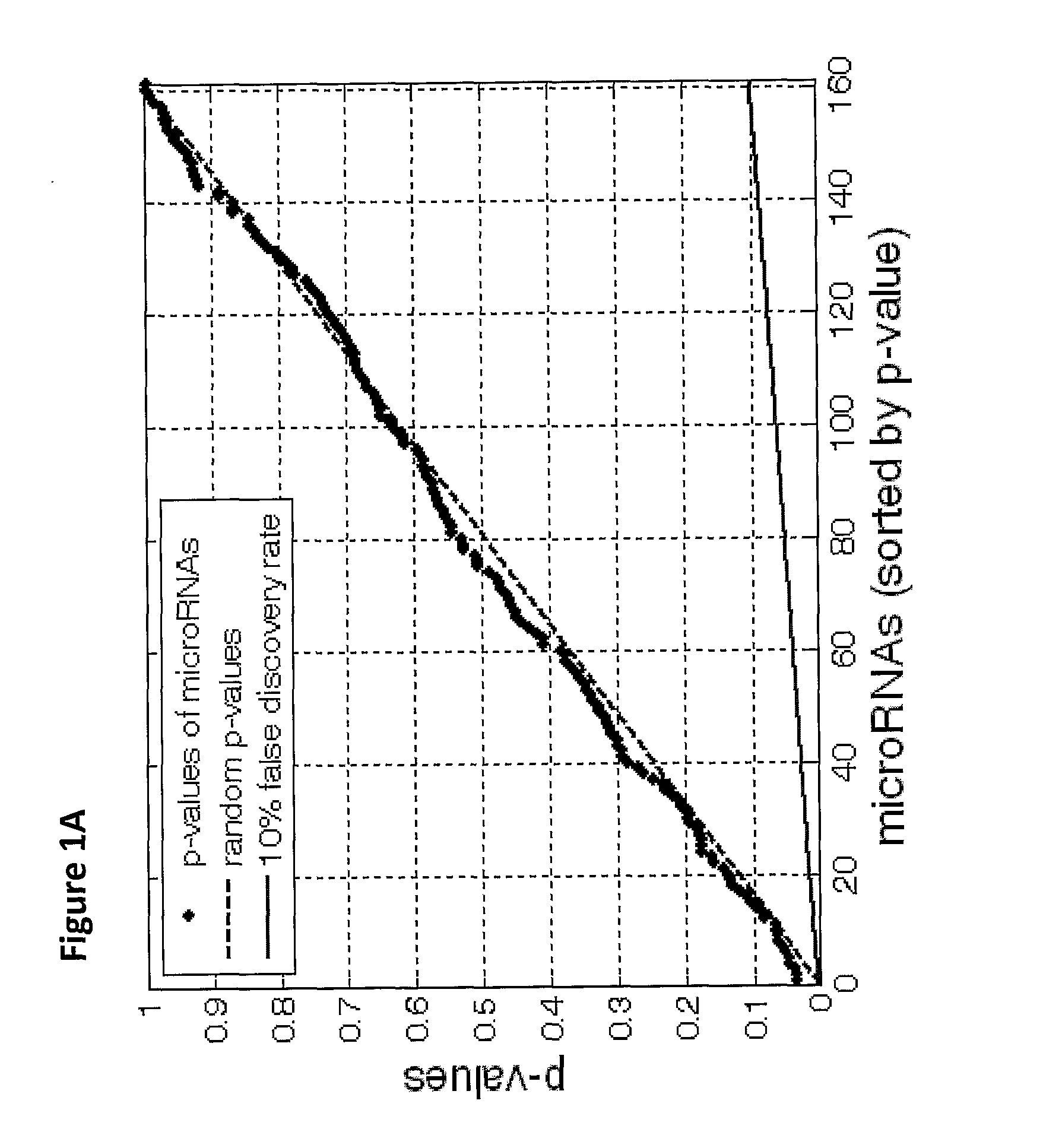
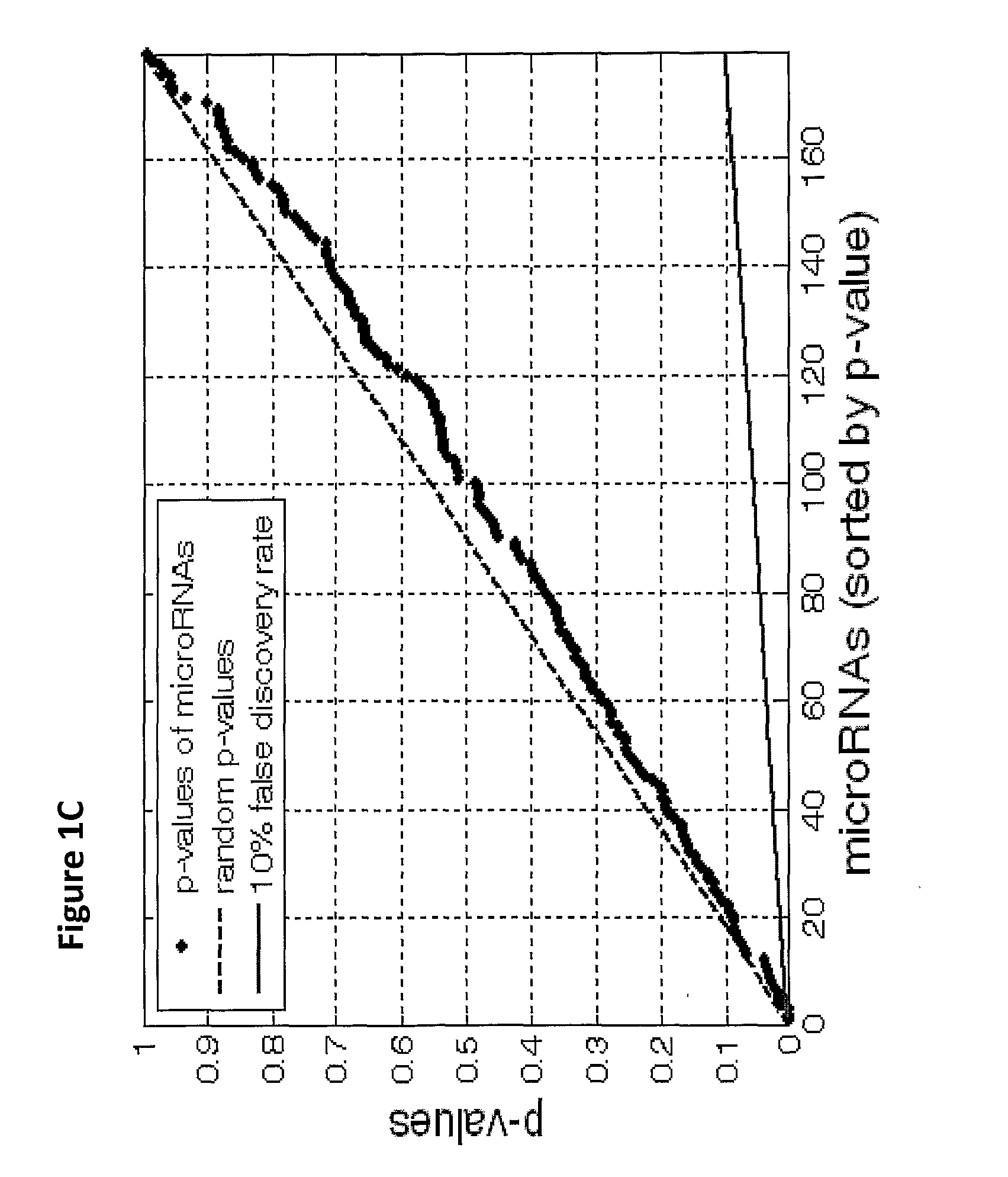
[0202]Due to the difficulty of obtaining sufficient numbers of metastatic samples, this study has relied on primary tumors to augment the sample set. Differences in expression profiles between primary and metastatic samples can be expected because of underlying biological differences in the tumors, or because of contamination from neighboring tissues. Such effects can hinder the performance of tumor classifiers on metastatic samples.

[0203]For most tissue origins, such as breast cancer or colon cancer (FIGS. 1A, B), no significant differences between primary and metastatic tumors were found. In other cases, a small set of microRNAs were differentially expressed. For example, in comparing stomach primary tumor samples to samples of stomach metastases to the lymph node, 3 microRNAs were significantly differentially expressed (FIGS. 1C, D). Hsa-miR-143 (SEQ ID NO: 99), characteristic of epithelial layers5, and hsa-miR-133a (SEQ ID NO: 97), whic...

example 3

Decision-Tree Classification Algorithm

[0204]A tumor classifier was built using the microRNA expression levels by applying a binary tree classification scheme (FIG. 2). This framework is set up to utilize the specificity of microRNAs in tissue differentiation and embryogenesis: different microRNAs are involved in various stages of tissue specification, and are used by the algorithm at different decision points or “nodes”. The tree breaks up the complex multi-tissue classification problem into a set of simpler binary decisions. At each node, classes which branch out earlier in the tree are not considered, reducing interference from irrelevant samples and further simplifying the decision (FIG. 3A). The decision at each node can then be accomplished using only a small number of microRNA biomarkers, which have well-defined roles in the classification (Table 2). The structure of the binary tree was based on a hierarchy of tissue development and morphological similarity18, which was modifi...


PUM

PropertyMeasurementUnit
Fractionaaaaaaaaaa

Abstract

The present invention provides a process for classification of cancers and tissues of origin through the analysis of the expression patterns of specific microRNAs and nucleic acid molecules relating thereto. Classification according to a microRNA tree-based expression framework allows optimization of treatment, and determination of specific therapy.

Description

FIELD OF THE INVENTION[0001]The present invention relates to methods for classification of cancers and the identification of their tissues of origin. Specifically the invention relates to microRNA molecules associated with specific cancers, as well as various nucleic acid molecules relating thereto or derived therefrom.BACKGROUND OF THE INVENTION[0002]microRNAs are a novel class of non-coding, regulatory RNA genes1-3 which are involved in oncogenesis4 and show remarkable tissue-specificity5-7. They have emerged as highly tissue-specific biomarkers2,5,6 postulated to play important roles in encoding developmental decisions of differentiation. Various studies have tied microRNAs to the development of specific malignancies4.[0003]Metastatic cancer of unknown primary (CUP) accounts for 3-5% of all new cancer cases, and as a group is usually a very aggressive disease with a poor prognosis10. The concept of CUP comes from the limitation of present methods to identify cancer origin, despit...

Claims


Application Information

Patent Timeline
IPC IPC(8): C12Q1/68C12N15/11C12N15/113
CPCC12N15/111C12N15/113C12N2310/14C12Q2600/178C12Q1/6886C12Q2600/112C12Q2600/158C12N2320/12
Inventor AHARONOV, RANITROSENFELD, NITZANROSENWALD, SHAIBARSHACK, IRIS
Owner TEL HASHOMER MEDICAL RES INFRASTRUCTURE & SERVICES
Why Eureka
Try Eureka