Optical Methods to Intraoperatively Detect Positive Prostate and Kidney Cancer Margins

a prostate and kidney cancer and intraoperative technology, applied in the field of in vivo cancer detection, can solve the problems of limiting tactile sensation, difficult procedure with significant time pressure to limit renal insult, and difficult to perform traditional diagnostic needle biopsy of small renal masses, etc., to reduce positive surgical margins and prostate cancer recurrence.

Inactive Publication Date: 2010-08-05
BOARD OF RGT THE UNIV OF TEXAS SYST
View PDF8 Cites 35 Cited by
  • Summary
  • Abstract
  • Description
  • Claims
  • Application Information

AI Technical Summary

Benefits of technology

[0017]The present invention includes a tri-modal optical spectroscopy using light-scattering reflectance, time-resolved auto-fluorescence and diffuse near infrared spectroscopy (NIRS). The present invention enables surgeons to intra-operatively demarcate prostate or kidney cancer over the entire resected prostate or kidney so as to significantly reduce positive surgical margins and prostate cancer recurrence after surgery

Problems solved by technology

However, the laparoscopic approach greatly limits tactile sensation during the procedure.
However, traditional diagnostic needle biopsy of small renal masses is not commonly performed due to its consistent and clinically unacceptable false negative rate.
However, both open and laparoscopic partial nephrectomies usually require renal ischemia to excise the tumors safely creating a unique, technically challenging procedure with significant time pressures to limit the renal insult.
Of course, complete excision with a negative margin is required to minimize the risk of tumor recurrence but unfortunately, the surgeon cannot evaluate the surgical specimen until the procedure is completed and specimen extracted.
Although intra-operative frozen section pathologic analysis of a few select tissue fragments from the resected specimen or renal parenchyma can be obtained, it is time consuming risking renal ischemic injury and costly.
Furthermore, concrete conclusions based on such samples are unreliable, as they do not reflect the entire surgical margin status.
For early clinically localized kidney tumors, it is not possible to reliably confirm benign versus malignant disease or identify the deep parenchymal tumor margin during surgery.
For early clinically localized prostate cancer, it is also not possible to visually identify the tumor during surgery, either within the prostate or at its capsular margin.
However, most of these techniques mainly target luminal malignancies, such as cervical, colon, and esophageal cancers.
However, for all of the technologies mentioned above, a surgeon cannot evaluate the surgical specimen until the procedure is completed and prostate extracted.
Though intraoperative frozen section pathologic analysis of a few select tissue fragments from the prostate or surgical site can be obtained, it is time consuming and costly.
Concrete conclusions based on such samples are unreliable, as they do not reflect the entire surgical margin status.

Method used


Image

  • Optical Methods to Intraoperatively Detect Positive Prostate and Kidney Cancer Margins
  • Optical Methods to Intraoperatively Detect Positive Prostate and Kidney Cancer Margins
  • Optical Methods to Intraoperatively Detect Positive Prostate and Kidney Cancer Margins

Examples


example

Prostate Cancer

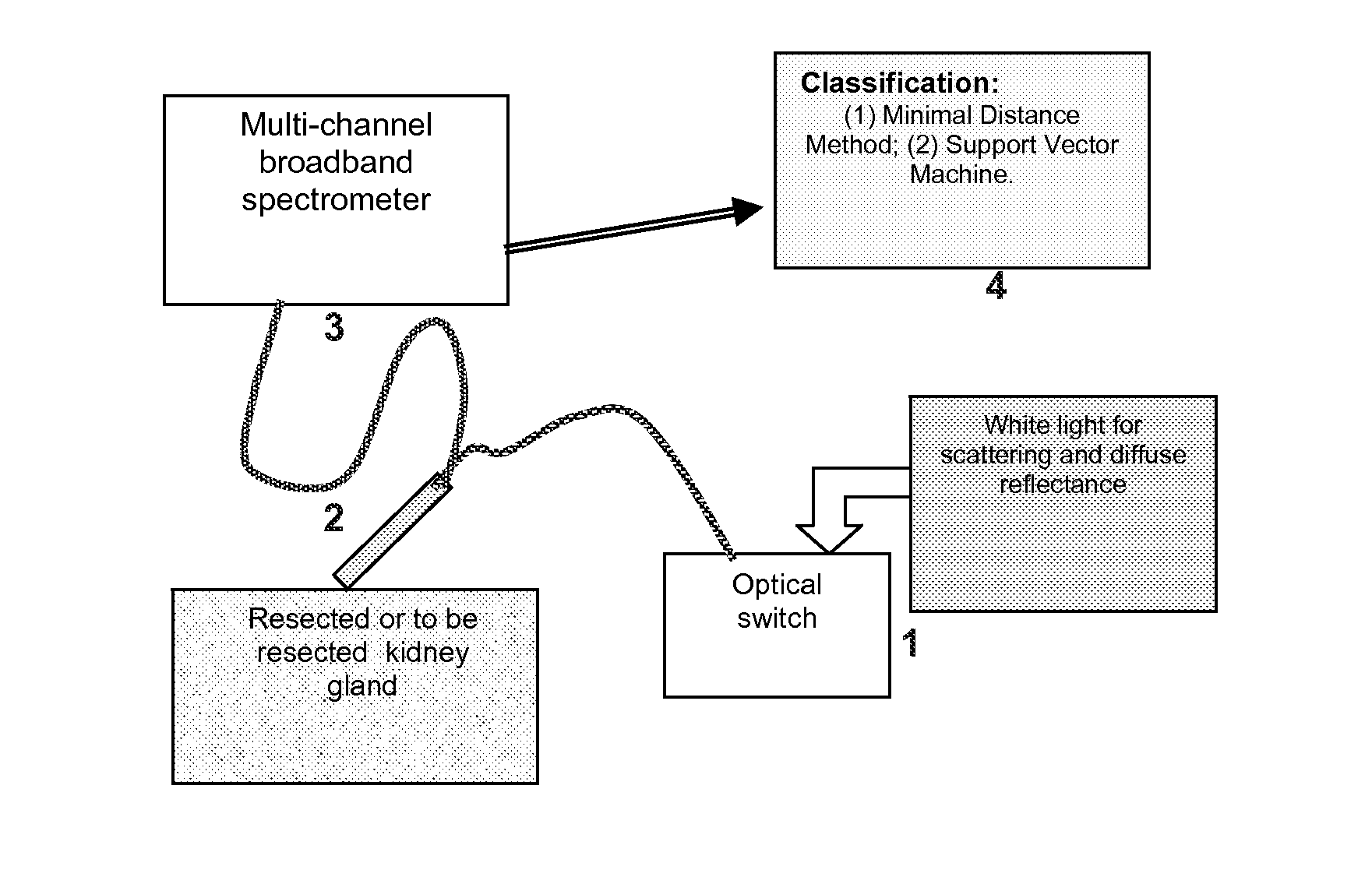
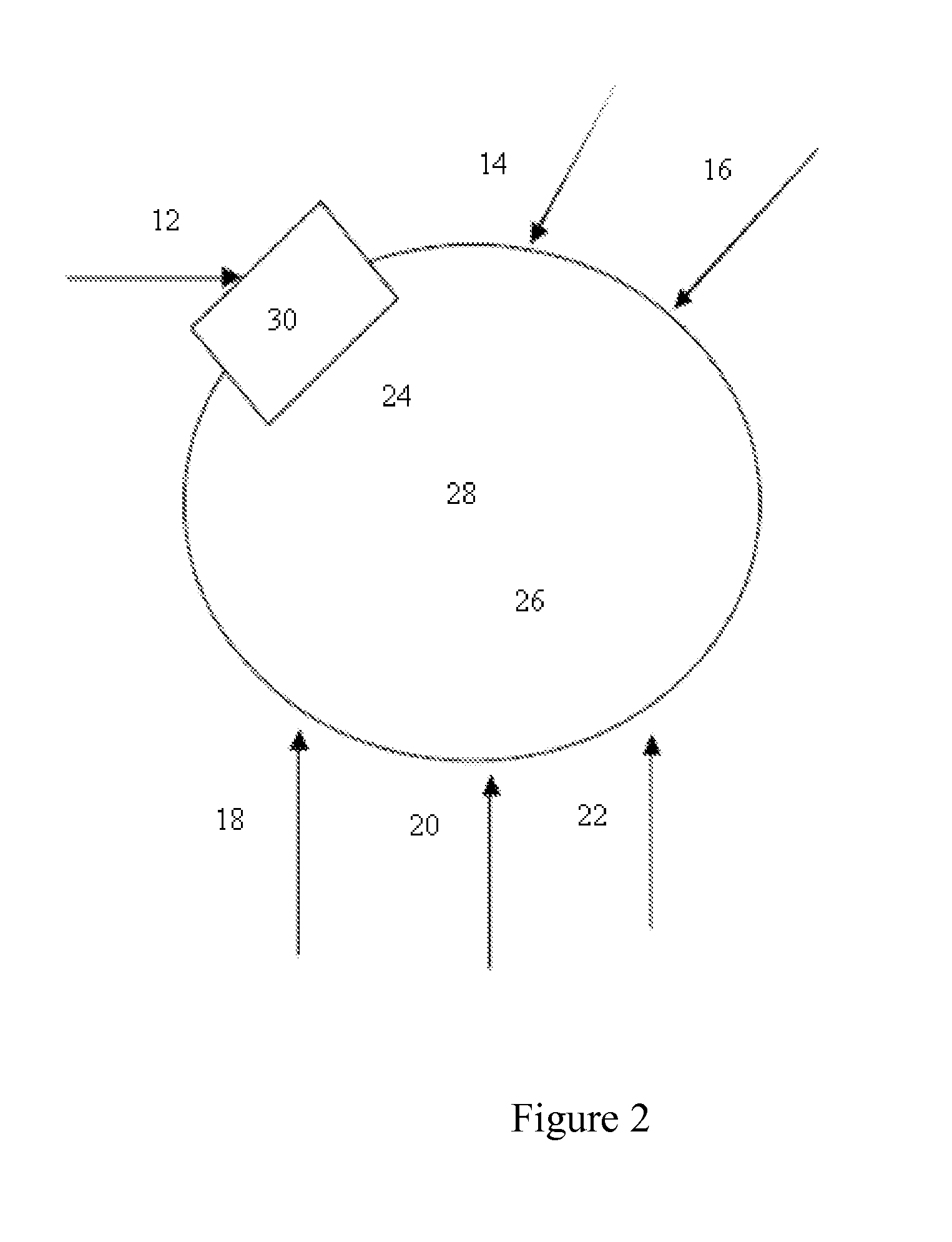
[0121]The present invention aims to develop a tri-modal optical spectroscopic approach that integrates three different techniques: light-scattering reflectance, time-resolved fluorescence, and diffuse near infrared spectroscopy (NIRS). The overall goal is that tri-modal optical spectroscopy (i.e. light-scattering reflectance, time-resolved auto-fluorescence and near infrared spectroscopy) enables surgeons to intraoperatively demarcate prostate cancer over the entire resected prostate so as to significantly reduce positive surgical margins and prostate cancer recurrence after surgery.

[0122]Near Infrared Spectroscopy (NIRS) and Optical Properties of Human Prostate: In spite of a significant amount of work have been conducted to quantify the optical and physiological properties of human breast and breast cancer using the non-invasive NIRS approach, limited information is available for the optical parameters of human prostate. With the help of MRI, it was reported that 9 ...


PUM


Abstract

The present invention includes using optical spectroscopy as an innovative technique for ex-vivo demonstration of renal and prostate tumors. The apparatus and methods disclosed herein demonstrate the ability of optical spectroscopy to reliably differentiate tumor from normal tissue in renal specimens.

Description

BACKGROUND OF THE INVENTION[0001]Without limiting the scope of the invention, its background is described in connection with the field of in vivo cancer detection, more particularly, optical methods to intra-operatively detect cancer margins in prostate and kidney cancers.[0002]Clinically localized prostate cancer is generally treated with either radiation therapy or surgery. Surgical treatment is currently undergoing a significant revolution; that is laparoscopic radical prostatectomy (LRP). This procedure permits complete removal of the prostate and seminal vesicles while minimizing pain and recovery. However, the laparoscopic approach greatly limits tactile sensation during the procedure. This is particularly true with robot-assisted LRP where no tactile feedback is available forcing the surgeon to rely solely on visual cues.[0003]Kidney cancer is increasingly detected at very early stages due to the wide spread use of axial imaging for a variety of complaints. Associated with th...

Claims


Application Information

Patent Timeline
Patent Type & Authority Applications(United States)
IPC IPC(8): A61B6/12G06N5/02
CPCA61B5/0071A61B5/4381A61B5/0086A61B5/0075
Inventor LIU, HANLICADEDDU, JEFFREY A.PESWANI, DISHA L.
Owner BOARD OF RGT THE UNIV OF TEXAS SYST
Why Eureka
Try Eureka