Methods and kits for diagnosis of cancer and prediction of therapeutic value

a cancer and kit technology, applied in the field of methods and kits for cancer diagnosis and prediction of therapeutic value, can solve the problems of limited treatment options for prostate cancer patients, many cancers had spread beyond the boundaries of operation, and limited systemic therapy, so as to improve the statistical significance of a clinical trial, reduce the number of patients required, and improve the ability of the trial

Active Publication Date: 2010-08-19
CORNELL UNIVERSITY
View PDF105 Cites 44 Cited by
  • Summary
  • Abstract
  • Description
  • Claims
  • Application Information

AI Technical Summary

Benefits of technology

[0016]In some aspects, the invention includes methods for improving statistical significance of a clinical trial. In one aspect, the invention includes methods for reducing a number of patients required for a clinical trial. In some embodiments, the methods include administering a diagnostic dose of a detectably labeled first binding agent to a plurality of patients. The detectably labeled first binding agent is capable of binding a cellular target. The patient or patients are selected based on the level of first binding agent in the patient or in a biological sample obtained from the patient. Certain patients having been administered the diagnostic

Problems solved by technology

There are currently very limited treatment options for prostate cancer patients once the cancer has metastasized (spread beyond the prostate).
Systemic therapy is primarily limited to various forms of androgen (male hormone) deprivation.
Historically, the drawback of this procedure is that many cancers had spread beyond the boundaries of the operation by the time the cancers were detected.
Patients with bulky, high-grade tumors ar

Method used


Image

  • Methods and kits for diagnosis of cancer and prediction of therapeutic value
  • Methods and kits for diagnosis of cancer and prediction of therapeutic value
  • Methods and kits for diagnosis of cancer and prediction of therapeutic value

Examples


example 1

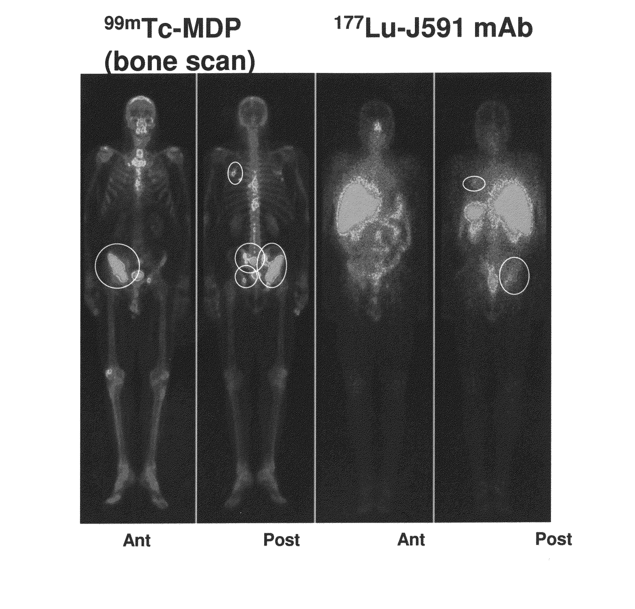
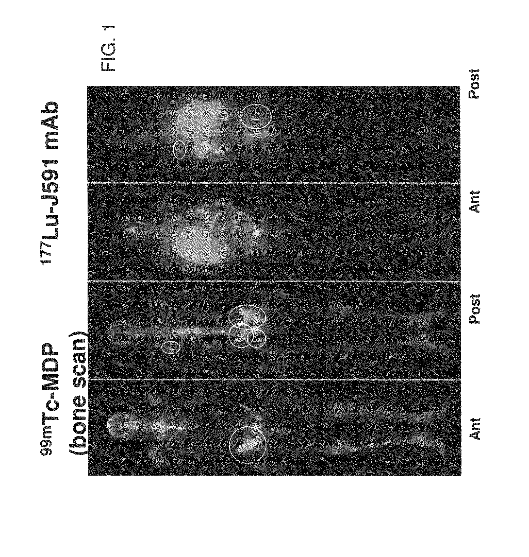
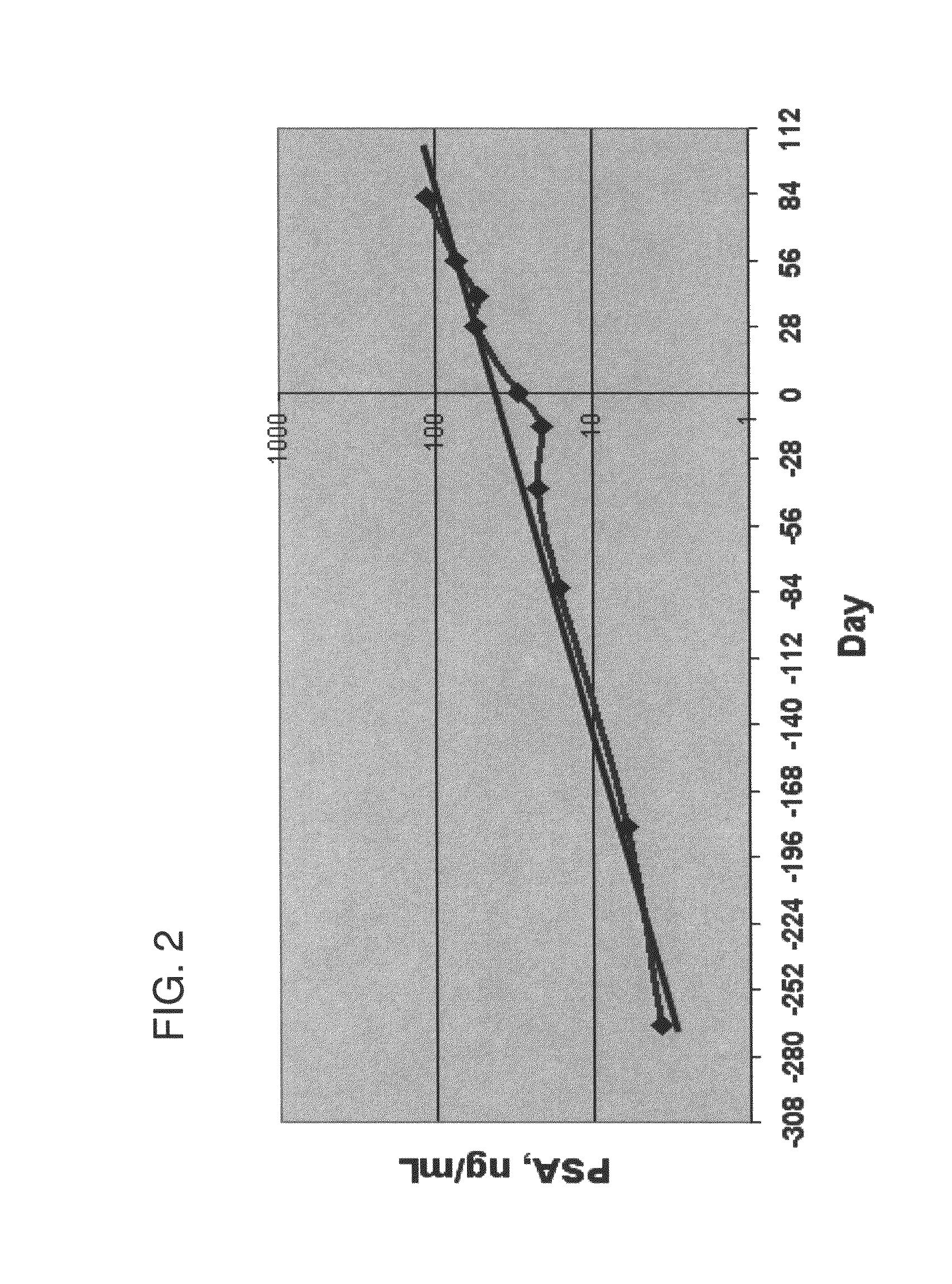
[0101]Thirty-two patients were selected for this study. The patients were administered 99mTc-MDP, for a standard bone scan, and 177Lu-J591 on different days. The study was divided into two cohorts. Cohort 1 (n=15) received a 177Lu-J591 dose of 65 mCi / m2. Cohort 2 (n=17) received a 177Lu-J591 dose 70 mCi / m2 (e.g., about the maximum tolerated dose). Approximately 3 hours after i.v. administration of 99mTc-MDP for a standard bone scan, patients underwent planar radionuclide imaging on a planar gamma camera. Radionuclide bone imaging procedures are well known in nuclear medicine and to those in the art.

[0102]Whole body images of were obtained of 177Lu-J591 and 99mTc-MDP for each patient. In addition, patients underwent either a CT scan and / or MR scan to visualize soft tissues not seen on the bone scan. The 177Lu-J591 images / scans of each patient were blindly scored / graded for targeting of the radiolabel to tumor sites 5-8 days post-treatment. The image grading system measured the intens...

example 2

[0110]One example of an alternative to the semi-quantitative 0-3+ scoring scale is to calculate a quantitative tumor targeting index (TTI). This can be done on a patient's J591 scan by measuring the count density over the most prominent tumor lesion / s divided by the pixel area of those respective lesions. The lesion count density is then corrected for background count density by doing the same calculation (count density divided by pixel area in the region of interest (ROI) using a lesion-negative area generally in the right anterior thigh). The whole body density is defined as the geometric mean of anterior and posterior imaging counts per pixel. TTI=(lesion ROI count density—background count density) / (total body count density). Tumor counts are calculated for a given tumor, over the area of the tumor:

Tcts / area=counts_per_regiontumorregion_area(px)tumor

Background counts are calculated for a given region, over the area of the region:

BGcts / area=counts_per_regionbackgroundregion_area(p...

example 3

Use of Positron Emission Tomography (PET) for Imaging

[0113]Similar to the use of planar imaging as described above, or SPECT (single photon emission computed tomography) imaging, one could also use positron emission tomography (PET) for imaging. PET imaging provides direct, quantitative data reflecting the uptake of the first binding agent. PET imaging can be done with a positron-emitting agent such as 124Iodine, 89zirconium, 86yttrium, or others positron-emitting agent known to those skilled in the art, coupled to the first binding agent. Similar to that described above, the PET-derived quantitative “standard uptake values” (SUVs) allow identification of patients whose lesions demonstrate higher uptake versus those with lower or no uptake. Patients with lesions demonstrating higher SUVs would be predicted to demonstrate higher uptake of the therapeutic second binding agent, in turn, followed by increased likelihood of therapeutic benefit. Use of a directly quantitative imaging moda...


PUM

PropertyMeasurementUnit
Molar densityaaaaaaaaaa
Capacitanceaaaaaaaaaa
Fractionaaaaaaaaaa

Abstract

A method for identifying a patient for cancer therapy can include administering a diagnostic dose of a detectably labeled first binding agent to a patient, the detectably labeled binding agent being capable of binding a molecular target. The method also includes selecting a patient for administration of a therapeutic dose of a second binding agent capable of binding a cellular target, wherein the selected patient exhibits a positive reading for the detectably labeled first binding agent. Furthermore, the method can include administering a therapeutic dose of the second binding agent to the patient.

Description

RELATED APPLICATIONS[0001]This application claims the benefit of U.S. Provisional Application No. 61 / 153,132, filed Feb. 17, 2009, which application is hereby incorporated by reference in its entirety. Reference is also made to the PCT International Application filed in the U.S. Receiving Office on Feb. 17, 2010, under Attorney Docket No. BZL-004PC, “Methods and Kits for Diagnosis of Cancer and Prediction of Therapeutic Value” by Bander et al., which also claims priority to U.S. Provisional Application No. 61 / 153,132, filed Feb. 17, 2009, and which is hereby also incorporated by reference in its entirety.FIELD OF THE INVENTION[0002]The invention relates generally to methods for identifying and / or selecting a patient for cancer therapy. The invention relates more particularly to using quantitative or semi-quantitative imaging to predict the therapeutic value of various cancer therapies.BACKGROUND OF THE INVENTION[0003]Prostate cancer is one of the most common causes of cancer deaths ...

Claims


Application Information

Patent Timeline
IPC IPC(8): A61K51/10A61K49/06A61K49/14G01N33/53A61P35/00
CPCA61K51/0482G01N2800/52A61K51/106A61P35/00
Inventor BANDER, NEILOSBORNE, JOSEPHGOLDSMITH, STANLEY J.VALLABHAJOSULA, SHANKAR
Owner CORNELL UNIVERSITY
Why Eureka
Try Eureka