
Mesenchymal stem cells, compositions, and methods for treatment of cardiac tissue damage
- Summary
- Abstract
- Description
- Claims
- Application Information
AI Technical Summary
Benefits of technology
Problems solved by technology
Method used
Image

Examples
example 1
Isolation of RS-MSCs Using PODXL and CD49f Epitope Expression
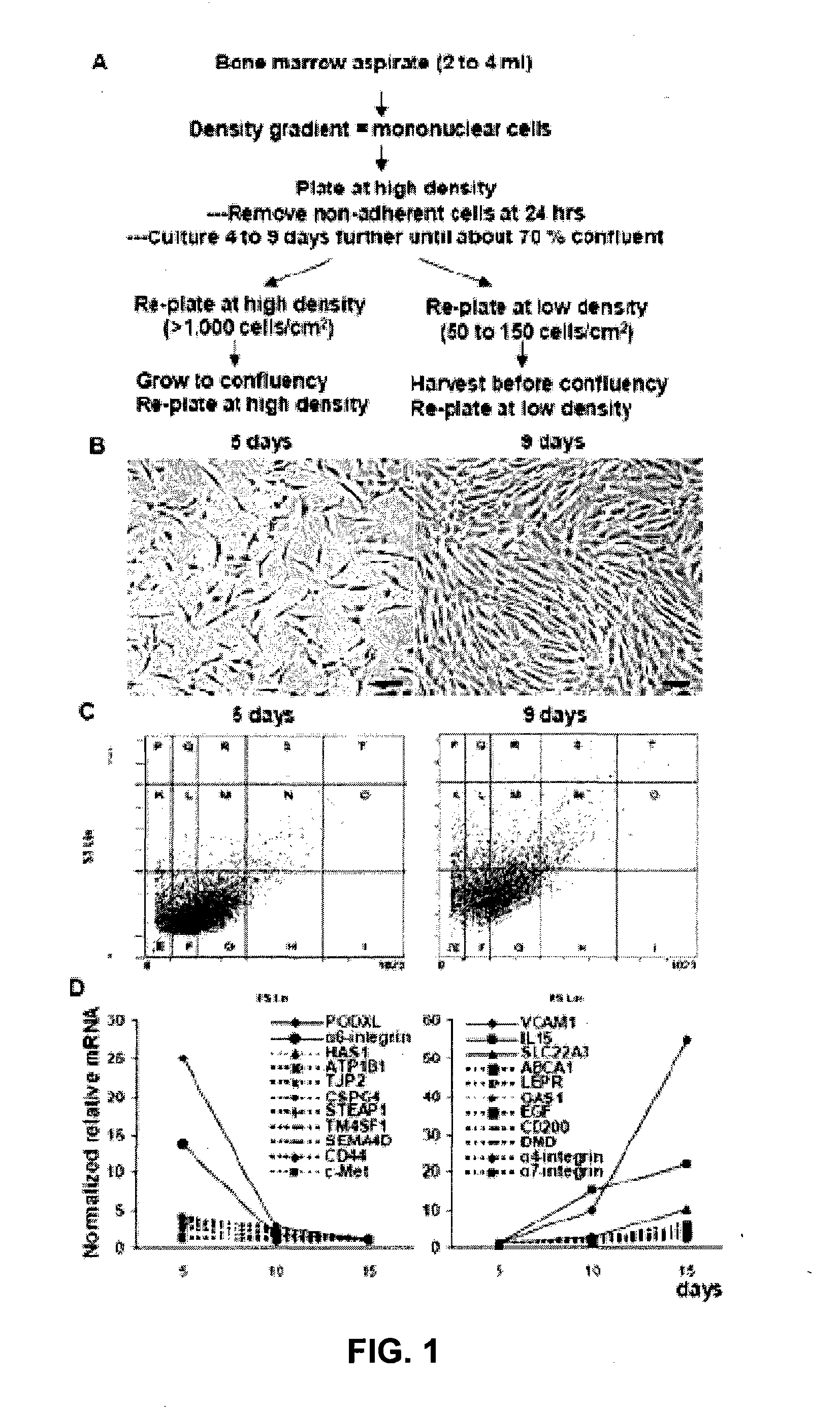
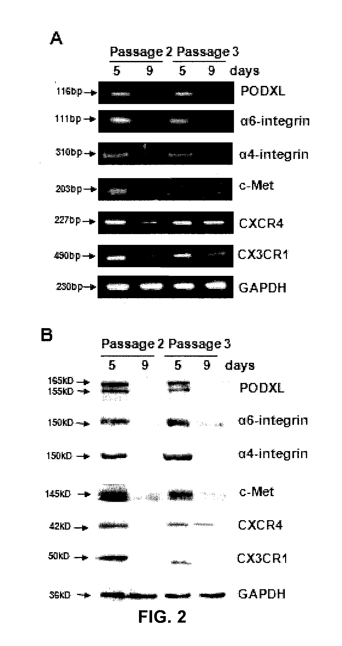
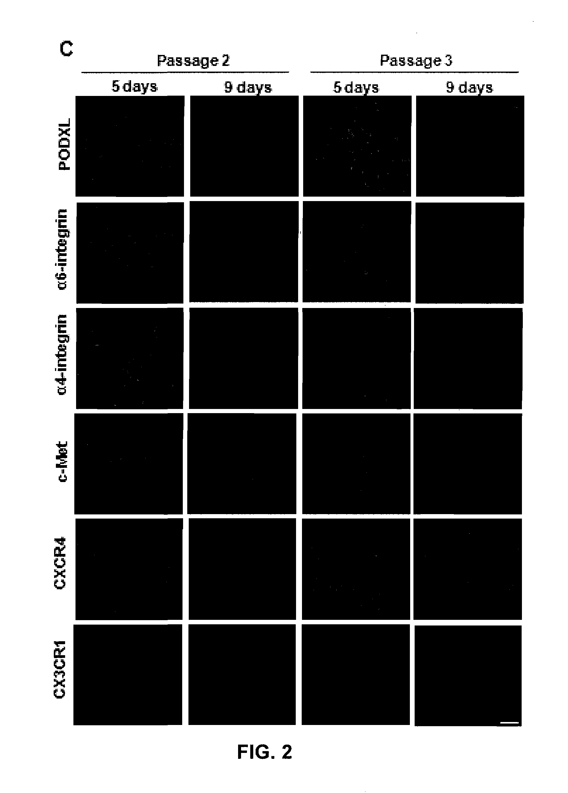
[0132]Recently, the inventors searched for antibodies to surface proteins that identify early progenitors in cultures of MSCs (FIGS. 1A and 1B). As an initial strategy, the inventors queried microarray data for changes in transcripts for surface proteins as hMSCs plated at low density and the cultures expanded. The results demonstrated that the steady-state levels of over 10 transcripts decreased (>2-fold) and an almost equal number increased (FIG. 2D). The two of the transcripts with the largest decreases coded for proteins previously shown to be linked to cell motility and tumor progression: PODXL (Furness and McNagny, 2006); and α6-integrin (CD49f) (Lipscomb and Mercurio 2005).
[0133]The inventors then used commercial antibodies to follow expression of PODXL, α6-integrin (CD49f), and several other epitopes related to cell trafficking and motility as cultures of MSCs were expanded (FIGS. 2 and 3). By the variety of assays...
example 2
[0135]More Efficient Engraftment of RS-MSCs into the Hearts of Mice Following MI
[0136]The inventors employed an improved PCR assay (see below) for human Alu to compare the engraftment of RS-MSCs (PODXLhi / CD49fhi) into mice with MIs. As indicated in FIG. 4, the RS-MSCs engrafted more efficiently into the hearts with MI. The cells also engrafted more efficiently into kidney, apparently as a result of secondary damage to the kidney, as reflected by an increase in serum creatinine levels (0.53 mg / del±0.08 SD vs 1.08±0.14 SD; n=4).
[0137]As demonstrated in FIG. 3D, cultures of MSCs from some donors remained PODXLhi / CD49fhi when plated at 100 cells / cm2 and expanded for up to 7 days. In order to conveniently obtain adequate numbers of cells, 7 day cultures enriched for RS-MSCs from such selected donors were used for subsequent experiments. Unless otherwise noted, they are referred to simply as MSCs.
example 3
[0138]Tissue Distribution of Infused hMSCs, Cancer Cells, and Human WBCs
[0139]After IV infusion into mice, hMSCs were cleared from the blood in less than 5 min (FIG. 5, Top Left). In confirmation of previous reports (Barbash et al., 2003; Gao et al., 2001b; Schrepfer et al., 2007), most of the infused cells were trapped in the lung (FIG. 5, Bottom Left). Similar observations were made with a human breast cancer cell line (MDA-MB-231) and with human WBCs, except a fraction of the human WBCs escaped trapping in the lung and were recovered in liver. hMSCs infused into the left ventricle of the heart (IC infused) were also cleared from the blood in less than 5 min (FIG. 5, Top Right), but compared to IV infusions larger numbers of hMSCs appeared in brain, heart, lung, liver, pancreas, spleen, kidney and bone marrow (FIG. 5, Bottom Right).
[0140]Alu sequences were assayed in blood, lung and seven tissues of mice for up to 4 days (100 hr) after IV infusions (FIG. 6, Top). There was no incr...
PUM
| Property | Measurement | Unit |
|---|---|---|
| Volume | aaaaa | aaaaa |
| Volume | aaaaa | aaaaa |
| Volume | aaaaa | aaaaa |
Abstract
Description
Claims
Application Information